Magnesium and Morphine in Chronic Neuropathic Pain: History
Please note this is an old version of this entry, which may differ significantly from the current revision.
Contributor:

The effectiveness of opioids in the treatment of neuropathic pain is limited. It was demonstrated that magnesium ions (Mg2+), physiological antagonists of N-methyl-D-aspartate receptor (NMDAR), increase opioid analgesia in chronic pain. Our study aimed to determine the molecular mechanism of this action. Early data indicate the cross-regulation of mu-opioid receptor (MOR) and NMDAR in pain control. Morphine acting on MOR stimulates protein kinase C (PKC), while induction of NMDAR (for example in a state of neuropathic pain) recruits protein kinase A (PKA) leading to disruption of the MOR-NMDAR complex and promoting functional changes in receptors. The level of phosphorylated NMDAR NR1 subunit (pNR1) and phosphorylated MOR (pMOR) in the periaqueductal gray matter was determined with the Western blot method. The activity of PKA and PKC was examined by standard enzyme immunoassays. Mg2+ administered alone significantly decreased the level of pNR1 and pMOR, and activity of both tested kinases. The results suggest that blocking NMDAR signaling by Mg2+ restores the MOR-NMDAR complex and thus enables morphine analgesia in neuropathic rats.

  • magnesium
  • morphine
  • N-methyl-D-aspartate receptor
  • µ-opioid receptor
  • receptor association
  • analgesia
  • neuropathic rats

1. Introduction

Despite the fact that the last few decades have brought progress in the treatment of many diseases, the relief of neuropathic pain is still one of the major challenges in medicine. The barrier in the alleviation of this type of pain is the limited efficacy of classic analgesics, including opioids [1][2][3]. A reduction in opioid analgesia appears to be associated with the increased activation of pronociceptive N-methyl-D-aspartate receptors (NMDARs) during the development of neuropathic pain [4][5].
One of the solutions is the administration of opioids in high doses [6], which unfortunately increases the risk of side effects, such as respiratory depression, opioid hyperalgesia, constipation, rapid development of tolerance, and spreading in recent years addiction risk [7][8][9][10].
Data available in the literature indicate that synthetic NMDAR antagonists not only alleviate neuropathic pain [11][12][13][14][15] but also enhance the analgesic effect of opioids [16][17][18]. It is worth noting that magnesium ions (Mg2+) are the physiological antagonists of the NMDAR channel, and their analgesic properties have been demonstrated both in preclinical [11][19][20] and clinical [21][22] studies in neuropathic pain. Mg2+ have relatively mild side effects at therapeutic doses and are considered safer and better-tolerated compounds than synthetic NMDAR antagonists. Importantly, several animal studies have reported that Mg2+ increase opioid analgesia in chronic neuropathic pain [23][24][25][26]. However, the cellular mechanism of this interaction has not been elucidated yet.
Considering the fact that µ-opioid receptor (MOR) is the primary and most widely studied mediator of opioid activity, including morphine (MRF), our investigations focus on this receptor. MOR belongs to the group of metabotropic G protein-coupled receptors (GPCRs). The signaling efficiency of this receptor is modulated and finally limited by phosphorylation of the appropriate intracellular amino acid residues. It was established that MOR phosphorylation precedes a process of receptor desensitization, uncoupling, and internalization [27]. It is noteworthy that MOR can undergo heterologous agonist-independent as well as homologous agonist-induced phosphorylation. Two protein families may phosphorylate the MOR residues: G protein-coupled receptor kinases (GRKs), as well as second messenger-dependent protein kinases including protein kinase C (PKC) and protein kinase A (PKA). It has been shown that serine 375 (Ser375) is the initiating residue in a hierarchical phosphorylation cascade [28][29]. In turn, NMDARs are ionotropic glutamate receptors, which form tetrameric complexes typically containing two essential NR1 subunits assembling with two modulatory NR2 subunits [30][31]. The function of NMDAR, including channel properties and localization at synapses, is also regulated by protein phosphorylation. It has been identified that NR1 C-terminus is phosphorylated via PKC on two serine residues (Ser890 and Ser896), while a neighboring site Ser897 is phosphorylated by PKA [32]. The C-terminus domains of NMDAR NR2 subunits (especially NR2A/B) are large, and they contain multiple amino acid sites that are phosphorylated by PKA, PKC, cyclin-dependent kinase-5 (Cdk5), calcium calmodulin-dependent kinase (CaMKII), casein kinase (CK2), Src and/or Fyn non-receptor tyrosine kinases [33]. What is important, studies have shown that activation of PKA and PKC potentiates the amplitude of NMDAR-mediated currents [34][35].
During recent years different groups have convincingly demonstrated the functional cross-regulation of MORs and NMDARs in pain control [36][37][38][39][40]. It was shown that NMDAR and MOR colocalize on the cell membranes of some postsynaptic central nervous system (CNS) structures, with particular concentration in the periaqueductal gray matter (PAG). In the resting state, MOR is linked to the NR1 subunit of NMDAR via the C1 segment of the NR1 C-terminus [39]. Importantly, MOR or NMDAR NR1 phosphorylation leads to a dissociation of these two proteins and NMDAR activation. In detail, MRF through MOR-Gβ/γ-PI3K-Akt-nNOS signaling pathway stimulates the production of NO [41]. Increased concentration of NO activates endogenous reserves of zinc ions, which are necessary for the recruitment of PKC [42][43]. Then, PKC phosphorylates serine residues in the C1 segment, separates the MOR-NMDAR complex, and produces the sustained potentiation of NMDAR calcium (Ca2+) currents [39]. Ca2+ ions influx through the NMDAR-activated ion channel stimulates CaMKII and PKA [39][44]. PKA stimulation, as well as NMDAR and MOR separation, can occur both indirectly through PKC activation and directly in response to NMDAR agonist binding. The activated PKA promotes MOR serine residues phosphorylation and uncoupling of G-proteins from MOR. As previously mentioned, Ser375 is phosphorylated first and, in some cases, exclusively [45]. MOR or NMDAR stimulation (when they are co-localized) disrupts the MOR-NMDAR complex and impair MOR reactivity, reducing the analgesic efficacy of MRF [39]. Importantly, in the case of neuropathic pain, the increased expression of NMDAR is observed, and MOR-NMDAR association is reduced.
Interactions between MOR and NMDAR described above represent a highly promising basis for the development of more effective pharmacotherapy of pain. Garzon et al. extensively investigated the molecular mechanism of opioid tolerance and dependence development [46]. Since NMDARs are involved during neuropathic pain and opioids are less efficacious in this kind of pain, it was interesting to evaluate the influence of Mg2+, as NMDAR physiological antagonists, on MOR activity. The level of activation and inactivation of MOR and NMDAR after the tested compounds administration was investigated by analyzing phosphorylation of MOR Ser375 and NMDAR NR1 subunit Ser896 using the Western blot method. The activity of PKA and PKC involved in the bidirectional interaction between tested receptors was determined using standard enzyme immunoassays.

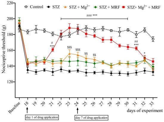
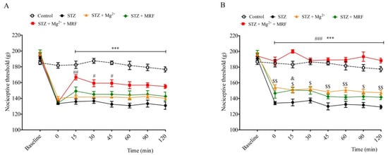
2. The Influence of Mg2+ on the Analgesic Effect of Morphine in Streptozotocin-Induced Hyperalgesia after Mechanical Stimulation

Streptozotocin (STZ)-treated rats developed mechanical hyperalgesia within 2 weeks after single intramuscular administration of STZ. In turn, we recorded no changes in the nociceptive threshold in the control (healthy animals) group (data not shown). Mg2+ and MRF were administered daily for seven days from day 18 to day 24. Mg2+ applied alone to neuropathic animals caused an increase in the nociceptive threshold observed from day 23 until day 26 of the experiment (Figure 1). On the 18th day of the experiment (the 1st day of application), Mg2+ alone did not modify STZ-induced hyperalgesia (Figure 2A). However, on day 24 of the experiment (the 7th day of application), Mg2+ increased the nociceptive threshold over a period of 120 min after administration (Figure 2B). In turn, MRF administered alone on seven consecutive days did not change STZ-induced hyperalgesia (Figure 1). Similarly, no significant changes in nociceptive thresholds were observed on the 1st day of the drug’s application (day 18 of the experiment) over a period of 120 min after MRF administration (Figure 2A). However, on the 7th day (day 24 of the experiment), MRF alone induced a slight statistically significant analgesic effect 15 min after injection (p < 0.05) (Figure 2B). As it is shown in Figure 1, the mechanical hyperalgesia was significantly reduced in rats receiving the combination of Mg2+ and MRF starting from day 21 of the experiment (the third day of drugs application) compared to MRF-treated animals. After cessation of tested drugs (Mg2+ and MRF) application, animals’ pain sensitivity was reported to increase. On day 18 of the experiment (the 1st day of administration), simultaneous injection of Mg2+ and MRF increased the nociceptive threshold in comparison to rats receiving only MRF starting from 15 min after administration (166.66 ± 3.33 g and 149.167 ± 5.23 g for STZ + Mg2+ + MRF and STZ + MRF, respectively; Figure 2A). It is noteworthy that this effect was significantly higher on day 24 of the experiment (the 7th day of application) at the same time point (200 ± 2.23 g and 150.83 ± 3.27 for STZ + Mg2+ + MRF and STZ + MRF, respectively; Figure 2B).
Figure 1. Influence of magnesium (Mg2+) on the analgesic activity of morphine (MRF) in streptozotocin (STZ)-treated rats. Days 18–24—drugs administration; days 19–25—measurements of prolonged activity of investigated drugs; days 26–33—measurements of pain threshold after discontinuation of drugs administration. Nociceptive thresholds, expressed in grams, were measured before STZ administration (baseline) and then before tested substances administration in rats with STZ-induced diabetic neuropathic pain from day 19 until day 25. Data are presented as means ± SEM. Statistical analysis was performed by two-way ANOVA followed by Bonferroni’s post-hoc test. *** p < 0.001, * p < 0.05 (STZ + Mg2+ + MRF vs. STZ), ### p < 0.001, ## p < 0.01, # p < 0.05 (STZ + Mg2+ + MRF vs. STZ + MRF), $$$ p < 0.001, $$ p < 0.01 (STZ + Mg2+ vs. STZ); n = 6 rats for each group.
Figure 2. Effect of magnesium (Mg2+) on the antinociceptive activity of morphine (MRF) on the 1st day (Panel A) and the 7th day (Panel B) of the drugs’ application in streptozotocin (STZ)-induced diabetic neuropathic pain. Nociceptive thresholds, expressed in grams, were measured before STZ administration (baseline) and then before (0) and after tested substances administration in rats with STZ-induced diabetic neuropathic pain for 2 h. Data are presented as means ± SEM. Statistical analysis was performed by two-way ANOVA followed by Bonferroni’s post-hoc test. *** p < 0.001 (STZ + Mg2+ + MRF vs. STZ), ### p < 0.001, ## p < 0.01, # p < 0.05 (STZ + Mg2+ + MRF vs. STZ + MRF), $$ p < 0.01, $ p < 0.05 (STZ + Mg2+ vs. STZ), & p < 0.05 (STZ + MRF vs. STZ); n = 6 rats for each group.

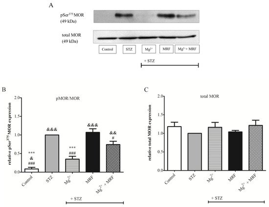
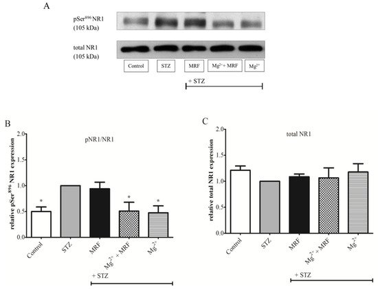
3. Changes in the Expression of Phosphorylated Proteins Measured at the Serine Residues of N-methyl-D-aspartate Receptor NR1 Subunit and µ Opioid Receptor

To determine changes in the level of protein phosphorylation at the Ser375 MOR (Figure 3) and at the Ser896 NMDAR NR1 subunit (Figure 4), we used the Western blot technique in PAG lysate. As shown in Figure 3B, phosphorylation of Ser375 at MOR for STZ-treated rats was significantly higher compared to the control group (healthy animals). Importantly, Mg2+ administered alone in rats with STZ-induced diabetic neuropathic pain resulted in a statistically significant decrease in the protein phosphorylation on Ser375 MOR in relation to the STZ group (0.35 ± 0.08 vs. 1 for STZ group; p < 0.001). MRF injected alone showed no changes in the phosphorylation of tested protein compared with the STZ group (1.07 ± 0.1 vs. 1 for the STZ group; p > 0.05). In turn, simultaneous administration of Mg2+ and MRF caused a significant reduction in the phosphorylation of MOR compared to the STZ group receiving MRF alone (0.74 ± 0.09 vs. 1.07 ± 0.1; p < 0.05). In the case of ionotropic NMDAR (Figure 4B), high phosphorylation of the Ser896 NR1 subunit was observed in STZ-treated rats compared to the control group (p < 0.05). Treating STZ animals with MRF did not affect the phosphorylation of the NR1 in relation to the STZ group (0.94 ± 0.13 vs. 1, p > 0.05). In contrast, coadministration of Mg2+ and MRF generated a statistically significant decrease in the level of NR1 phosphorylation in rats with STZ-induced diabetic neuropathic pain in comparison to the non-treated STZ group (0.51 ± 0.17 vs. 1; p < 0.05). A similar effect was observed after Mg2+ administered alone (0.48 ± 0.13 vs. 1; p < 0.05). The expression of total MOR (Figure 3C) and total NR1 (Figure 4C) did not change in all tested groups.
Figure 3. The effect of chronic administration of magnesium (Mg2+), morphine (MRF) and combination of these substances (Mg2+ + MRF) in periaqueductal gray matter (PAG) lysate from streptozotocin (STZ)-treated rats on the level of serine 375 (Ser375) µ opioid receptor (MOR) phosphorylation. (A) Representative result of the Western blot analysis showing phosphorylated serine 375 (pSer375) MOR (top panel) and total MOR (bottom panel). (B) The level of protein phosphorylation at the Ser375 residue expressed as the ratio of the phosphorylated form to the total form of the tested proteins (pMOR/MOR). (C) Changes in the expression of total MOR. Statistical analysis was performed using one-way ANOVA followed by Dunnett’s post-hoc test. *** p < 0.001 vs. STZ; &&& p < 0.001; && p < 0.01; & p < 0.05 vs. STZ + Mg2+; ### p < 0.001; # p < 0.05 vs. STZ + MRF (graph B). There were no statistically significant differences in total MOR expression (p > 0.05; graph C); n = 6 rats/group.
Figure 4. The effect of chronic administration of morphine (MRF), magnesium and morphine cotreatment (Mg2+ + MRF), and magnesium (Mg2+) in periaqueductal gray matter (PAG) lysate from streptozotocin (STZ)-treated rats on the expression of N-methyl-D-aspartate receptor (NMDAR) NR1 subunit phosphorylation. (A) Representative results of the Western blot assay showing phosphorylated serine 896 (pSer896) NR1 (top panel) and total NR1 (bottom panel). (B) The level of Ser896 phosphorylation expressed as the ratio of the phosphorylated form to the total form of the tested proteins (pNR1/NR1). (C) Changes in the expression of total NR1. Statistical analysis was performed using one-way ANOVA followed by Dunnett’s post-hoc test. * p < 0.05 vs. STZ (graph B). There were no statistically significant differences in total NR1 (p > 0.05; graph C); n = 6 rats/group.

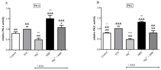
4. Changes in the Activity of Protein Kinase A and Protein Kinase C in the Streptozotocin-Treated Rats after Administration of Tested Compounds

As shown in Figure 5B, an increase in the activity of PKC was observed in STZ-treated rats compared to the control animals. In turn, no statistically significant changes were recorded in levels of PKA in these two groups (Figure 5A). However, for both PKA and PKC, the 7-day application of Mg2+ led to a reduction in their activities (0.49 ± 0.08 vs. 1, p < 0.01 for PKA and 0.48 ± 0.05 vs. 1, p < 0.001 for PKC). Importantly, the application of MRF to animals with chronic neuropathic pain resulted in an increase in the activity of both kinases compared to the STZ group (1.51 ± 0.16 vs. 1 for PKA and 1.31 ± 0.04 vs. 1 for PKC; p < 0.01). Simultaneous administration of Mg2+ and MRF showed no changes in the level of PKA and PKC (1.08 ± 0.1 for PKA and 0.8 ± 0.11 for PKC, p > 0.05) in relation to the STZ group. However, statistically significant changes in the activity of both enzymes were observed after injection of Mg2+ and MRF in relation to MRF alone (1.08 ± 0.1 vs. 1.51 ± 0.16 for PKA, p < 0.05 and 0.8 ± 0.11 vs. 1.31 ± 0.04 for PKC, p < 0.001).
Figure 5. Changes in the level of (A) protein kinase A (PKA) and (B) protein kinase C (PKC) in response to magnesium (Mg2+), morphine (MRF) and the combination of these substances (Mg2+ + MRF) in periaqueductal gray matter (PAG) lysate from streptozotocin (STZ)-treated rats. Results are presented as means ± SEM. Statistical analysis was performed using one-way ANOVA followed by Dunnett’s post-hoc test. *** p < 0.001; ** p < 0.01; * p < 0.05 vs. STZ; ### p < 0.001; ## p <0.01, # p < 0.05; vs. STZ + MRF; &&& p < 0.001, && p < 0.01 vs. STZ + Mg2+n = 6 rats/group.

This entry is adapted from the peer-reviewed paper 10.3390/ijms222413599

References

  1. S. Arnér; B. A. Meyerson; Lack of analgesic effect of opioids on neuropathic and idiopathic forms of pain. Pain 1988, 33, 11-23, 10.1016/0304-3959(88)90198-4.
  2. Luigi Morrone; Damiana Scuteri; Laura Rombola; Hirokazu Mizoguchi; Giacinto Bagetta; Opioids Resistance in Chronic Pain Management. Current Neuropharmacology 2017, 15, 444-456, 10.2174/1570159x14666161101092822.
  3. Ajay S. Yekkirala; David P. Roberson; Bruce P. Bean; Clifford J. Woolf; Breaking barriers to novel analgesic drug development. Nature Reviews Drug Discovery 2017, 16, 545-564, 10.1038/nrd.2017.87.
  4. A. H. Dickenson; NMDA receptor antagonists: interactions with opioids. Acta Anaesthesiologica Scandinavica 1997, 41, 112-115, 10.1111/j.1399-6576.1997.tb04624.x.
  5. Jianren Mao; NMDA and opioid receptors: their interactions in antinociception, tolerance and neuroplasticity. Brain Research Reviews 1999, 30, 289-304, 10.1016/s0165-0173(99)00020-x.
  6. Russell K. Portenoy; Kathleen M. Foley; Charles E. Inturrisi; The nature of opioid responsiveness and its implications for neuropathic pain: new hypotheses derived from studies of opioid infusions. Pain 1990, 43, 273-286, 10.1016/0304-3959(90)90025-9.
  7. Patricia Lavand'Homme; Arnaud Steyaert; Opioid-free anesthesia opioid side effects: Tolerance and hyperalgesia. Best Practice & Research Clinical Anaesthesiology 2017, 31, 487-498, 10.1016/j.bpa.2017.05.003.
  8. Sebastiano Mercadante; Edoardo Arcuri; Angela Santoni; Opioid-Induced Tolerance and Hyperalgesia. CNS Drugs 2019, 33, 943-955, 10.1007/s40263-019-00660-0.
  9. J. Nee; V. Rangan; A. Lembo; Reduction in pain: Is it worth the gain? The effect of opioids on the GI tract. Neurogastroenterology & Motility 2018, 30, e13367, 10.1111/nmo.13367.
  10. Eugene A. Kiyatkin; Respiratory depression and brain hypoxia induced by opioid drugs: Morphine, oxycodone, heroin, and fentanyl. Neuropharmacology 2019, 151, 219-226, 10.1016/j.neuropharm.2019.02.008.
  11. Sophie Begon; Gisèle Pickering; Alain Eschalier; Claude Dubray; Magnesium and MK-801 have a similar effect in two experimental models of neuropathic pain. Brain Research 2000, 887, 436-439, 10.1016/s0006-8993(00)03028-6.
  12. Jianren Mao; Donald Price; Ronald L. Hayes; Juan Lu; David J. Mayer; Hanan Frenk; Intrathecal treatment with dextrorphan or ketamine potently reduces pain-related behaviors in a rat model of peripheral mononeuropathy. Brain Research 1993, 605, 164-168, 10.1016/0006-8993(93)91368-3.
  13. Hirokazu Mizoguchi; Chizuko Watanabe; Akihiko Yonezawa; Shinobu Sakurada; Chapter 19 New Therapy for Neuropathic Pain. International Review of Neurobiology 2009, 85, 249-260, 10.1016/s0074-7742(09)85019-8.
  14. Christine N Sang; NMDA-Receptor Antagonists in Neuropathic Pain: Experimental Methods to Clinical Trials. Journal of Pain and Symptom Management 2000, 19, 21-25, 10.1016/s0885-3924(99)00125-6.
  15. Maarten Swartjes; Aurora Morariu; Marieke Niesters; Leon Aarts; Albert Dahan; Nonselective and NR2B-selective N -methyl-d-aspartic Acid Receptor Antagonists Produce Antinociception and Long-term Relief of Allodynia in Acute and Neuropathic Pain. Anesthesiology 2011, 115, 165-174, 10.1097/aln.0b013e31821bdb9b.
  16. Valéria Martinez; Dennis Christensen; Valérie Kayser; The glycine/NMDA receptor antagonist (+)-HA966 enhances the peripheral effect of morphine in neuropathic rats. Pain 2002, 99, 537-545, 10.1016/s0304-3959(02)00270-1.
  17. Michael L Nichols; Yvan Lopez; Michael H Ossipov; Di Bian; Frank Porreca; Enhancement of the antiallodynic and antinociceptive efficacy of spinal morphine by antisera to dynorphin A (1–13) or MK-801 in a nerve-ligation model of peripheral neuropathy. Pain 1997, 69, 317-322, 10.1016/s0304-3959(96)03282-4.
  18. Tatsuo Yamamoto; Tony L. Yaksh; Studies on the spinal interaction of morphine and the NMDA antagonist MK-801 on the hyperesthesia observed in a rat model of sciatic mononeuropathy. Neuroscience Letters 1992, 135, 67-70, 10.1016/0304-3940(92)90137-v.
  19. L. J. Rondón; A. M. Privat; L. Daulhac; N. Davin; A. Mazur; J. Fialip; A. Eschalier; C. Courteix; Magnesium attenuates chronic hypersensitivity and spinal cord NMDA receptor phosphorylation in a rat model of diabetic neuropathic pain. The Journal of Physiology 2010, 588, 4205-4215, 10.1113/jphysiol.2010.197004.
  20. Wen-Hua Xiao; Gary J. Bennett; Magnesium suppresses neuropathic pain responses in rats via a spinal site of action. Brain Research 1994, 666, 168-172, 10.1016/0006-8993(94)90768-4.
  21. S Brill; P M Sedgwick; W Hamann; P P Di Vadi; Efficacy of intravenous magnesium in neuropathic pain.. British Journal of Anaesthesia 2002, 89, 711-4, .
  22. A. A. Yousef; A. E. Al‐Deeb; A double‐blinded randomised controlled study of the value of sequential intravenous and oral magnesium therapy in patients with chronic low back pain with a neuropathic component. Anaesthesia 2012, 68, 260-266, 10.1111/anae.12107.
  23. Sophie Begon; Gisèle Pickering; Alain Eschalier; Claude DuBray; Magnesium Increases Morphine Analgesic Effect in Different Experimental Models of Pain. Anesthesiology 2002, 96, 627-632, 10.1097/00000542-200203000-00019.
  24. Magdalena Bujalska.; Helena Makulska-Nowak.; Stanisław W. Gumułka; Magnesium ions and opioid agonists in vincristine-induced neuropathy. Pharmacological Reports 2009, 61, 1096-1104, 10.1016/s1734-1140(09)70172-0.
  25. Magdalena Bujalska; Ewelina Malinowska; Helena Makulska-Nowak; Stanisław Witold Gumułka; Magnesium Ions and Opioid Agonist Activity in Streptozotocin-Induced Hyperalgesia. Pharmacology 2008, 82, 180-186, 10.1159/000151346.
  26. Ahmet Ulugol; Aysegul Aslantas; Yesim Ipci; Alev Tuncer; Cetin Hakan Karadag; Ismet Dokmeci; Combined systemic administration of morphine and magnesium sulfate attenuates pain-related behavior in mononeuropathic rats. Brain Research 2002, 943, 101-104, 10.1016/s0006-8993(02)02618-5.
  27. John T. Williams; Susan L. Ingram; Graeme Henderson; Charles Chavkin; Mark Von Zastrow; Stefan Schulz; Thomas Koch; Christopher J. Evans; Macdonald J. Christie; Regulation of µ-Opioid Receptors: Desensitization, Phosphorylation, Internalization, and Tolerance. Pharmacological Reviews 2013, 65, 223-254, 10.1124/pr.112.005942.
  28. Anika Mann; Susann Illing; Elke Miess; Stefan Schulz; Different mechanisms of homologous and heterologous μ-opioid receptor phosphorylation. British Journal of Pharmacology 2014, 172, 311-316, 10.1111/bph.12627.
  29. Stefan Schulz; Dana Mayer; Manuela Pfeiffer; Ralf Stumm; Thomas Koch; Volker Höllt; Morphine induces terminal μ-opioid receptor desensitization by sustained phosphorylation of serine-375. The EMBO Journal 2004, 23, 3282-3289, 10.1038/sj.emboj.7600334.
  30. Kasper B. Hansen; Feng Yi; Riley Perszyk; Hiro Furukawa; Lonnie P. Wollmuth; Alasdair Gibb; Stephen F. Traynelis; Structure, function, and allosteric modulation of NMDA receptors. Journal of General Physiology 2018, 150, 1081-1105, 10.1085/jgp.201812032.
  31. Pierre Paoletti; Jacques Neyton; NMDA receptor subunits: function and pharmacology. Current Opinion in Pharmacology 2007, 7, 39-47, 10.1016/j.coph.2006.08.011.
  32. Whittemore G. Tingley; Michael D. Ehlers; Kimihiko Kameyama; Carol Doherty; Janine B. Ptak; Clark T. Riley; Richard L. Huganir; Characterization of Protein Kinase A and Protein Kinase C Phosphorylation of the N-Methyl-D-aspartate Receptor NR1 Subunit Using Phosphorylation Site-specific Antibodies. Journal of Biological Chemistry 1997, 272, 5157-5166, 10.1074/jbc.272.8.5157.
  33. John Q. Wang; Ming-Lei Guo; Dao-Zhong Jin; Bing Xue; Eugene E. Fibuch; Li-Min Mao; Roles of subunit phosphorylation in regulating glutamate receptor function. European Journal of Pharmacology 2013, 728, 183-187, 10.1016/j.ejphar.2013.11.019.
  34. Jian-Yu Lan; Vytenis A. Skeberdis; Teresa Jover; Sonja Y. Grooms; Ying Lin; Ricardo Araneda; Xin Zheng; Michael V. L. Bennett; R. Suzanne Zukin; Protein kinase C modulates NMDA receptor trafficking and gating. Nature Neuroscience 2001, 4, 382-390, 10.1038/86028.
  35. Xiaoju Zou; Qing Lin; William D. Willis; Effect of protein kinase C blockade on phosphorylation of NR1 in dorsal horn and spinothalamic tract cells caused by intradermal capsaicin injection in rats. Brain Research 2004, 1020, 95-105, 10.1016/j.brainres.2004.06.017.
  36. L.-M Kow; K.G Commons; S Ogawa; D.W Pfaff; Potentiation of the excitatory action of NMDA in ventrolateral periaqueductal gray by the μ-opioid receptor agonist, DAMGO. Brain Research 2002, 935, 87-102, 10.1016/s0006-8993(02)02532-5.
  37. Susumu Koyama; Norio Akaike; Activation of μ-opioid receptor selectively potentiates NMDA-induced outward currents in rat locus coeruleus neurons. Neuroscience Research 2008, 60, 22-28, 10.1016/j.neures.2007.09.003.
  38. Gilles Martin; Zhiguo Nie; George Robert Siggins; μ-Opioid Receptors Modulate NMDA Receptor-Mediated Responses in Nucleus Accumbens Neurons. The Journal of Neuroscience 1997, 17, 11-22, 10.1523/JNEUROSCI.17-01-00011.1997.
  39. María Rodríguez-Muñoz; Pilar Sánchez-Blázquez; Ana Vicente-Sánchez; Esther Berrocoso; Javier Garzón; The Mu-Opioid Receptor and the NMDA Receptor Associate in PAG Neurons: Implications in Pain Control. Neuropsychopharmacology 2011, 37, 338-349, 10.1038/npp.2011.155.
  40. Pilar Sánchez-Blázquez; Maria Rodríguez-Muñoz; Esther Berrocoso; Javier Garzón; The plasticity of the association between mu-opioid receptor and glutamate ionotropic receptor N in opioid analgesic tolerance and neuropathic pain. European Journal of Pharmacology 2013, 716, 94-105, 10.1016/j.ejphar.2013.01.066.
  41. Pilar Sánchez-Blázquez; María Rodríguez-Muñoz; Javier Garzón; Mu-Opioid Receptors Transiently Activate the Akt-nNOS Pathway to Produce Sustained Potentiation of PKC-Mediated NMDAR-CaMKII Signaling. PLOS ONE 2010, 5, e11278, 10.1371/journal.pone.0011278.
  42. María Rodríguez-Muñoz; Elena de la Torre-Madrid; Pilar Sánchez-Blázquez; Javier Garzón; NO-released Zinc Supports the Simultaneous Binding of Raf-1 and PKCγ Cysteine-Rich Domains to HINT1 Protein at the Mu-Opioid Receptor. Antioxidants & Redox Signaling 2011, 14, 2413-2425, 10.1089/ars.2010.3511.
  43. María Rodríguez-Muñoz; Elena De La Torre-Madrid; Pilar Sánchez-Blázquez; Jia Bei Wang; Javier Garzón; NMDAR-nNOS generated zinc recruits PKCγ to the HINT1–RGS17 complex bound to the C terminus of Mu-opioid receptors. Cellular Signalling 2008, 20, 1855-1864, 10.1016/j.cellsig.2008.06.015.
  44. Pilar Sánchez-Blázquez; María Rodríguez-Muñoz; Carlos Montero; Elena De La Torre-Madrid; Javier Garzón; Calcium/calmodulin-dependent protein kinase II supports morphine antinociceptive tolerance by phosphorylation of glycosylated phosducin-like protein. Neuropharmacology 2008, 54, 319-330, 10.1016/j.neuropharm.2007.10.002.
  45. Sascha Just; Susann Illing; Michelle Trester-Zedlitz; Elaine K. Lau; Sarah J. Kotowski; Elke Miess; Anika Mann; Christian Doll; Jonathan C. Trinidad; Alma L. Burlingame; et al. Differentiation of Opioid Drug Effects by Hierarchical Multi-Site Phosphorylation. Molecular Pharmacology 2012, 83, 633-639, 10.1124/mol.112.082875.
  46. Javier Garzón; María Rodríguez-Muñoz; Pilar Sánchez-Blázquez; Do pharmacological approaches that prevent opioid tolerance target different elements in the same regulatory machinery?. Current Drug Abuse Reviewse 2008, 1, 222-238, 10.2174/1874473710801020222.
More
This entry is offline, you can click here to edit this entry!
Video Production Service