Augmented Reality and Image-Guided Robotic Liver Surgery: History
Please note this is an old version of this entry, which may differ significantly from the current revision.
Subjects: Surgery
Contributor:

Robotic surgery has gained much attention in liver resection for its potential to increase surgical dexterity in a minimally invasive scenario. In liver surgery, robotic systems help surgeons to localize tumors and improve surgical results with well-defined preoperative planning or increased intraoperative detection. Furthermore, they can balance the absence of tactile feedback and help recognize intrahepatic biliary or vascular structures during parenchymal transection. In addition, the robotic system presents the advantage of creating a hybrid interface in which pre- and intra-operative imaging tools could be exploited alone or together in order to guide surgical resection.

  • robotic
  • liver surgery
  • augmented reality

1. Introduction

Since its presentation, the robotic platform has drawn much attention in liver resections for its possibility to combine the multiple advantages of laparoscopic surgery and the dexterity of an open approach [1,2]. Laparoscopy has widely demonstrated its benefits in terms of shortened recovery and reduced post-operative pain, morbidity, and blood loss, with similar results to open surgery in oncologic outcomes [3,4,5,6,7]. However, some issues are still present when considering technically challenging hepatectomies and most of these disadvantages are related to a limited maneuverability, the presence of rigid instruments, and a restricted field or the quality of vision [8]. Robotic technology is able to overcome some of these limitations and more series and meta-analysis are present in the literature, highlighting the well-known robotic advantages with comparable post-operative and oncological results [9,10,11,12,13,14,15]. Beyond the technical upgrades, one of the improvements of the DaVinci system is the possibility to create an interactive visual interface rather than a simple operative field in which, through some dedicated software, surgeons can be guided by preoperative and/or intraoperative imaging during parenchymal resection [16]. The robot must be considered as a means of putting between the surgeon and the patient a computer and its computing power. Related imaging technologies, such as Augmented Reality (AR), have been developed in order to assist the operator and limit the intrinsic drawbacks of a minimally invasive approach, such as the lack of a tactile feedback, which can hamper tumor detection or pedicle dissection.

2. Augmented Reality

Implementing technologies of robotic hepatic surgery through AR means overcoming the limitations widely described when performing liver resections through this approach. Despite the sense of depth achieved by the 3D visualization and the improved video resolution and magnification, the robotic system presents all the drawbacks of a minimally invasive view compared to an open hepatectomy. The absence of the tactile feedback and the unavailability of an ultrasonic dissector are undoubtedly the main issues found in this context. The importance of a tactile sensation is twofold in this kind of surgery. Firstly, from an oncological point of view, it helps locate the tumor and thus guide parenchymal transection. AR is effective in planning preoperatively the strategy through 3D rendering and, intraoperatively, targeting the lesion and resection margins (Figure 1) [17,18,19,20,21]. Buchs et al. [22], for example, overlaid the 3D preoperative images onto the endoscopic stream and added distance information in order to obtain a real-time guided transection. After projecting the tumor onto the liver surface and defining the margin width, a bar and a dartboard appeared on the screen to guide the tip of the instrument, with colors ranging from green to red to alert the operator if the distance to the tumor was respected or not. Ultrasound guidance or an intraoperative CT scan could be equally used, with the disadvantage of radiation, shifting the instruments constantly, or, occasionally, having difficulty localizing the nodules. In fact, AR has been described in open hepatectomies in the detection of vanishing metastasis, with the superimposition of lesions pre-existent to chemotherapy and undetectable at preoperative images or intraoperative US [23,24]. Secondly, the sense of touch aids the operator to orient him/herself in relation to some intra hepatic landmarks. Arteries, veins, and biliary structures, especially in the first-orders pedicles, present a thickened fibrotic sheath and the robotic “insensibility” can disorient surgeons during the dissection, with possible vascular injuries. Furthermore, lesions can be located in critical areas such as the hepatic confluence, and robotic dissection could thus be more complex [25]. AR-based intraoperative reconstructions and tracking systems may be used to map resection planes and show vascular structures during liver transection (Figure 1). Moreover, 3D planning can enable the identification of anatomical variants, as demonstrated in laparoscopic cholecystectomy [26]. These reconstructions could also represent a solution to the absence of some familiar devices in robotic liver surgery.
Figure 1. (A) Surgical preoperative planning through 3D reconstruction of an anatomical S5 segmentectomy. The tumor is colored in green and the theorical resection plane in red. (BD) Intraoperative superimposition of planned resection area rendering. Vascular and biliary structures are projected during different phases of parenchymal transection, with the identification of the S5 vascular pedicle.
Another advantage of AR described for robotic liver resections is port placement (Figure 2) [28]. This phase is a key step in minimally invasive resections and even more in robotic surgery. Projecting a virtual image of the liver parenchyma on the skin surface in relation to some external landmarks allows targeting the lesion and liver structures at the beginning of the operation for camera placement and, after CO2 insufflation, for other ports. This results in an improved manageability of the operator, above all for posterior segment approach, which can lead to fewer operative times and lower intraoperative complications.
Figure 2. Projection of a virtual liver 3D reconstruction on the skin surface in relation to some external landmarks. The positioning of the optical port (left) is guided by the inferior border of the liver and the resection planned. After the first trocar is inserted, the “see-through” view will aid the operator to place other robotic ports (right).

3. Image-Guided Robotic Liver Surgery

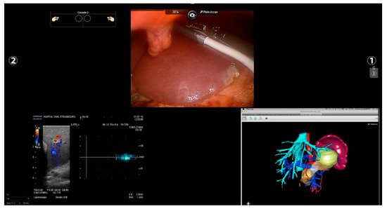
Other imaging strategies are more frequently used by surgeons during robotic liver resections in order to improve lesion detection and evaluate intraparenchymal biliary and vascular structures. Before exploring these modalities, a special mention goes to TilePro (Intuitive Surgical Inc., Sunnyvale, CA, USA). This is a multi-input display software integrated into the robotic platform, which shows more video sources simultaneously on the same screen (Figure 2). By simply connecting an external source to the Da Vinci console, surgeons and other operating room assistants can easily switch from the operating field to other input such as Intraoperative Ultrasound (IOUS) or preoperative cross-sectional imaging and 3D reconstructions.

3.1. Preoperative Imaging and 3D Rendering

A correct planification of surgical strategy is essential in liver surgery and it usually includes a 2D cross-sectional preoperative evaluation. However, complex liver intraparenchymal anatomy and anatomical variations can hinder a correct assessment made by a simple CT scan or MRI. Since its first description in 1998 [33], 3D liver reconstruction is nowadays performed more often during preoperative surgical planning. It consists of creating a 3D model starting from preoperative CT scan or MRI, which allows an easier identification of the stereoscopic relationships between the tumor and intrahepatic structures to calculate the hepatic volumes for the risk of post-operative liver failure and create resection plans to follow intraoperatively [34]. 

3.2. Intra-Operative Robotic Ultrasound Application

IOUS is nowadays an essential tool for hepatobiliary surgeons. Several series emphasize its utility in tumor identification, the recognition of its spatial relationship with intrahepatic structures, and, therefore, in guiding parenchymal resection [38]. Furthermore, IOUS is demonstrated to be able to modify preoperative strategy in up to a quarter of cases because of different vascular relationships or new nodules found during surgical exploration, despite the performance of a liver-specific MRI [39,40,41]. 
Recently, a specific transducer was introduced to the robotic ecosystem with consequent better manageability, higher precision, and the creation of a multi-input environment. Surgeons can now easily control the probe, grabbing its dorsal fin with a forceps, and a highly flexible cable and a small transducer surface make the access to the posterior segments or to the inferior surface easier compared to the rigid laparoscopic counterpart [46,47]. Moreover, thanks to software such as TilePro, the operator can shift from the 3D camera view to the ultrasound directly from the console, or even create a split-view with both intraoperative and ultrasound images (Figure 4). 
Figure 4. Robotic “split-view”. Through a dedicated probe and a specific software, the surgeon can shift from to the endoscopic to the ultrasound view or create a split-view with both the images. In this figure, a 3D model was added intraoperatively at the same time to check the tumoral vascular relationship studied preoperatively.

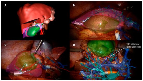
3.3. Indocyanine Green Fluorescence

Indocyanine green (ICG) is a fluorescent dye with a rapid hepatic clearance largely used in hepato-biliary surgery thanks to its pharmacokinetics features [53,54]. After portal vein or intravenous injection [55], it allows the identification of anatomical liver vessels and biliary ducts by providing a rapid parenchymal mapping [54]. This enables anatomical resections with lower risks of vascular injuries or bile leaks. Moreover, it increases tumor detectability, helps differentiate hepatic lesions based on their vascular patterns, and allows the detection of additional superficial hepatic lesions [56,57,58,59]. Fluorescence software was incorporated into the Da Vinci system in 2010 and its application enhances robotic advantages in liver surgery from a different point of view. In fact, when a tumor-bearing portal vein injection is chosen—the so-called “negative staining” technique—this could be cumbersome in laparoscopic resections due to the impaired dexterity and lack of ergonomics, and, at the same time, the presence of a rigid linear transducer. The robotic system, through its delicate movements and endowrist instruments, ensures a fine and safe dissection of the hepatic pedicle, allowing it to reachthe hilum and the portal bifurcation easier in the case of a direct portal injection [57,58,59,60,61]. Furthermore, through the dedicated probe, the transhepatic needle insertion could be also less demanding.
Another aspect to consider in ICG-guided resections is tumor clearance. Minimally invasive approaches—and robotic, in particular—lack a tactile feedback, and achieving a parenchymal free margin or performing an anatomical resection could be challenging. Furthermore, an IOUS exclusive evaluation could be insufficient because it is a user-dependent procedure and presents a heterogeneous detection rate according to tumor size and location and parenchymal stiffness [62,63,64]. In this context, fluorescence is a precious tool in robotic surgery, with some authors reporting an enlargement of the resection area after ICG application, both in benign ad malignant lesions, in order to achieve a R0 resection [57,65,66,67], and a significantly higher rate of margin-free specimens when comparing robotic hepatectomies with and without ICG [67]. As in open surgery, even in robotic surgery, some series described the detection of newer superficial lesions that the dye injection missed before [57]. This high sensitivity found is, however, limited to the liver surface because of the low penetration of the dye under 8 mm of depth, thus requiring the use of other imaging tools such as IOUS. Although no long-term results have been published, these findings have a significant impact in terms of oncological outcomes. ICG is a promising instrument of intraoperative navigation surgery, allowing rapid and easy identification of the resection plane without the inconveniences mentioned for other image-guided techniques. It can be used in combination with IOUS or AR as an additional aid rather than as a replacement [61] and with its features, it seems to fill some gaps found in robotic surgery, making tailored and oncological surgery less challenging.

4. Future Prospective

The application of the above-described technologies is nowadays limited in the robotic
liver experience, mainly due to some technical limitations and to a relatively newborn and still debated approach [15]. AR, for example, is a time-consuming procedure, not only for the intraoperative installation, but also for preoperative planning and liver rendering [68]. In the context of an atypical or less demanding hepatic resection, which represent the first steps of a necessary learning curve, this time could appear exaggerated. Furthermore, AR in hepatic surgery has showed a delayed distribution compared to other surgical fields as neurosurgery, otolaryngology, orthopedics, and maxillofacial surgery [69,70,71]. This difference comes from anatomical obstacles, such as working with a deformable soft organ that is constantly moving during operation because of respiratory cycles as well as pneumoperitoneum creation [72]. Although some strategies have been described in this context [23,73,74], these features make the development of AR more complex, and new software are needed for shortening modeling creation and improving the accuracy of manual, semiautomatic, and automatic images overlapping.
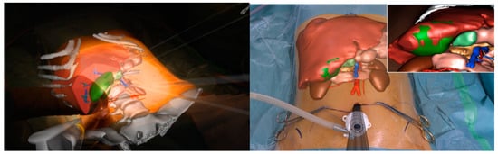
All the imaging techniques described must be seen, however, as a part of a puzzle rather than an independent solution towards a guided surgery; an example comes from registration accuracy in AR. IOUS and ICG have been proposed to improve overlapping quality through fluorescent markers and 3D ultrasounds used for intraoperative landmarks [75,76]. In this scenario, the robotic platform fits perfectly by creating a unique merged environment with the possibility of using and visualizing preoperative reconstruction and intraoperative images simultaneously within the operative field (Figure 4).
Another potential benefit of image-guided technology is minimally invasive training.
In laparoscopy, telementoring based on AR seems to speed up simple skills acquisition such as suturing [77] or even reduce the learning curve in more complex procedures such as cholecystectomy [78]. Similar applications in robotic training are lacking, with only a few experiences described [79]. Hepato-biliary surgery lacks standards of training and learning curves in robotic procedures [80], but recently, an expert panel of HPB surgeons agreed that a correct training path in hepatobiliary procedures needs different steps, starting from basic robotic skills before performing a liver resection [81]. In this context, AR could be a useful tool to support less-experienced surgeons performing simple procedures and lower their learning curve.

5. Conclusions

The application of pre- and intra-operative imaging modalities in guiding hepatic surgery presents promising results, and the robotic ecosystem can facilitate their use and magnify their benefits. Potential advantages include reduced morbidity and improvements in oncological outcomes. However, some limitations are still present, related to limited robotic diffusion and still insufficient technological development, and most of the data in the literature come from preclinical studies or small series.

This entry is adapted from the peer-reviewed paper 10.3390/cancers13246268

This entry is offline, you can click here to edit this entry!