Microbiota Alterations in Pancreatic Cancer: History
Please note this is an old version of this entry, which may differ significantly from the current revision.
Subjects: Microbiology | Oncology
Contributor:

The human microbiome is a key factor in many malignancies, having the ability to alter host metabolism and immune responses and participate in tumorigenesis. Gut microbes have an influence on physiological functions of the healthy pancreas and are themselves controlled by pancreatic secretions. An altered oral microbiota may colonize the pancreas and cause local inflammation by the action of its metabolites, which may lead to carcinogenesis. The mechanisms behind dysbiosis and pancreatic cancer (PC) development are not completely clear. An altered microbiota may induce oncogenomic changes, or, on the other hand, cancer mutations may have an impact on microbiota composition. Altered microbiota can also influence drug efficacy in PC chemo- and immunotherapies. Possible future scenarios are the intentional manipulation of the gut microbiota in combination with therapy or the utilization of microbial profiles for the noninvasive screening and monitoring of PC.

  • Pancreatic
  • Cancer
  • Microbiota
  • Dysbiosis
  • Oncogenomics
  • Drug Response
  • Bacterial Metabolites
  • Inflammation

1. The Microbiome in Pancreatic Health

The pancreas plays an important role in metabolism through the secretions of its exocrine and endocrine glands. While the exocrine gland controls digestion by producing pancreatic juice (digestive enzymes and sodium bicarbonate), the endocrine gland secretes islet peptide hormones to maintain glucose homeostasis [27]. A healthy pancreas is pivotal in controlling the gut microbiota and reciprocally the gut microbiota has a key impact on pancreatic function [28]. Studies on mouse models have shown that antimicrobial peptides secreted by the pancreas control the composition and diversity of the gut microbiota, and, as a consequence, protect against inflammation. The cathelicidin-related antimicrobial peptide (CRAMP), produced by pancreatic endocrine β-cells, destroys unwanted intestinal microbes by permeabilizing the bacterial membrane [29]. A preclinical study demonstrated that the absence of CRAMP due to impaired exocytosis leads to an alteration in the gut microbiota and bacterial overgrowth in pancreatic cells, and further, to intestinal inflammation and death. Supplementation of CRAMP, however, prevented this bacterial overgrowth and inflammation, which confirms the influence of the pancreas on the gut microbiota [30].
On the other hand, certain metabolites produced by gut bacteria affect pancreatic function. In a study on mice, butyrate, a short-chain fatty acid (SCFA) produced by intestinal bacteria, has been shown to induce the expression of CRAMP in pancreatic β-cells [31]. Likewise, acetate, another SCFA metabolite of gut bacteria, induced insulin secretion in rats via a microbiome–brain β-cell axis [32]. This mutual interaction between the gut microbiota and the pancreas is thus important both in health and disease. Bacterial imbalance or dysbiosis may lead to a dysfunctional pancreas and result in disease, and reciprocally pancreatic disease may cause intestinal dysbiosis.
The gut microbiota is, nevertheless, not the only potential influencer of pancreatic health. Contrary to previous assumptions, the pancreas itself is not sterile and has its own microbiota. The presence of bacterial DNA in pancreatic tissue has been reported in 76% of PDAC patients and 15% of healthy individuals [33]. Microbes are thought to migrate from the duodenum to the pancreas through the pancreatic duct. A comparison of the microbiota from different gastrointestinal sites has shown an overlap of the pancreatic and duodenal microbiome both in Pancreatic cancer (PC) patients and in healthy controls, affirming that pancreatic bacteria may migrate from the intestine. The pancreatic microbiota have been reported to be very diverse and include certain taxa typically detected in the oral cavity. Moreover, the pancreatic bacterial diversity in PC patients has been found to vary significantly from that of healthy controls [34]. The presence of bacterial DNA in pancreatic tissue samples was also confirmed by Thomas et al., but they did not find significant differences in genus richness or diversity between cancerous and noncancerous tissue [35].
Pushalkar et al. have demonstrated migration of oral fluorescently labeled Enterococcus faecalis to the pancreas via the intestine, with a higher level of migration in PDAC mice compared to noncancerous mice [17]. Interestingly, in an experiment with germ-free mice, the pancreas was not colonized by bacteria under normal physiological conditions [35]. Besides the pancreatic duct, alternative ways of colonization of the pancreas have been suggested, including oral, mesenteric venous drainage, and mesenteric lymphatic drainage routes [36]. These partly contradictory findings illustrate that the questions regarding the origin and significance of the intrapancreatic microbiome are not fully resolved. Further studies on larger cohorts are needed for clarification.

2. Microbiota Alterations in Pancreatic Cancer (PC)

In addition to influencing the physiological functions of the pancreas, microbial dysbiosis can also enhance inflammation and affect tumorigenic processes, such as cellular proliferation, invasion, metastasis, angiogenesis, and immune modulation [37]. Besides genetic and environmental factors, the oncogenic microbiome, or oncobiome, is one of the regulators of the hallmarks of cancer [38,39]. In addition to the local tumorigenic effects, alterations in microbiota may also exert hormone-like, long-distance effects on different organs [40]. The following subsections provide an overview of microbial alterations in different parts of the gastrointestinal tract and their possible significance in PC.

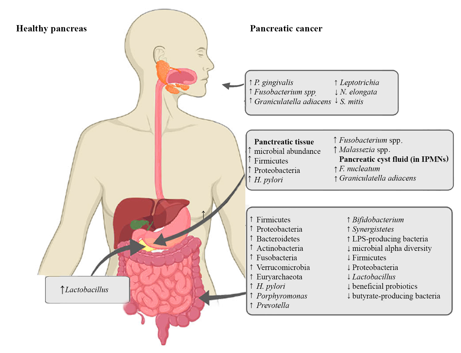
Figure 1. Microbiota alterations in PC


2.1. Oral Microbiota and PC

Several studies have shown an association of oral bacterial dysbiosis with PC tumorigenesis [41,42,43]. The spread of oral microbes to the pancreas via translocation or dissemination has been discussed in the previous section [44,45]. Periodontal disease, a condition linked to alterations in oral bacteria, has been related to an increased risk of PC [46]. Poor oral health, pathogenic oral flora, periodontal disease, and tooth loss are well-established independent risk factors for PDAC [41].
Unique oral microbiota profiles have been associated with PDAC in several studies. PC was associated with significantly increased abundances of the oral bacteria Porphyromonas gingivalis [47,48,49], Fusobacterium [49], Graniculatella adiacens [50] and Leptotrichia [48,49] and significantly decreased abundances of Neisseria elongata and Streptococcus mitis [50], amongst others. Combinations of certain microbes, like N. elongata and S. mitis, or the increased ratio of Leptotrichia to Porphyromonas were found to significantly differentiate PC patients from healthy controls and were thus suggested as potential predictive biomarkers for the early detection of PC, especially since saliva sampling is noninvasive and very easy to arrange [48,50].

2.2. Pancreatic Microbiota and PC

Preclinical models have shown that the microbial abundance in pancreatic tumor tissue is up to a thousand times higher than in healthy pancreatic tissue [17,51]. The PDAC microbiota has been profiled in several studies, with partly similar, and partly differing results [17,34,42,52,53,54,55]. The first pathogen to be detected in pancreatic tumor tissue and associated with PC was H. pylori [56]. H.pylori DNA was reported in the pancreatic tissue of 75% of PDAC patients, in 60% of patients with chronic pancreatitis, but in none of the healthy controls [57]. In addition, the abundance of Fusobacterium spp., an oral pathogen, was found to be significantly increased in PDAC tissue compared to controls and was associated with a worse prognosis [34,42]. Lactobacillus, on the other hand, was more common in healthy controls than in PDAC patients [34]. An increased abundance of Firmicutes and Proteobacteria, which are also the most prominent phyla of the healthy gut, has been observed in several studies in PC tissue as compared to healthy pancreatic tissue [36,55,56]. An enrichment of the fungal microbe Malassezia spp. in PDAC tumor tissue has also been reported [52]. In addition to that, pancreatic cyst fluid was found to contain its own unique microbiome [58]. An increased abundance of the oral bacteria Fusobacterium nucleatum and Granulicatella adiacens was detected in the pancreatic cyst fluid of IPMNs compared to non-IPMN pancreatic cystic neoplasia. Since IPMNs can develop into invasive PC, the results point to the possible pathogenicity of these species and underscores the likelihood of bacterial colonization from the oral cavity [59].

2.3. The Intestinal Microbiota and PC

As the pancreas is connected to the intestine through the pancreatic duct, it is obvious that the gut microbiota can influence the pancreas and vice versa [36]. To investigate the relationship between gut microbial dysbiosis and PC, the intestinal tissue microbiota, as well as the fecal microbiota of PC patients have been studied. Clear associations of a unique gut microbiome profile with PDAC were shown in several studies [56,60,61,62,63], and a significant decrease in the microbial alpha diversity was observed in PDAC cancer patients compared to healthy controls [56,61,64].
Summarizing the main findings, H. pylori infection in the upper gastrointestinal tract has been associated with an increased risk of developing PDAC [65,66]. H. pylori is believed to have an impact on carcinogenesis by promoting cell proliferation [67]. Significantly increased abundances of bacteria belonging to the phyla Bacteroidetes, Firmicutes [61,68], Proteobacteria, Actinobacteria, Fusobacteria, Verrucomicrobia [34,36,69], the genera Porphyromonas, Prevotella, Bifidobacterium [34] and Synergistetes, as well as the archaeal phylum Euryarchaeota [17], have been reported in PC in comparison to healthy controls. Other changes in the gut microbiota reported in PC, were decreased abundances of Firmicutes, Proteobacteria [61] and Lactobacillus [34]. Ren et al. noted an increase in the abundance of potentially pathogenic lipopolysaccharide (LPS)-producing bacteria and a decrease in the abundance of beneficial probiotics and butyrate-producing bacteria in PC patients [61]. Results of different studies are partly similar, but partly contradictory, indicating that larger studies will be needed for establishing a clearer profile of the PDAC gut microbiome.

3. Microbiota in Pancreatic Inflammation, Oncogenesis and Tumor Immunity

Inflammation of the pancreas plays a key role in the development of pancreatic cancer. One cause of pancreatic inflammation is a dysbiotic oral, gastric, or intestinal microbiota that can cause an overgrowth of harmful bacteria. This can lead to epithelial barrier breaches and the migration of bacteria to the pancreas. Continual colonization of the pancreas by dysbiotic bacteria results in persistent inflammation and promotes cancer development [24].
Microbial products or metabolites support tumor growth by maintaining inflammation and by immune modulation [37]. Bacterial products such as LPS, SCFAs, lipoproteins, lipopeptides, as well as CpG DNA and single- or double-stranded DNA, can induce immune suppression by binding to pattern recognition receptors (PPR) and by activating Toll-like receptors (TLR). This promotes tumor growth by immune evasion, especially during early carcinogenesis [70]. LPS-induced TLR signaling is also thought to help in maintaining inflammation in PC [37].
Inflammation can also contribute to PC development through its oncogenic effect. Chronic inflammation in pancreatic tissue can trigger KRAS oncogenic mutation in insulin-positive endocrine cells and induce the differentiation of epithelial cells, resulting in PDAC [69]. KRAS is mutated in 93% of PC cases [15] and despite being a common mutation, the activation of KRAS can still require hyperstimulation from LPS-driven inflammation [61]. The activated KRAS can further advance carcinogenesis by activating the nuclear factor kappa B (NF-κB) pathway [71].
A distinct tumor microbial profile (Pseudoxanthomonas/Streptomyces/Saccharopolyspora/Bacillus clausii) has been linked with longer survival of PC patients. Moreover, a higher diversity of tumor microbiota was found associated with higher tumor infiltration of T-cells. Notably, the tumor bacterial profile of long-term survivors was associated with higher infiltrating CD8+ T-cells expressing granzyme B and better cytotoxic T-cell responses [53].

4. The Microbiota and Drug Response in PC

PC patients are often treated with gemcitabine-based chemotherapy [72] and they frequently develop chemoresistance and reduced drug sensitivity [73]. It has been shown that the microbiota plays an important role in the therapeutic efficacy in PC [74]. In CRC mouse models, Geller and colleagues observed that the enzyme cytidine deaminase, produced by Gammaproteobacteria (especially Mycoplasma hyorhinis), metabolizes gemcitabine into its inactive form. Gemcitabine resistance was induced by intratumor Gammaproteobacteria and abrogated by antibiotic treatment [33]. Moreover, they detected bacterial DNA, mostly belonging to Gammaproteobacteria in 76% of the tumors of PDAC patients. Therefore, they suggested that antibiotics could be coadministered with gemcitabine therapy to prevent the development of drug resistance [33]. Similarly, Fusobacterium nucleatum was shown to promote chemoresistance in CRC [75]. However, the gut microbiota can also have positive effects in chemotherapy. For example, Lactobacillus plantarum culture supernatant had a favorable influence on the treatment of colorectal cancer cells with 5-fluorouracil by increasing its chemosensitivity [76]. Likewise, Enterococcus hirae and Barnesiella intestinihominis improved the therapeutic efficacy of cyclophosphamide by facilitating immunomodulatory effects [77].
Immunotherapeutic approaches in PC that are currently under investigation include immune checkpoint inhibitors (ICIs), vaccine therapy, adoptive cell transfer, myeloid-targeted therapy, immune agonist therapy and combinations with chemoradiotherapy or other molecularly targeted agents [78,79]. Bacteria can exert both positive or negative influences on the immune response and immunotherapies. For example, Bacteroidetes spp. were shown to activate Th1 immune responses, and Listeria monocytogenes changed tumor-associated macrophages from the immunosuppressive M2 phenotype to the antitumor M1 phenotype [74]. The immune response in cancer therapy was improved by the inhibition of regulatory T cells (Tregs) through Bifidobacterium adolescentis, Enterococcus faecium, Collinsella aerofaciens and Parabacteroides merdae [74]. The gut microbiota has been shown to increase the efficacy of blockade therapy of programmed cell death 1 (PD-1) protein and its ligand, programmed cell death ligand 1 (PD-L1) [80]. On the contrary, the anticancer immune response increased and the tumor burden was reduced by depletion of the gut microbiota through oral gavage antibiotics treatment in a mouse model of PC [81].
The use of gut microbes in combination with immunotherapies has been suggested for the future [74]. However, their mechanisms in enhancing or attenuating the efficacy of immunotherapies need to be identified. Through fecal microbiota transplant (FMT) or supplementation with certain prebiotics, probiotics, or antibiotics, the gut microbial composition could be manipulated to enhance host anticancer immunity and combat drug resistance [80]. Moreover, the gut microbiota could be used as a biomarker for drug efficacy, treatment response and drug side effects [74].

This entry is adapted from the peer-reviewed paper 10.3390/ijms222312978

This entry is offline, you can click here to edit this entry!