Multifunctional Polymeric Nanogels for Biomedical Applications: History
Please note this is an old version of this entry, which may differ significantly from the current revision.
Contributor:

Currently, research in nanoparticles as a drug delivery system has broadened to include their use as a delivery system for bioactive substances and a diagnostic or theranostic system. Nanogels, nanoparticles containing a high amount of water, have gained attention due to their advantages of colloidal stability, core-shell structure, and adjustable structural components. These advantages provide the potential to design and fabricate multifunctional nanosystems for various biomedical applications. Modified or functionalized polymers and some metals are components that markedly enhance the features of the nanogels, such as tunable amphiphilicity, biocompatibility, stimuli-responsiveness, or sensing moieties, leading to specificity, stability, and tracking abilities. 

  • polymeric nanogels
  • stimuli-responsive
  • functionalized polymer
  • core-shell nanogels

1. Introduction

Drug delivery systems (DDSs are platforms that protect loaded active ingredients from degradation due to physiological conditions, as well as allowing the payloads to perform their desired activities at the target sites. Furthermore, some chemicals and biological drugs are cytotoxic to some extent, which is referred to as an undesired off-target effect. DDSs, such as inorganic nanotubes [1], liposomes [2], and inorganic [3] or polymeric nanoparticles [4], have been developed to overcome these disadvantages. A polymeric nanogel is classified as containing small nanoparticles (ca. 20–200 nm) that is fabricated using hydrophilic polymers as the main component. The crosslinked networks of the polymeric chains encapsulate and enhance the colloidal stability of the loaded molecules, and the hydrophilicity of the polymers entraps large amounts of water, which facilitate the diffusion and mass-exchange with physiological milieu, resulting in a controlled or sustained release of the payload.
Most drugs exhibit low solubility and stability; polymer modification, i.e., a smart polymer, has been used to overcome these limitations. Smart polymers refer to a modification or functionalization of the polymers with side chains or ligands to adjust the polymer hydrophobicity [5][6][7]. Additionally, smart polymers can be designed to display distinct stimuli-responsive behaviors, e.g., thermo-, pH-, and redox-responsiveness [8][9][10][11][12][13][14]. These environmentally sensitive features have been extensively investigated because site-specific interactions of the DDS can increase specificity, targeted drug release [15][16][17][18], and diminish side effects [19][20]. Moreover, the improved properties of smart nanogels can be used as diagnostic or theranostic devices [21][22][23][24]. Smart polymers can be designed to specifically respond to various stimuli, light, radiation, an electric field, or temperature, which could be advantageous for target-specific applications [25][26][27]. Therefore, smart polymeric nanogel systems are promising drug delivery platforms with specificity and safety that can be used in a wide range of biomedical applications, such as an effective therapy or an accurate diagnostic system.

2. Core-Shell Structure of Polymeric Nanogels

The core-shell structure of nanogels is composed of inner and outer layers that contribute to the functionality of the delivery system. The properties of the core or the inner compartment can be adjusted to protect a loaded substance from incompatible environments or provide a hydrophilic cavity for the hydrophilic therapeutic substances. Nanogels can be fabricated using two or more distinct polymers, and thus the ratio of hydrophilicity and hydrophobicity can be adjusted to meet the requirements of various types of substances. Moreover, the amphiphilicity of the nanogel can transport substances with a low aqueous solubility through the circulatory system. Thus, the inner layer is typically fabricated to enhance the capability of the nanogel to hold and/or stabilize the payload, while the outer compartment, the so-called shell, covers the core, and acts as a protective layer exposed to the surrounding environment. Furthermore, the shell can be modified with specialized functional groups for specific features, such as targetability, stimuli-responsiveness, colloidal stability, or increased retention time in the circulatory system.
In 2011, He F. et al. [28] developed multi-responsive semi-interpenetrating network (semi-IPN) hydrogels whose nanostructure comprised core-shell spherical nanoparticles. The stimuli-responsive semi-IPN hydrogels were modified by magnetic nanoparticles with Fe3O4 nanoparticles as a core structure together with the combination of poly-N-isopropylacrylamide (PNIPAM, thermo-responsive) and polyacrylic acid (PAA, pH-responsive). This study demonstrated the successful fabrication of multifunctional materials using core-shell structure nanoparticles [28]. Gonzalez-Urias A. et al. [29] introduced pH-sensitive core-shell nanoparticles using a poly (N,N-diethylaminoethyl methacrylate) (PDEAEMA; anionic polymer) or poly (2-methacryloyloxi benzoic acid) (P2MBA; cationic polymer) core shielded with polyethylene glycol (PEG) that can be used for treating cancer. These nanogels can specifically deliver cisplatin, a cancer drug, to a tumor by targeting its acidic environment. The results indicated that the charge of the inner layer influenced nanogel drug release, cell viability, and cell internalization. Additionally, core-shell nanogels composed of gold nanoparticles as a core and biodegradable chitosan as the outer segment were developed to enhance curcumin cytotoxicity to cancer cells [30]. The findings demonstrated an improved cell uptake of the nanogels in breast cancer cells, which resulted in increased toxicity to the cancer cells. This study revealed that polymer and metal materials could be combined to form core-shell nanoparticles for cancer therapy. The findings of these studies indicate that the core-shell structured nanoparticles can be developed from a wide variety of materials to modify the materials with characteristics specific to the required therapeutic properties of the nanogel. In this review, the nanogels systems were categorized into two groups based on the major components of the core or shell of the nanoparticles. Additionally, hollow sphere drug delivery systems with a functionalized shell and empty core are discussed. Illustrations of the three types of nanogels are presented in Figure 1A.
Figure 1. (A) Core-shell structures of nanogels (B) TEM image of hyaluronic acid-based polymeric nanogels (unpublished data).
The homogeneity of the nanogel is critical for the stability of the system, and thus the nanogel macrostructure was frequently investigated using transmission electron microscope (TEM). Figure 1B illustrates hyaluronic acid-based nanogel’s TEM images. Likewise, nanogel microstructure was also observed after freeze drying that demonstrated the polymer’s porous structure and interconnected network [31]. The porosity of the microscale structure can increase the mass encapsulation of specific substances, such as oxygen or nutrients [32]. Moreover, the void space between the polymer network increases the diffusion of the nanogels compared with hydrogels [33].

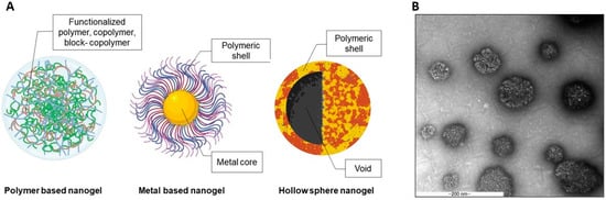
2.1. Polymer Based Polymeric Nanogels

A nanogel is a colloidal system consisting of a crosslinked, water-swellable, 3-dimensional polymeric network whose size can reach ~1000 nm in a fully-swelled state [34]. The crosslinked network of a nanogel can swell or shrink based on external physical or chemical stimuli. This morphological reversibility leads to the use of nanogels as a smart drug carrier because the site-specific drug release is controllable. Furthermore, several materials possess unique characteristics that are beneficial for the functional design of nanogels. Hydrophobically modified polymers are used to cross the lipid bilayer structure of the cell membrane for an effective cell-targeted delivery system. Liechty WB. et al. [35] introduced the tunability of the hydrophobicity and polymer charge by incorporating hydrophobic moieties, tert-butyl methacrylate (TBMA) and 2-(tert-butylamino)ethyl methacrylate (TBAEMA), to the pH-responsive polymer, P(DEAEMA-g-PEGMA; PDET) [36].
The presence of cationic moieties can destabilize the integrity of the cell membrane, which possesses a negative charge surface, leading to increased cell internalization. In addition, amphiphilic polymers were shown to be internalized by fungal cells in Horvat S. et al. [37]. Thiol-functionalized poly (glycidol) nanogels were fabricated, and the amount of alkyl chain conjugated with the thiol groups was varied to optimize the amphiphilicity of the polymers. These nanogels demonstrated an enhanced antifungal effect of entrapped amphotericin B by lowering the minimal inhibitory concentration or MIC, and reduced biofilm formation after treatment.
A polymeric nanogel was used as a DDS for gene delivery. Costa D. et al. used a conjugated polyamine (spermine, protamine sulfate, or polyethylenimine) on an ethylene glycol diglycidyl ether (EGDE) backbone to encapsulate plasmid DNA. The polymeric nanogels facilitated the delivery of plasmid DNA and anti-cancer drugs, e.g., doxorubicin, epirubicin, and paclitaxel, by increasing the drug-loading capacity because the active substance chemically bonded with EGDE. Moreover, the EGDE crosslinking is broken by ultraviolet light, resulting in drug release [38].
A hollow sphere, a unique structure that contains an empty space surrounded by a polymer has also been reported as a smart delivery system, because the core can facilitate drug encapsulation and controlled release [39]. Together with the functionalized polymeric shell, hollow sphere nanogels can be site specific or stimuli-responsive. Many research groups have used the distinctive properties of a hollow sphere in biomedical applications because the inner void provides a large space for drug encapsulation and the shell thickness is tunable [40].
A combination of thermo-responsive polymers that were chosen to increase the advantages of the hollow sphere structure was described by Li G. et al. The thermo-sensitive polymeric hollow spheres were assembled using sodium alginate-graft-poly(N-isopropylacrylamide) (ALG-g-PNIPAM) and β-cyclodextrin (β-CD) for 5-fluorouracil (5-FU) controlled release. The nanoparticles in the nanorods and coils were prepared using β-CD/PNIPAM and sodium alginate, respectively. The fabricated hollow sphere nanoparticles were expected to increase 5-FU loading. At temperatures above the lower critical solution temperature (LCST) of PNIPAM or in an acidic environment, loaded 5-FU release was enhanced due to the morphological change of the nanoparticles [39]. Furthermore, the characteristics of the hollow sphere can be optimized by modified shell polymers.
The fabrication of a hollow shell-shell nanocontainer composed of thermo-responsive PNIPAM and poly(N-isopropylmethacrylamide) or PNIPMAM as an inner and outer shell was reported by Schmid AJ. et al. [39]. An in silico study of the shrink-swell behavior of the inner shell demonstrated a controllable uptake and drug release, while the outer shell facilitated colloidal stability and maintains the hollow sphere’s void. More recently, hollow sphere nanogels were used as a skin hydration and penetration enhancer. Osorio-Blanco ER. et al. fabricated a thermo-responsive hollow sphere nanocapsule using PNIPAM and PNIPMAM as shell polymers, which provided thermo-responsive behavior to the system. The ratio of the polymers was varied to optimize the volume phase transition temperature and size of the nanogels. After being triggered by heat, this nanocarrier demonstrated improved skin penetration because it could move through the stratum corneum to the viable epidermis. Interestingly, the nanocapsule exhibited the penetration of a high molecular weight Atto oxa12 (MW = 835 g/mol) into the viable epidermis, which indicated increased skin penetration compared with DMSO, which was used as the positive control [41].
Wang Z. et al. introduced gold hollow/nanoshells conjugated with small interference RNA (siRNA) designed to interfere with the expression of heat shock protein 70 (hsp70) that is produced during photothermal therapy (PTT) of a tumor and is associated with tumor resistance and tumor recurrence. In this study, siRNA was entrapped by the gold hollow/nanoshells that protected the siRNA from enzymatic degradation and effectively delivered it to the cell cytosol. Together with the light absorption and light-heat transformation potential of gold hollow/nanoshells, hsp70 was down-regulated, resulting in increased PTT efficacy [42]. An inorganic substance was also incorporated with hollow sphere nanogels. Han H. et al. used PS-PAA (polystyrene- poly(acrylic acid)) core-shell nanogels as a template for hollow silica nanoparticles. After removing the PS-PAA core, a pH-responsive silica hollow sphere was obtained and demonstrated successful pH-triggered drug release [43].
Functionalized polymers have also been fabricated using layer-by-layer techniques, the so-called nanofilms [44]. In these core-shell particles, nanofilms were used as a polymeric shell coated on particles or microspheres to facilitate multifunctionality [45][46], specificity [47], controlled release [48], or cytoprotective effects [49]. The various uses of the functionalized polymers represent their advantages as a smart drug delivery material, especially the tunability of the polymers that influence efficacy, specificity, or stimuli-responsiveness of the fabricated carriers.

2.2. Metal Based Polymeric Nanogels

Numerous studies have demonstrated wide applications of metal-based nanomaterials, such as catalysts or nanoconductors. The distinct features of metal-based materials, quantum dots and metallic hybrid nanogels, have resulted in their use in biomedical fields [50]. Several metallic substances are light-responsive or emit fluorescence when in an excited state. Hence, the nanogels loaded or conjugated with these materials are used as diagnostic devices, such as optical nanosensors, contrast imaging, and cellular tracking.
Wu W. et al. developed silver (Ag)/gold (Au) dual-metal nanoparticles coated with a hydrophobic polystyrene (PS) layer as an inner core and shieled with PEG. These bimetallic nanoparticles emitted intense fluorescence, which could be used for cell imaging. Moreover, near-infrared (NIR) light can be absorbed by these nanogels, leading to photothermal conversion that can be beneficial for drug release [51]. Au has photothermal conversion activity that can enhance the effectiveness of anti-cancer therapeutics [50], and the use of other metals were also reported.
Selenium (Se) was conjugated to sulfhydryl groups as Se-S to create reduction-responsive linkages. The linkages were designed to prepare a doxorubicin (DOX)-loaded superparamagnetic nanogel with reduction and pH-responsiveness for drug release. The release of DOX from the magnetic Se-S nanogels coated with alginate was enhanced in the high glutathione (GSH) tumor environment [52].
Nanoclusters embedded in nanogels were reported by Gao X. et al. [53]. Copper, as copper nanoclusters (CuNCs) with cysteine ligands, were incorporated into glycol chitosan to form nanocomposites. The modified nanoparticles were designed to detect physiological Zn2+, because the level of Zn2+ in the human body impacts several biological mechanisms, such as growth and neurotransmission. These nanogels can be localized due to their increased photoluminescence intensity from the transition between the dispersed and aggregated state of CuNCs@GC in the presence of Zn2+. This feature provides an interesting tool for live cell imaging.
Quantum dots or QDs exhibit outstanding properties, such as photo-luminescence with less dye fading compared with traditional dyes. This property makes QDs efficient tools for in vitro and in vivo imaging. Adipic acid dihydrazide-modified QDs were conjugated to the carboxyl groups of hyaluronic acid as an initial nanogel polymer. The obtained nanogels demonstrated an intense fluorescence signal after cell uptake, which was related to the affinity of the modified hyaluronic acid-based nanogels to CD44 receptors on the cell surface [54]. Colloidal semiconductor QDs display a unique physical property where the size of the particle is dependent on the optical properties owing to their electronic state limit [55].
In multiple QD studies, a QD-modified core–shell structure was designed to replace metals, which significantly reduced the cytotoxicity of the materials. QDs-chitosan core-shell nanogels as an “OFF”/“ON” biosensor were fabricated as a cancer cell targeted probe. This system was composed of Mn2+-doped CdS/ZnS, which was a glutathione sensitive QD core, attached to dopamine as an intracellular simulation moiety and model drug. The complex core was coated with chitosan functionalized folic acid as a targeted ligand for cancer cells and fluorescein isothiocyanate (FITC). The interaction between this QD nanogel and cells that expressed different levels of folic acid was demonstrated along with being nontoxic up to 100 mg/L. [56]
Cancer has become a worldwide health issue because it exhibits complicated pathologies [57] and can metastasize through the blood to distal parts of the human body [58]. Various therapeutic substances and delivery systems were investigated to overcome the challenges of this disease. To estimate the disease severity and efficacy of cancer treatments, tracking nanogels were designed and evaluated in vitro and in vivo.
Tan L. et al. reported that ZnO nanoparticles decorated with 2,3-dimethylmaleic anhydride (DMMA) and loaded with DOX exhibited a synergistic cytotoxicity to cancer cells under acidic conditions. The ZnO QD system prolonged the retention time in the blood and an accumulated at the tumor that improved the efficacy of the loaded chemotherapeutics [59]. Notably, in Liu Y. et al., the tumor-suppressor gene p53 was effectively delivered using dextran-QD nanogel to elevate its stability and imaging property. The dextran-QD nanogels were functionalized with polycation PGEA (ethanolamine-functionalized poly (glycidyl methacrylate)), and this nanoparticle demonstrated improved gene transfection efficiency and real-time in vivo fluorescent imaging [60].
QDs can also be modified by a stimuli-responsive polymer as described in Gui R. et al. In this study, nanospheres of QDs-embedded mesoporous silica nanoparticles (Q-MS) coated with chitosan-functionalized PNIPAM or PNIPAM-g-CS shell were fabricated. The results suggested the potential of nanospheres as a cell imaging agent in Hep2 cells along with the thermo-responsive behavior of PNIPAM, which can further facilitate the specificity to particular target sites [61].

3. Biomedical Applications

As smart drug carriers, the modified polymeric nanogel systems are expected to enhance the efficiency of drug delivery, which can reduce side effects by enhancing the specificity to target sites and improve the drug’s pharmacokinetics that can prolong the circulation time, leading to a greater bioavailability and an elevated therapeutic efficacy. Moreover, with the stimuli-sensitiveness of polymers, polymeric nanogels can act as diagnostic tools for tracking specific organs or tissue with a higher precision. Furthermore, there are various modifications based on a combination of therapeutic and diagnosis, the so-called theranostic systems, which indicate the multiple functionalities of the modified nanogels.

3.1. Therapeutics

To enhance the therapeutic properties of the nanogels, nano-carriers were designed to release their payloads upon the application of a stimulus, known as stimuli-responsive nanogels. The nanogels formulated as a therapeutic eye drop were typically made of chitosan, which was known for its mucoadhesive properties [62]. A chitosan derivative, N-succinyl-chitosan, was synthesized with self-crosslinking properties that stabilize the nanogel system. The results suggested the potential of this nanogel as a mucoadhesive enhancer due to the higher affinity to mucous epithelium compared with native chitosan. Additionally, the nanogels displayed a double-step release; an initial burst that results in a high therapeutic concentration in a short period, followed by a consequent sustained release to maintain the therapeutic level of the drug in a prolonged fashion.
The development of biopharmaceutical products containing macromolecules, i.e., proteins, has increased. The major limitation of protein delivery is maintaining its stability during storage or exposure to physiological enzymes, which affects its structure. Hemoglobin (Hb), a functional protein that transports oxygen, carbon dioxide, and other gases to support normal body functions in mammals, was used as a model protein in Wei X. et al. [63] Hb was loaded into a functionalized nanogel containing dextran grafted with succinic anhydride to promote self-assembly, and dopamine (DA) to modify the hydrophobicity of the nanoparticles. A gas-binding capacity study revealed that the nanogels demonstrated a positive trend in binding and releasing CO, N2, or O2 without the loss of Hb’s inherent bioactivities. Higher oxygen affinity compared with free Hb and increased stability of the protein in physiological buffers were observed. The nanoparticles also showed an enhanced biocompatibility and hemo-compatibility features because there were no signs of significant cell death or hemorrhagic abnormalities in the tested conditions.
Dandan Li et al. [64] used reducing-sensitive nanogels loaded with ovalbumin (OVA) as a model antigen for vaccine applications. Methacrylated dextran and trimethyl aminoethyl methacrylate (TMAEMA)-based nanogels were conjugated with pyridyldisulfide groups to facilitate the response to a presence of a reducing agent. In the circulatory system or extracellular matrices, which are considered to be non-reducing conditions, the OVA antigen was entrapped in the nanoparticles, enhancing its stability against physiological enzymes. The OVA release from nanogels occurred as a response to the reductive urokinase-type plasminogen activator (uPA) environment after cell internalization. Therefore, the antigen-presenting property can be enhanced by this reducing-responsive nanogel, because dendritic cells, a potent antigen presenting cell, can exploit this stimuli-responsiveness to promote MHC class I cascades.
Another intriguing example is multifunctional polymeric nanogels that were developed for improving the specificity of a rescue drug for stroke and decrease fatal side effects, including pulmonary edema and intracranial hemorrhage. PEG-conjugated glycol chitosan hollow nanogels were used to stimulate drug release after ultrasonic stimulation, which limited their specific target release and enhanced uPA thrombolysis activity and stability in circulatory system. The developed nanogels with ultrasonic stimulation showed a decreased risk of hemorrhage because the loaded uPA did not penetrate the blood-brain barrier [65][66]. In addition, PNIPAM core-acrylic acid shell nanogels were fabricated to deliver fibrin to enhance the efficacy of tissue-type plasminogen activator (tPA) for treating disseminated intravascular coagulation [67]. The findings indicated that the dual functionalities of the nanogels, the thrombolytic effect on microthrombi that can reduce risk of organ failure and the ability to ameliorate blood clots at the injured sites, provide a promising tool for targeted tPA treatment together with a restoration the hemostatic balance in patients.
The tumor microenvironment displays distinctive conditions that allows for wide functional design of drug delivery systems. A co-release system of DOX and cisplatin made of a tri-coblock polymer, poly (acrylic acid-b-PNIPAM-b-acrylic acid), or PNA, was fabricated [68]. DOX was loaded into the core and electrostatically bound to the carboxyl groups of PNA, and cisplatin was incorporated into the shell. Due to the different loading compartments of the drugs, the DOX and cisplatin exhibited different release behaviors, resulting in synergistic tumor suppression. The DOX and cisplatin ratio can be optimized to achieve different release patterns. Moreover, the thermo-responsiveness of PNIPAM contributed to the sol-gel transition of the nanogels that was beneficial for targeted release at the tumor site.
Topical nanogel formulations have also been widely investigated. Zhu J. et al., fabricated chlorhexidine diacetate-loaded nanogels enclosed in aminoethyl methacrylate hyaluronic acid and methacrylated methoxy polyethylene glycol crosslink system, referred to as Gel@CLN hydrogels as a wound dressing. The results demonstrated that Gel@CLN hydrogels reduced infection by E. coli and S. aureus with prolonged inhibition for up to 10 days. Gel@CLN hydrogels treatment also accelerated wound healing due to its high water content, which enhanced its moisturizing effects and supported rapid wound closure [69].

3.2. Diagnotics and Theranostics

Apart from using nanogels as drug delivery systems to improve their therapeutic effects, the nanogels also possess multi-functional features that can be used for diagnosis. QD-containing polymeric nanogels that emitted a specific light/fluorescent signal were used as biological dyes for live cells or in vivo imaging. Multi-responsive biosensing nanoparticles containing Cadmium-Selenium (CdSe) QDs shielded with chitosan-poly (methacrylic acid) were fabricated to combine the optical properties of QDs and the pH-responsiveness of the modified chitosan [70]. Therefore, the controlled release of loaded drugs occurred at a pH of 5.0–7.4, and the internalization of the nanogels can be visually observed due to the optical activities of CdSe QDs. Thermo-responsive copolymers were used as a diagnostic device for high contrast MRI, by Kolouchova K. et al. [71]. Two block co-polymers, PHPMA or PMeOx, and a thermo-responsive PDFEA, were fabricated, and the materials self-assembled into nanoparticles at human body temperature. The nanogels were then used to encapsulate fluorine atoms as a probe for high sensitivity MRI.
Various targeted diagnostic nanogel platforms have been evaluated for their potential biomedical application; however, for those containing metals, their toxicity is a concern. Hence, several core-shell structured nanogels were used to reduce the cytotoxicity by shielding the metals from being exposed to the cell surface. Gold nanoparticles (AuNPs)-based chitin-MnO2 ternary composite nanogels (ACM-TNGs) were fabricated as a radio-assisted cancer therapy [72]. This system was designed to reduce the toxicity of MnO2 nanorods that absorbed low frequency radio waves. To minimize the toxicity from the metals, chitin nanogels were used as a biocompatible shell that wrapped the AuNPs and MnO2 nanorods.
A cytotoxicity study using L929 HDF, MG63, T47D, and A375 cell lines demonstrated the cytocompatibility of ACM-TNGs and that their cell internalization did not alter cell morphology. Moreover, this nano-system also killed breast cancer cells at a 100 watts/2 min radio frequency. The potential of the nanogel as a cell tracker was investigated using NIR light-triggered drug release [73].
Kimura A. et al. used ultra-small Galodium-gelatin nanogels as an MRI contrast agent because the coated gelatin shell minimizes the risk of nanoparticles leaking through the blood-brain barrier and blood-cerebrospinal fluid barrier [74]. The developed nanogels provided high contrast MRI images with low toxicity to cultured cells. The in vivo biodistribution in tumor-bearing mice demonstrated rapid renal clearance with no accumulation in the kidney and liver, including over long-term observation. Moreover, no nanoparticles were found in the blood stream and cerebrospinal fluid, which confirmed the high safety profile of the developed nanoparticles as a MRI contrast agent.
Theranostic is a term used to describe tools, devices, or systems that have combined diagnostic and therapeutic features. For a drug delivery system, these platforms were designed to improve the therapeutic potential and decrease side effects, along with being able to diagnose or indicate the drug’s biodistribution after administration. Wu W. et al. used pH-responsive polymers, PNIPAM-co-acrylic acid with embedded Au nanoparticles, as an imaging probe [75]. The nanogels exhibited pH-sensitive behavior due to the protonation of their acrylic acid moieties, and increased hydrophobic drug loading capacity from the temperature-sensitive hydrophobic NIPAM groups. In another study, folate-terminated poly (ethylene glycol)-modified hyaluronic acid crosslinked with carbon dots was synthesized to target tumor cells that highly express folate receptors [76]. This system provided pH- and acidic-sensitive drug release that should be specific to tumor tissue conditions, and the carbon dots could be used for bioimaging.
A graphene-DOX-entrapped hyaluronic acid nanogel was evaluated as a theranostic system [77]. The nanogels emitted light from the entrapped graphene when exposed to red-light radiation, which is advantageous for cancer-cell imaging. Due to the high expression of CD44, hyaluronic acid receptors, DOX-targeted release via pH/red light-responsiveness was seen in cultured non-small cell lung cancer A549 cells. The specificity of the nanogels decreased the side effects of the cancer drug and increased the accuracy of the cancer identification.
Peng N. et al. formulated novel hybrid nanogels composed of a disulfide-modified alginate shield covering superparamagnetic iron oxide nanoparticles (SPIONs, an MRI probe, for theranostic applications [78]. The nanogels can be used as an imaging agent in acidic and reductive environments, which was beneficial for tumor-targeted release and diagnostic targetability. The combination of SPIONs and biocompatible alginate derivative as a delivery and cell imaging system also exhibited high DOX loading and high toxicity to tumor cells.
In conclusion, the biomedical uses of nanogels require the biocompatibility and specific designs of the nanogels to minimize the side effects to surrounding tissue and enhance the efficacy and stability of active drugs, respectively. The core-shell structure of the nanogels protects the encapsulated drugs and increases the specificity to the nanocarriers because its components influence the responsiveness to various stimuli (physiological and external environment). In addition, the adjustable design of polymers results in the stability of active compounds in the physiological environment and having feature-specific functions. Moreover, the stimuli-responsive behavior of nanogels allows them to be used as biocompatible diagnostic tools that provide precise results. The practical biomedical use of nanogels is being increasingly investigated to develop efficient and specific medical tools and advance the use of nanotechnology in drug delivery systems.

This entry is adapted from the peer-reviewed paper 10.3390/gels7040228

References

  1. Son, S.J.; Bai, X.; Lee, S.B. Inorganic hollow nanoparticles and nanotubes in nanomedicine: Part Drug/gene delivery applications. Drug Discov. Today 2007, 12, 650–656.
  2. Lee, Y.; Thompson, D.H. Stimuli-responsive liposomes for drug delivery. WIREs Nanomed. Nanobiotechnol. 2017, 9, e1450.
  3. Jamkhande, P.G.; Ghule, N.W.; Bamer, A.H.; Kalaskar, M.G. Metal nanoparticles synthesis: An overview on methods of preparation, advantages and disadvantages, and applications. J. Drug Deliv. Sci. Technol. 2019, 53, 101174.
  4. George, A.; Shah, P.A.; Shrivastav, P.S. Natural biodegradable polymers based nano-formulations for drug delivery: A review. Int. J. Pharmaceut. 2019, 561, 244–264.
  5. Sunshine, J.C.; Akanda, M.I.; Li, D.; Kozielski, K.L.; Green, J.J. Effects of base polymer hydrophobicity and end-group modification on polymeric gene delivery. Biomacromolecules 2011, 12, 3592–3600.
  6. Wang, Y.; Li, J.; Chen, Y.; Oupický, D. Balancing polymer hydrophobicity for ligand presentation and siRNA delivery in dual function CXCR4 inhibiting polyplexes. Biomater. Sci. 2015, 3, 1114–1123.
  7. Wang, C.; Zhou, D.-D.; Gan, Y.-W.; Zhang, X.-W.; Ye, Z.-M.; Zhang, J.-P. A partially fluorinated ligand for two super-hydrophobic porous coordination polymers with classic structures and increased porosities. Natl. Sci. Rev. 2020, 8.
  8. Liu, D.; Ma, L.; An, Y.; Li, Y.; Liu, Y.; Wang, L.; Guo, J.; Wang, J.; Zhou, J. Thermoresponsive Nanogel-Encapsulated PEDOT and HSP70 Inhibitor for Improving the Depth of the Photothermal Therapeutic Effect. Adv. Funct. Mater. 2016, 26, 4749–4759.
  9. Ji, Y.; Winter, L.; Navarro, L.; Ku, M.-C.; Periquito, J.S.; Pham, M.; Hoffmann, W.; Theune, L.E.; Calderón, M.; Niendorf, T. Controlled Release of Therapeutics from Thermoresponsive Nanogels: A Thermal Magnetic Resonance Feasibility Study. Cancers 2020, 12, 1380.
  10. Shi, X.; Ma, X.; Hou, M.; Gao, Y.-E.; Bai, S.; Xiao, B.; Xue, P.; Kang, Y.; Xu, Z.; Li, C.M. pH-Responsive unimolecular micelles based on amphiphilic star-like copolymers with high drug loading for effective drug delivery and cellular imaging. J. Mater. Chem. B 2017, 5, 6847–6859.
  11. Liu, S.; Ono, R.J.; Yang, C.; Gao, S.; Ming Tan, J.Y.; Hedrick, J.L.; Yang, Y.Y. Dual pH-Responsive Shell-Cleavable Polycarbonate Micellar Nanoparticles for in Vivo Anticancer Drug Delivery. ACS Appl. Mater. Interfaces 2018, 10, 19355–19364.
  12. Cazotti, J.C.; Fritz, A.T.; Garcia-Valdez, O.; Smeets, N.M.B.; Dubé, M.A.; Cunningham, M.F. Graft modification of starch nanoparticles with pH-responsive polymers via nitroxide-mediated polymerization. J. Polym. Sci. 2020, 58, 2211–2220.
  13. Maiti, C.; Parida, S.; Kayal, S.; Maiti, S.; Mandal, M.; Dhara, D. Redox-responsive core-cross-linked block copolymer micelles for overcoming multidrug resistance in cancer cells. ACS Appl. Mater. Interfaces 2018, 10, 5318–5330.
  14. Aramoto, H.; Osaki, M.; Konishi, S.; Ueda, C.; Kobayashi, Y.; Takashima, Y.; Harada, A.; Yamaguchi, H. Redox-responsive supramolecular polymeric networks having double-threaded inclusion complexes. Chem. Sci. 2020, 11, 4322–4331.
  15. Vicario-de-la-Torre, M.; Forcada, J. The Potential of Stimuli-Responsive Nanogels in Drug and Active Molecule Delivery for Targeted Therapy. Gels 2017, 3, 16.
  16. Zhang, Y.; Dosta, P.; Conde, J.; Oliva, N.; Wang, M.; Artzi, N. Prolonged Local In Vivo Delivery of Stimuli-Responsive Nanogels That Rapidly Release Doxorubicin in Triple-Negative Breast Cancer Cells. Adv. Healthc. Mater. 2020, 9, 1901101.
  17. Li, S.; Zhang, T.; Xu, W.; Ding, J.; Yin, F.; Xu, J.; Sun, W.; Wang, H.; Sun, M.; Cai, Z. Sarcoma-targeting peptide-decorated polypeptide nanogel intracellularly delivers shikonin for upregulated osteosarcoma necroptosis and diminished pulmonary metastasis. Theranostics 2018, 8, 1361.
  18. Pan, G.; Guo, Q.; Cao, C.; Yang, H.; Li, B. Thermo-responsive molecularly imprinted nanogels for specific recognition and controlled release of proteins. Soft Matter 2013, 9, 3840–3850.
  19. Liu, P. 1—Redox—and pH-responsive polymeric nanocarriers. In Stimuli Responsive Polymeric Nanocarriers for Drug Deliv. Applications; Makhlouf, A.S.H., Abu-Thabit, N.Y., Eds.; Woodhead Publishing: Cambridge, UK, 2019; pp. 3–36.
  20. Badeau, B.A.; DeForest, C.A. Programming Stimuli-Responsive Behavior into Biomaterials. Annu. Rev. Biomed. Eng. 2019, 21, 241–265.
  21. Caro, C.; García-Martín, M.L.; Pernia Leal, M. Manganese-Based Nanogels as pH Switches for Magnetic Resonance Imaging. Biomacromolecules 2017, 18, 1617–1623.
  22. Zhang, X.; Chen, X.; Wang, H.-Y.; Jia, H.-R.; Wu, F.-G. Supramolecular Nanogel-Based Universal Drug Carriers Formed by “Soft–Hard” Co-Assembly: Accurate Cancer Diagnosis and Hypoxia-Activated Cancer Therapy. Adv. Ther. 2019, 2, 1800140.
  23. Wang, Y.; Zu, M.; Ma, X.; Jia, D.; Lu, Y.; Zhang, T.; Xue, P.; Kang, Y.; Xu, Z. Glutathione-Responsive Multifunctional “Trojan Horse” Nanogel as a Nanotheranostic for Combined Chemotherapy and Photodynamic Anticancer Therapy. ACS Appl. Mater. Interfaces 2020, 12, 50896–50908.
  24. Zhang, H.; Ba, S.; Lee, J.Y.; Xie, J.; Loh, T.-P.; Li, T. Cancer Biomarker-Triggered Disintegrable DNA Nanogels for Intelligent Drug Delivery. Nano Lett. 2020, 20, 8399–8407.
  25. Cazares-Cortes, E.; Espinosa, A.; Guigner, J.-M.; Michel, A.; Griffete, N.; Wilhelm, C.; Ménager, C. Doxorubicin Intracellular Remote Release from Biocompatible Oligo(ethylene glycol) Methyl Ether Methacrylate-Based Magnetic Nanogels Triggered by Magnetic Hyperthermia. ACS Appl. Mater. Interfaces 2017, 9, 25775–25788.
  26. Khan, A.R.; Yang, X.; Du, X.; Yang, H.; Liu, Y.; Khan, A.Q.; Zhai, G. Chondroitin sulfate derived theranostic and therapeutic nanocarriers for tumor-targeted drug delivery. Carbohydr. Polym. 2020, 233, 115837.
  27. Fusco, L.; Gazzi, A.; Peng, G.; Shin, Y.; Vranic, S.; Bedognetti, D.; Vitale, F.; Yilmazer, A.; Feng, X.; Fadeel, B.; et al. Graphene and other 2D materials: A multidisciplinary analysis to uncover the hidden potential as cancer theranostics. Theranostics 2020, 10, 5435–5488.
  28. He, F.; Zhang, Y.; Li, J.; Liu, S.; Chi, Z.; Xu, J. Preparation and properties of multi-responsive semi-IPN hydrogel modified magnetic nanoparticles as drug carrier. J. Control. Rel. 2011, 152, e119–e121.
  29. Gonzalez-Urias, A.; Zapata-Gonzalez, I.; Licea-Claverie, A.; Licea-Navarro, A.F.; Bernaldez-Sarabia, J.; Cervantes-Luevano, K. Cationic versus anionic core-shell nanogels for transport of cisplatin to lung cancer cells. Colloids Surf. B Biointerfaces 2019, 182, 110365.
  30. Amanlou, N.; Parsa, M.; Rostamizadeh, K.; Sadighian, S.; Moghaddam, F. Enhanced cytotoxic activity of curcumin on cancer cell lines by incorporating into gold/chitosan nanogels. Mater. Chem. Phys. 2019, 226, 151–157.
  31. Qiu, M.; Wu, H.; Cao, L.; Shi, B.; He, X.; Geng, H.; Mao, X.; Yang, P.; Jiang, Z. Metal–Organic Nanogel with Sulfonated Three-Dimensional Continuous Channels as a Proton Conductor. ACS Appl. Mater. Interfaces 2020, 12, 19788–19796.
  32. Tang, J.; Cui, X.; Caranasos, T.G.; Hensley, M.T.; Vandergriff, A.C.; Hartanto, Y.; Shen, D.; Zhang, H.; Zhang, J.; Cheng, K. Heart Repair Using Nanogel-Encapsulated Human Cardiac Stem Cells in Mice and Pigs with Myocardial Infarction. ACS Nano. 2017, 11, 9738–9749.
  33. Shoueir, K.R.; Sarhan, A.A.; Atta, A.M.; Akl, M.A. Macrogel and nanogel networks based on crosslinked poly (vinyl alcohol) for adsorption of methylene blue from aqua system. Environ. NanoTechnol. Monitor. Manag. 2016, 5, 62–73.
  34. Lou, S.; Gao, S.; Wang, W.; Zhang, M.; Zhang, J.; Wang, C.; Li, C.; Kong, D.; Zhao, Q. Galactose-functionalized multi-responsive nanogels for hepatoma-targeted drug delivery. Nanoscale 2015, 7, 3137–3146.
  35. Liechty, W.B.; Scheuerle, R.L.; Vela Ramirez, J.E.; Peppas, N.A. Uptake and function of membrane-destabilizing cationic nanogels for intracellular drug delivery. Bioeng. Transl. Med. 2019, 4, 17–29.
  36. Liechty, W.B.; Scheuerle, R.L.; Peppas, N.A. Tunable, responsive nanogels containing t-butyl methacrylate and 2-(t-butylamino)ethyl methacrylate. Polymer 2013, 54, 3784–3795.
  37. Horvat, S.; Yu, Y.; Bojte, S.; Tessmer, I.; Lowman, D.W.; Ma, Z.; Williams, D.L.; Beilhack, A.; Albrecht, K.; Groll, J. Engineering Nanogels for Drug Delivery to Pathogenic Fungi Aspergillus fumigatus by Tuning Polymer Amphiphilicity. Biomacromolecules 2020, 21, 3112–3121.
  38. Costa, D.; Valente, A.J.; Queiroz, J. Plasmid DNA nanogels as photoresponsive materials for multifunctional bio-applications. J. Biotechnol. 2015, 202, 98–104.
  39. Schmid, A.J.; Dubbert, J.; Rudov, A.A.; Pedersen, J.S.; Lindner, P.; Karg, M.; Potemkin, I.I.; Richtering, W. Multi-Shell Hollow Nanogels with Responsive Shell Permeability. Sci. Rep. 2016, 6, 22736.
  40. Yasun, E.; Gandhi, S.; Choudhury, S.; Mohammadinejad, R.; Benyettou, F.; Gozubenli, N.; Arami, H. Hollow micro and nanostructures for therapeutic and imaging applications. J. Drug Deliv. Sci. Technol. 2020, 60.
  41. Osorio-Blanco, E.R.; Rancan, F.; Klossek, A.; Nissen, J.H.; Hoffmann, L.; Bergueiro, J.; Riedel, S.; Vogt, A.; Ruhl, E.; Calderon, M. Polyglycerol-Based Thermoresponsive Nanocapsules Induce Skin Hydration and Serve as a Skin Penetration Enhancer. ACS Appl. Mater. Interfaces 2020, 12, 30136–30144.
  42. Wang, Z.; Li, S.; Zhang, M.; Ma, Y.; Liu, Y.; Gao, W.; Zhang, J.; Gu, Y. Laser-Triggered Small Interfering RNA Releasing Gold Nanoshells against Heat Shock Protein for Sensitized Photothermal Therapy. Adv. Sci. 2017, 4, 1600327.
  43. Han, H.; Li, L.; Tian, Y.; Wang, Y.; Ye, Z.; Yang, Q.; Wang, Y.; von Klitzing, R.; Guo, X. Spherical polyelectrolyte nanogels as templates to prepare hollow silica nanocarriers: Observation by small angle X-ray scattering and TEM. RSC Adv. 2017, 7, 47877–47885.
  44. Ranzoni, A.; Cooper, M.A. Chapter One—The Growing Influence of Nanotechnology in Our Lives. In Micro and NanoTechnol. in Vaccine Development; Skwarczynski, M., Toth, I., Eds.; William Andrew Publishing: Norwich, NY, USA, 2017; pp. 1–20.
  45. Roberts, J.R.; Ritter, D.W.; McShane, M.J. A design full of holes: Functional nanofilm-coated microdomains in alginate hydrogels. J. Mater. Chem. B 2013, 1, 3195–3201.
  46. Pan, H.M.; Yu, H.; Guigas, G.; Fery, A.; Weiss, M.; Patzel, V.; Trau, D. Engineering and Design of Polymeric Shells: Inwards Interweaving Polymers as Multilayer Nanofilm, Immobilization Matrix, or Chromatography Resins. ACS Appl. Mater. Interfaces 2017, 9, 5447–5456.
  47. Bornhoeft, L.R.; Biswas, A.; McShane, M.J. Composite hydrogels with engineered microdomains for optical glucose sensing at low oxygen conditions. Biosensors 2017, 7, 8.
  48. Choi, D.; Heo, J.; Park, J.H.; Jo, Y.; Jeong, H.; Chang, M.; Choi, J.; Hong, J. Nano-film coatings onto collagen hydrogels with desired drug release. J. Ind. Eng. Chem. 2016, 36, 326–333.
  49. Kim, M.; Kim, H.; Lee, Y.-s.; Lee, S.; Kim, S.-E.; Lee, U.-J.; Jung, S.; Park, C.-G.; Hong, J.; Doh, J.; et al. Novel enzymatic cross-linking–based hydrogel nanofilm caging system on pancreatic β cell spheroid for long-term blood glucose regulation. Sci. Adv. 2021, 7, eabf7832.
  50. Lu, S.; Neoh, K.G.; Huang, C.; Shi, Z.; Kang, E.T. Polyacrylamide hybrid nanogels for targeted cancer chemotherapy via co-delivery of gold nanoparticles and MTX. J. Colloid Interface Sci. 2013, 412, 46–55.
  51. Wu, W.; Shen, J.; Banerjee, P.; Zhou, S. Water-dispersible multifunctional hybrid nanogels for combined curcumin and photothermal therapy. BioMaterials 2011, 32, 598–609.
  52. Xue, Y.; Xia, X.; Yu, B.; Tao, L.; Wang, Q.; Huang, S.W.; Yu, F. Selenylsulfide Bond-Launched Reduction-Responsive Superparamagnetic Nanogel Combined of Acid-Responsiveness for Achievement of Efficient Therapy with Low Side Effect. ACS Appl. Mater. Interfaces 2017, 9, 30253–30257.
  53. Gao, X.; Zhuang, X.; Tian, C.; Liu, H.; Lai, W.-F.; Wang, Z.; Yang, X.; Chen, L.; Rogach, A.L. A copper nanocluster incorporated nanogel: Confinement-assisted emission enhancement for zinc ion detection in living cells. Sens. Actuat. B Chem. 2020, 307, 127626.
  54. Jiang, G.; Park, K.; Kim, J.; Kim, K.S.; Hahn, S.K. Target Specific Intracellular Delivery of siRNA/PEI−HA Complex by Receptor Mediated Endocytosis. Molec. Pharmaceut. 2009, 6, 727–737.
  55. Nagahama, K.; Sano, Y.; Kumano, T. Anticancer drug-based multifunctional nanogels through self-assembly of dextran-curcumin conjugates toward cancer theranostics. Bioorg. Med. Chem. Lett. 2015, 25, 2519–2522.
  56. Maxwell, T.; Banu, T.; Price, E.; Tharkur, J.; Campos, M.G.; Gesquiere, A.; Santra, S. Non-Cytotoxic Quantum Dot-Chitosan Nanogel Biosensing Probe for Potential Cancer Targeting Agent. NanoMater. 2015, 5, 2359–2379.
  57. The global challenge of cancer. Nat. Cancer 2020, 1, 1–2.
  58. Martin, T.A.; Ye, L.; Sanders, A.J.; Lane, J.; Jiang, W.G. Cancer invasion and metastasis: Molecular and cellular perspective. In Madame Curie BioSci. Database ; Landes Bioscience: Austin, TX, USA, 2013.
  59. Tan, L.; He, C.; Chu, X.; Chu, Y.; Ding, Y. Charge-reversal ZnO-based nanospheres for stimuli-responsive release of multiple agents towards synergistic cancer therapy. Chem. Eng. J. 2020, 395.
  60. Liu, Y.; Zhao, N.; Xu, F.J. pH-Responsive Degradable Dextran-Quantum Dot Nanohybrids for Enhanced Gene Delivery. ACS Appl. Mater. Interfaces 2019, 11, 34707–34716.
  61. Gui, R.; Wang, Y.; Sun, J. Embedding fluorescent mesoporous silica nanoparticles into biocompatible nanogels for tumor cell imaging and thermo/pH-sensitive in vitro drug release. Colloids Surf. B Biointerfaces 2014, 116, 518–525.
  62. Nasr, F.H.; Khoee, S. Design, characterization and in vitro evaluation of novel shell crosslinked poly(butylene adipate)-co-N-succinyl chitosan nanogels containing loteprednol etabonate: A new system for therapeutic effect enhancement via controlled drug delivery. Eur. J. Med. Chem. 2015, 102, 132–142.
  63. Wei, X.; Xiong, H.; He, S.; Wang, Y.; Zhou, D.; Jing, X.; Huang, Y. A facile way to prepare functionalized dextran nanogels for conjugation of hemoglobin. Colloids Surf. B Biointerfaces 2017, 155, 440–448.
  64. Li, D.; Chen, Y.; Mastrobattista, E.; van Nostrum, C.F.; Hennink, W.E.; Vermonden, T. Reduction-Sensitive Polymer-Shell-Coated Nanogels for Intracellular Delivery of Antigens. ACS Biomater. Sci. Eng. 2017, 3, 42–48.
  65. Jin, H.; Tan, H.; Zhao, L.; Sun, W.; Zhu, L.; Sun, Y.; Hao, H.; Xing, H.; Liu, L.; Qu, X.; et al. Ultrasound-triggered thrombolysis using urokinase-loaded nanogels. Int. J. Pharm. 2012, 434, 384–390.
  66. Teng, Y.; Jin, H.; Nan, D.; Li, M.; Fan, C.; Liu, Y.; Lv, P.; Cui, W.; Sun, Y.; Hao, H.; et al. In vivo evaluation of urokinase-loaded hollow nanogels for sonothrombolysis on suture embolization-induced acute ischemic stroke rat model. Bioact. Mater. 2018, 3, 102–109.
  67. Mihalko, E.P.; Sandry, M.; Mininni, N.; Nellenbach, K.; Deal, H.; Daniele, M.; Ghadimi, K.; Levy, J.H.; Brown, A.C. Fibrin-modulating nanogels for treatment of disseminated intravascular coagulation. Blood Adv. 2021, 5, 613–627.
  68. Zhao, H.; Zhao, Y.; Xu, J.; Feng, X.; Liu, G.; Zhao, Y.; Yang, X. Programmable co-assembly of various drugs with temperature sensitive nanogels for optimizing combination chemotherapy. Chem. Eng. J. 2020, 398.
  69. Zhu, J.; Li, F.; Wang, X.; Yu, J.; Wu, D. Hyaluronic Acid and Polyethylene Glycol Hybrid Hydrogel Encapsulating Nanogel with Hemostasis and Sustainable Antibacterial Property for Wound Healing. ACS Appl Mater. Interfaces 2018.
  70. Wu, W.; Mitra, N.; Yan, E.C.; Zhou, S. Multifunctional hybrid nanogel for integration of optical glucose sensing and self-regulated insulin release at physiological pH. Am. Chem. Soc. 2010, 4, 9.
  71. Kolouchova, K.; Sedlacek, O.; Jirak, D.; Babuka, D.; Blahut, J.; Kotek, J.; Vit, M.; Trousil, J.; Konefal, R.; Janouskova, O.; et al. Self-Assembled Thermoresponsive Polymeric Nanogels for (19)F MR Imaging. Biomacromolecules 2018, 19, 3515–3524.
  72. Rejinold, N.S.; Ranjusha, R.; Balakrishnan, A.; Mohammed, N.; Jayakumar, R. Gold–chitin–manganese dioxide ternary composite nanogels for radio frequency assisted cancer therapy. RSC Adv. 2014, 4.
  73. Zan, M.; Li, J.; Huang, M.; Lin, S.; Luo, D.; Luo, S.; Ge, Z. Near-infrared light-triggered drug release nanogels for combined photothermal-chemotherapy of cancer. Biomater. Sci. 2015, 3, 1147–1156.
  74. Kimura, A.; Jo, J.I.; Yoshida, F.; Hong, Z.; Tabata, Y.; Sumiyoshi, A.; Taguchi, M.; Aoki, I. Ultra-small size gelatin nanogel as a blood brain barrier impermeable contrast agent for magnetic resonance imaging. Acta Biomater. 2021.
  75. Wu, W.; Zhou, T.; Berliner, A.; Banerjee, P.; Zhou, S. Smart Core−Shell Hybrid Nanogels with Ag Nanoparticle Core for Cancer Cell Imaging and Gel Shell for pH-Regulated Drug Delivery. Chem. Mater. 2010, 22, 1966–1976.
  76. Jia, X.; Han, Y.; Pei, M.; Zhao, X.; Tian, K.; Zhou, T.; Liu, P. Multi-functionalized hyaluronic acid nanogels crosslinked with carbon dots as dual receptor-mediated targeting tumor theranostics. Carbohydr. Polym. 2016, 152, 391–397.
  77. Khatun, Z.; Nurunnabi, M.; Nafiujjaman, M.; Reeck, G.R.; Khan, H.A.; Cho, K.J.; Lee, Y.K. A hyaluronic acid nanogel for photo-chemo theranostics of lung cancer with simultaneous light-responsive controlled release of doxorubicin. Nanoscale 2015, 7, 10680–10689.
  78. Peng, N.; Ding, X.; Wang, Z.; Cheng, Y.; Gong, Z.; Xu, X.; Gao, X.; Cai, Q.; Huang, S.; Liu, Y. Novel dual responsive alginate-based magnetic nanogels for onco-theranostics. Carbohydr. Polym. 2019, 204, 32–41.
More
This entry is offline, you can click here to edit this entry!