Chitosan is a nontoxic natural antimicrobial polymer and is approved by GRAS (Generally Recognized as Safe by the United States Food and Drug Administration). Chitosan and chitosan derivatives can kill microbes by neutralizing negative charges on the microbial surface. Besides, chemical modifications give chitosan derivatives better water solubility and antimicrobial property.
1. Introduction
Ingestion of pathogens and disruption of normal microbiota cause enteric infections [
1,
2]. Enteric infections mainly manifest as fairly distinct clinical syndromes, including acute vomiting, acute watery diarrhea, profuse watery diarrhea, invasive or bloody diarrhea (dysentery), persistent diarrhea, and enteric fever [
3]. Enteric infections are induced by viruses, bacteria, protozoa, or parasites, e.g., norovirus,
Shigella spp.,
Vibrio cholerae,
Listeria,
Shiga toxin-producing
Escherichia coli (STEC),
Clostridium difficile,
Salmonella typhimurium, and
Giardia lamblia [
4,
5]. Pathogens contain virulence factors such as enterotoxins and flagella, which increase intestinal intracellular cyclic nucleotides and activate Cl
− channels in the apical membrane of enterocytes, resulting in increased fluid secretion and decreased fluid absorption [
6]. Such a mechanism explains the cause of microbial diarrhea.
Rehydration is the backbone of the treatment of enteric infections. Numerous cases show that oral rehydration salts (ORS) can effectively rehydrate patients [
7]. Severe sufferers require intravenous fluids and antimicrobial therapy [
7]. Antibiotics demonstrated benefits in randomized controlled trials (RCT) in the treatment of infection with
Shigella [
8],
Vibrio cholerae [
9], and Enterotoxigenic
E. coli (ETEC) [
10], especially in moderate or severe cases. Recently, there is a consensus that antibiotics overuse contributed to increased drug resistance and the resulting dysbiosis [
2]. Novel antibiotics are massively produced, but the continuous fear of the resulting antibiotic resistance and dysbiosis elicited researchers to concentrate on the application of nonantibiotic compounds as antimicrobial agents [
1].
Chitin is a biocompatible and biodegradable polysaccharide extracted from crustaceans, fungi, and insects [
11]. Chitin can be converted into its deacetylated derivative, chitosan [
1]. Conversion process includes enzymatic and chemical conversions; however, lower cost of chemical conversion contributes to its dominance in mass production in chitosan extraction [
12]. In the chemical deacetylation, high NaOH concentrations (50–60%) are used at above 80 °C in the treatment of chitin. Under the most drastic conditions, high NaOH concentrations (50–60%) and high temperatures (130–150 °C) shorten the deacetylation time to less than 2 h [
13]. Chitosan, a promising natural polymer possessing antimicrobial properties, shows bactericidal activity. Furthermore, antimicrobial properties, biocompatibility, and nontoxicity made chitosan an ideal compound in medical science [
1,
14]. The United States Food and Drug Administration (FDA) approved that chitosan is GRAS (Generally Recognized as Safe by FDA) [
15,
16]. Also, a variety of antimicrobial dressings and drug vehicles using chitosan were approved by the FDA [
17].
Positive charges from amino groups of chitosan interact electrostatically with negatively charged components on the microbial membrane, creating antimicrobial properties. The antimicrobial properties are primarily confined to a pH below 6 [
18]. This behavior can limit applications and investigations under neutral and alkaline conditions; chemical modification of functional groups can give chitosan derivatives enhanced bioactivity and improved aqueous solubility, which maintains the inherent properties of chitosan as well as magnifies the scope of application [
18]. Furthermore, chitosan’s mucoadhesive properties and a capacity to promptly open epithelial tight junctions were exploited in drug delivery across various epithelia [
19,
20,
21,
22,
23,
24]. Apart from the above features, biodegradability and biocompatibility add to chitosan usage as an antimicrobial agent and drug delivery vehicle. Recently, chitosan derivatives were prepared as novel materials to expand the scope of application and drug absorption rates, e.g., nanoparticles, microspheres, and polyelectrolyte complexes [
25]. Those materials can be utilized in intestine-targeted drug delivery by protecting drugs from being absorbed and degraded in the upper gastrointestinal tract [
26]. As chitosan derivatives can transiently open epithelial tight junctions to facilitate the transmembrane transport of drugs, chitosan derivatives can serve as adjuvants or delivery vehicles for mucosal vaccines [
27].
Chitosan can serve as prebiotics to increase the colonic mucosal populations of beneficial bacteria and decrease proinflammatory bacteria, improving imbalanced gut microbiota and mucosal inflammation [
28]. Also, it gained widespread popularity as a dietary supplement due to the lipid-lowering effects and anti-diabetic properties in the absence of adverse reactions [
11]. Besides, healthy gut microbiota can occupy binding sites in the mucus layer covering enterocytes and prevent pathogens from colonizing the human intestine [
29]. Infection with conditional pathogens can occur in dysregulated gut microbiota [
2]. Therefore, chitosan can be considered a potential and promising prebiotic to improve gut microbiota against colonization of pathogens.
Although there is an increasing interest in the antimicrobial properties of chitosan and chitosan derivatives were reported [
30,
31,
32], there was no agreement on the role of chitosan in antimicrobial agents or therapy in the treatment of enteric infections. There are limited reviews to summarize and analyze mechanisms directly or indirectly related to antimicrobial properties against enteric pathogens.
2. General Properties of Chitosan
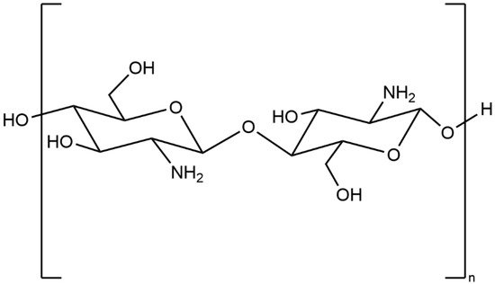
Chitosan is a random copolymer (
Figure 1), composed of
d-glucosamine (the deacetylated ones) and
N-acetyl-
d-glucosamine units linked through β-(1−4) glycosidic linkages [
18]. Chitosan can be obtained through partial deacetylation of chitin, which is the primary commercial source of chitosan [
33]. Chitin is the second most abundant natural polysaccharide following cellulose [
18]. The proportion of the deacetylated glucosamine units defines the deacetylation degree (DD). A DD value from 50 to 100% illustrates that the polymer consists of more
d-glucosamine units, implying the generation of chitosan. A DD value from 0 to 50% indicates that the polymer comprises more
N-acetyl-
d-glucosamine units, which indicates the copolymer is still chitin [
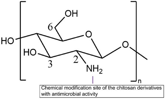
33]. Functional groups on the chitosan molecules consist of three nucleophilic functional groups (C3–OH, C6–OH, and C2–NH
2), acetyl amino, and glycoside bonds (
Figure 1). Among them, the stable acetyl amino bond and the glycosidic bond are not easy to modify; the enlarged steric hindrance of C3–OH decreases chemical reactivity significantly [
18]. Thus, C6–OH and C2–NH
2 groups are more likely to introduce other groups to chitosan derivatives to improve the physical and chemical properties [
34]. The antimicrobial effect of chitosan and chitosan derivatives was observed in a multitude of research [
35,
36,
37]. When chitosan is dissolved, the amino groups (–NH
2) of glucosamine units are protonated to –NH
3+. The polycationic charge of chitosan is generally thought to be the most significant factor bringing about antimicrobial effects due to electrostatic interaction between the polycationic chitosan and microbial cell surface [
38]. Chitosan is only active in the acidic medium; however, it was reported that the limited activity above pH 6 may originate from the poor solubility above pH 6. Chitosan was observed to be equally active against
S. aureus at pH 5.5 and 7.2 (MIC = 256 μg/mL) [
39]. Further investigations are required to fully understand the importance of the polycationic charge of chitosan.
Figure 1. Structure of chitosan.
Therefore, chemical modification of chitosan can increase the solubility of chitosan in water medium and enhance chitosan’s antimicrobial properties.
3. Chemical Modification of Chitosan
Modification methods primarily consist of
N-substitution in C2–NH
2,
O-substitution in C6–OH, and free radical graft copolymerization [
34]. Amino groups demonstrate higher reactivity than hydroxyl groups. Hence,
O-substitution requires the protection of primary amino groups before modification [
40]. There are other chemical modification methods of chitosan. For instance, K. Zhang has utilized “click chemistry” and single-electron-transfer nitroxide-radical-coupling (SET-NRC) reaction to synthesize chitosan-g-(PEO-PLLA-PEO) polymer [
41]. Different methods of chemical modification define different properties. Amino groups or hydroxyl groups require protecting functional groups introduced in chitosan. The widely used protecting groups for amino groups are phthaloyl [
42] and acetyl [
18], while triphenylmethyl [
43], trimethylsilyl [
44], tertiarybutyldimethylsilyl [
45], and acetyl [
18]. The positive charge from the NH
3+ group of the glucosamine units in chitosan and chitosan derivatives generates antimicrobial effects and reacts electrostatically with negatively charged microbial cell membranes and biofilms components [
46,
47]. For alleviating limitations that antimicrobial properties exist in acidic medium (pH below 6), trimethyl, 2-hydroxy-3-trimethylammoniumyl, guanidinyl, trimethyl ammmoniumyl, pyridiniumyl, and quaternary alkyl groups are introduced in chitosan to obtain a permanent positive charge, which enables chitosan to gain antimicrobial properties in the neutral and basic medium [
18]. An overview of types of chemical modification is shown in
Figure 2. The preparation and biological activities of chitosan and chitosan derivatives with antimicrobial properties are shown in
Table 1.
Figure 2. Schematic diagram of chitosan chemical modification.
Table 1. Table showing preparation methods and biological activities of chitosan and its derivatives.
|
Polymer
|
Preparation
|
Biological Activities
|
Citation
|
|
Hydroxypropyl chitosan
|
Reacting with propylene epoxide under alkaline medium (NaOH)
|
Water solubility, film-forming property, antibacterial property
|
[39,48]
|
|
Thioglycolic Chitosan
|
Reacting with 1-ethyl-3-(3-dimethyl aminopropyl) carbodiimide and thioglycolic acid
|
High antimicrobial property and good mucoadhesive property
|
[49,50]
|
|
N-(2-(N,N,N-trimethylammoniumyl)acetyl)-chitin
|
A combination of Boc and tert-butyldimethylsilyl (TBDMS) protection strategies
|
Antimicrobial property and enhancing permeation
|
[51,52]
|
|
Carboxymethyl chitosan
|
Reacting with 2-chloroacetic acid with NaOH
|
Enhanced antimicrobial property and water solubility
|
[39]
|
|
N,N,N-trimethyl chitosan
|
Reacting with methylation reagents
|
Antimicrobial property and enhanced solubility in alkaline medium
|
[53,54]
|
|
N-(2-Hydroxyl) Propyl-3-
Trimethyl Ammonium
Chitosan
|
Reacting with glycidyl trimethyl ammonium chloride
|
Antimicrobial property and good aqueous solubility in acidic, neutral, and alkaline medium
|
[39,55]
|
|
Chitosan- polyethylene glycol-peptide (PEG)-peptide conjugate
|
The chitosan was PEGylated by a carboxyl and azideterminated polyethylene glycol; peptide was
conjugated onto chitosan-PEG through a click reaction
|
Antimicrobial property and antibiofilm activity against Pseudomonas aeruginosa
|
[56]
|
|
Thiosemicarbazone O-Carboxymethyl-chitosan
(TCNCMCs) derivatives
|
Preparation of O-Carboxymethyl chitosan (CMCs): alkalized chitosan reacting with monochloroacetic, acetic acid, and methanol; TCNCMCs preparation: CMC reacting with ammonium hydroxide and carbon disulfide, followed by adding sodium chloroacetate and hydrazine hydrate
|
High antimicrobial and antifungal properties
|
[57]
|
4. Action Modes of Chitosan against Pathogen Microorganisms
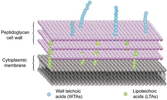
Chitosan and chitosan derivatives exhibit different action modes towards the Gram-positive and Gram-negative bacteria. This difference in mechanisms can be attributed to the difference in the component of the cell wall. As shown in
Figure 3, the cell wall of Gram-positive bacteria is composed of peptidoglycan, wall teichoic acids (WTAs) covalently linked to peptidoglycan, and lipoteichoic acids (LTAs) tied to the microorganism cell membrane [
82]. WTAs and LTAs contain a negatively charged anionic backbone [
82,
83]. The teichoic acids can provide arranged uniform high-density negative charges in the cell wall, thereby inhibiting the passage of ions across the membrane [
82].
Figure 3. Teichoic acid polymers are located within Gram-positive cell wall.
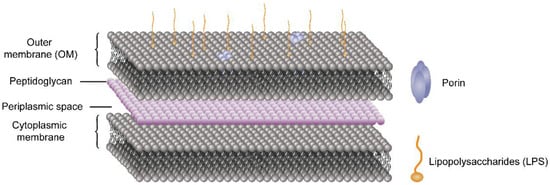
In the case of Gram-negative bacteria, the cell envelope consists of two membranes divided by a periplasmic space comprising a thin peptidoglycan layer [
84]. As shown in
Figure 4, the lipid composition of the outer membrane (OM) of Gram-negative bacteria is asymmetric: the outer leaflet contains lipopolysaccharide (LPS), whereas the inner leaflet comprises a variety of phospholipids.
Figure 4. Cell envelope of Gram-negative bacteria.
The surface of Gram-negative bacteria comprises negative charges from the phosphate and pyrophosphate groups of LPS in the outer layer of the OM [
85]. The widely accepted mechanisms of antimicrobial effects of chitosan can be explained in four models.
5. Current Treatment of the Enteric Infections
Rehydration is the primary treatment of enteric infections. Most cases can be efficiently treated with oral rehydration salts (ORS) [
7]. WHO and the United Nations Children’s Fund have recommended a reduced osmolarity ORS solution (245 mOsm/L), containing 75 mmol/L of sodium, 10 mmol/L citrates, 20 mmol/L potassium, and 75 mmol/L of glucose, for reducing stool output and the incidence of vomiting [
130]. Digestible food is usually recommended for patients with diarrhea [
131]. Severe suffers require intravenous fluids. Apart from normal saline infusion, lactated Ringer solution is also needed [
132]. Considering serum electrolyte level and urinary excretion, a potassium supplement can correct electrolyte disorders [
130].
Antibiotics demonstrated benefits, including the reduction of the duration of the condition as well as the alleviation of symptoms and complications, in randomized, controlled trials (RCT) in the treatment of enteric infections [
3]. In terms of
Shigella spp., enteroinvasive
Escherichia coli, enterotoxigenic
E. coli,
Vibrio cholerae,
Aeromonas, and
Plesiomonas, antibiotics have exhibited benefits for patients with moderate-to-severe disease in RCTs [
3]. For other pathogens (e.g.,
Campylobacter), antibiotics make a modest reduction in the duration of symptoms [
133]. Antibiotics are recommended for patients with the severe conditions or risk factors for severe illness, such as the elderly, pregnancies, and the immunocompromised [
133]. Nevertheless, for patients with nontyphoidal
Salmonella infection, antibiotics should not be given except in particularly severe cases, namely, in patients older than 50 years (who are at risk for a mycotic aneurysm), in infants younger than 12 months (who are at risk for
Salmonella meningitis), in individuals with cardiac or joint prostheses, and the immunocompromised [
134]. Furthermore, RCTs showed a prolonged load of the microorganisms in the stool [
134]. Antibiotics for
Shiga toxin-producing
E. coli (STEC) infection can increase the risk for hemolytic-uremic syndrome (HUS), particularly in children younger than 10 years [
135].
Moreover, antimotility agents may exacerbate the condition without effective antibiotics, resulting in absorption of enterotoxin and subsequently increased risks for HUS [
3]. Considering the progressively common antimicrobial resistance against enteric infection, researchers have focused applying nonantibiotic compounds as antimicrobial agents [
1]. Chitosan and chitosan derivatives showed antimicrobial activities against Gram-positive, Gram-negative, and fungi [
91]. Besides such direct bactericidal and fungicidal effects, chitosan and chitosan derivatives have other mechanisms for treating enteric infections.
6. Antibiofilm Properties of Chitosan and Chitosan Derivatives
Biofilm is an assemblage of microbial cells imbedded in a matrix of extracellular polymeric substances (EPS) produced by microbial cells [
136]. Biofilm contains extracellular polysaccharides, extracellular DNA (e-DNA), and proteins in the matrix [
137,
138,
139]. Biofilm can serve as a defense mechanism against antibiotics and microbicides [
140]. The polymeric matrix present in the biofilm impedes the access of antimicrobial compounds to the surface of bacterial cells [
47]. At present, researchers concentrated on combating the pathogenesis by inhibiting biofilm formation and uprooting mature biofilm by several reactive agents [
141,
142,
143,
144,
145,
146,
147,
148]. However, several current antibiofilm agents comprise cytotoxicity properties [
149,
150,
151,
152]. Therefore, a biocompatible, biodegradable, innocuous, nonallergenic, cost-effective, and environmentally-friendly antibiofilm compound from natural origin, chitosan can be a potential antimicrobial and antibiofilm agent [
153,
154,
155].
Antibiofilm property of chitosan is primarily attributed to the polycationic nature donated by amino groups of
N-acetylglucosamine monomers [
156,
157,
158]. Positive charges of chitosan can interact electrostatically with negatively charged biofilm compositions such as EPS, extracellular proteins, and e-DNA, inducing an inhibitory effect on microbial biofilm [
159,
160]. Thenceforth, chitosan has access to the passage through the biofilm, killing microorganisms subsequently via the above-mentioned action modes. Moreover, the conjugation of chitosan with antimicrobial peptides (AMPs) can create cationic peptide-polysaccharide to enhance electrostatic interactions between the copolymer and microbial cell membrane components as well as inhibit the growth of Gram-positive and Gram-negative bacteria [
161,
162]. Chitosan and chitosan derivatives in different structural forms (e.g., chemical modified ones, nanoparticles, conjugation with other polymers, conjugation with antibiofilm nanoparticles, and vehicles for drugs) exhibit antibiofilm effects against microbial biofilm [
47]. Such materials in food industry or for personal use can prevent the intake of pathogens which is responsible for enteric infections, especially methicillin-resistant
Staphylococcus aureus [
163], vancomycin-resistant
Staphylococcus aureus [
164], and
Listeria [
165,
166].
7. Gut Microbiota and Colonization Resistance against Enteric Infections
A variety of microorganisms colonize the human gastrointestinal (GI) tract, collectively termed gut microbiota, including bacteria, viruses, fungi, archaebacteria, and protozoa [
167]. The majority of bacteria colonize the colon (about 10
14) [
168], which is much larger than the concentration of microbes in the stomach and the upper part of the intestine owing to the acidic gastric ambient and the rapid passage of the food through the upper GI tract [
167].
Discoveries of the Human Microbiome Project (HMP) boosted the understanding of the host-microbe interactions in the intestine [
169,
170]. The microbiome contains 3.3 million nonredundant microbial genes, which is 150-times larger than the human genome [
169]. Healthy gut microbiota is dominated by
Bacteroidetes and
Firmicutes [
171], followed by
Actinobacteria,
Proteobacteria, and
Verrucomicrobia [
172], as well as methanogenic archaea (e.g.,
Methanobrevibacter smithii), eukaryote (e.g., yeasts), and various phages [
173]. Environmental factors interact with endogenous factors to form individuals’ unique microbial phenotypes [
174]. The gut microbiota in early life is shaped by types of delivery for pregnancy [
175], host immune system [
176], maternal microbiome [
177], environmental microbes [
178,
179], as well as the solid food after birth [
180]. Multiple host-endogenous and host-exogenous factors shape the gut microbiota to form a resilient and balanced gut microbiota [
181]. However, once there are changes in those influencing factors, the dysbiosis can induce gut barrier dysfunction, invasion of pro-inflammatory contents (e.g., LPS), and low-grade chronic inflammation [
182]. Various studies showed the association between dysbacteriosis and obesity [
183], type 2 diabetes [
184], and inflammatory bowel disease [
185].
Healthy gut microbiota can prevent enteric infections via a variety of mechanisms, including the production of antimicrobial agents, nutrient competition, aid to intestinal mucosal barrier integrity, and immune response activation [
186]. These mechanisms collectively contribute to colonization resistance (CR) against pathogens. The metabolites of the gut microbiota contain antibacterial properties, including short-chain fatty acids (SCFAs), secondary bile acids (BAs), and bacteriocins [
187].
8. The Role of Chitosan in the Treatment of Enteric Infections
In the treatment of enteric infections, chitosan, oligo-chitosan, and their derivatives can serve as antimicrobial drug delivery vehicles, as prebiotics to improve the colonization resistance against enteric infections, and as antimicrobial agents independently, and conjugate with other reactive agents to increase antimicrobial activities.
8.1. Chitosan as Drug Delivery System
Apart from the antimicrobial properties, chitosan can increase the pH sensitivity of the drug release of antimicrobial agents, enabling intestine-targeted antimicrobial effects [
237]. Formulated chitosan-coated amphotericin-B-loaded nanostructured lipid carriers (ChiAmp NLC) can prevent Amphotericin-B from exposure outside the intestine, decrease toxicity to enterocytes and erythrocytes, and thus enhance the bioavailability of Amphotericin-B [
237]. Bovine serum albumin (BSA)-chitosan core (CS)-nano-delivery-systems (BSA-CS-NDS) enables the effective delivery of carvacrol, a natural antimicrobial agent, to the intestine for successful removal of
Salmonella enterica [
238]. Carla Mura and her coworkers prepared two chitosan amide conjugates of metronidazole, metronidazole-glutaryl- and metronidazole-succinyl-chitosan conjugates. The results have shown adequate stability of the two conjugations in the acidic environment as well as a potential as colon-targeted delivery systems of metronidazole [
239]. Chitosan nanoparticle intracellular delivery system of ceftriaxone sodium can reduce the count of
Salmonella typhimurium in intestinal cells and macrophages [
240]. Chitosan- coated alginate microparticle system of lactoferrin, a protein delaying
Clostridioides difficile growth and inhibiting toxin production, can assist in the stability of lactoferrin and protection from
C. difficile-induced intestinal epithelial damage [
241]. Albendazole-associated chitosan nanoparticles (ABZ-CS-NPs) can improve the stability of albendazole in acidic ambient and absorption of albendazole in the intestine, suggesting improved effects of killing enteric parasites [
242]. Chitosan nanoparticles can serve as carriers for supernatant of mesenchymal stem cells for the treatment of multidrug-resistant (MDR)
Vibrio cholerae infections [
1].
8.2. Chitosan as Antimicrobial Agents
Chitosan oligosaccharide sensitizes multidrug resistant
Staphylococcus aureus to antibiotic formulations by electrostatically interacting with multidrug efflux pumps [
243]. Chitosan nanoparticles can serve as a good candidate among natural giardiacidal agents [
244]. Mohamed Mammeri and his coworkers have observed the anti-cryptosporidium properties of chitosan in vitro and in vivo [
244]. Chitosan and chitosan nanoparticles were observed to contain antimicrobial activity against gastrointestinal pathogens such as
Salmonella spp. and
E. coli [
245]. Chitosan nanoparticles can facilitate the inhibition of norovirus, the most frequent cause of nonbacterial diarrhea [
246]. A study investigated the potential effect of chitosan particles to enhance the immune response against
Hymenolepis nana, the most common intestinal cestode [
247]. Moreover, Aleksandra Milewska and her coworkers have prepared a cationically modified chitosan,
N-(2-hydroxypropyl)-3-trimethylammonium chitosan chloride (HTCC), which can be utilized as potential inhibitors of currently circulating highly pathogenic coronaviruses, namely severe acute respiratory syndrome coronavirus 2 [SARS-CoV-2] and Middle East respiratory syndrome coronavirus [MERS-CoV] [
248].
8.3. Chitosan Conjugation with Other Polymers or Nanoparticles
As chitosan can reduce the numbers of
E. coli O157:H7 in feces, remain nontoxic to host, and possess antimicrobial properties against
E. coli, antibody-conjugated chitosan nanoparticles are utilized to selectively kill
Shiga toxin-producing
Escherichia coli (STEC) without inhibiting the growth of beneficial bacteria [
249]. Cranberry proanthocyanidin-chitosan composite nanoparticles (PAC-CHT NPs), which are formulated using 10:1 to 30:1 proanthocyanidin to chitosan weight ratio, can form stable and bioactive nanoparticles for potential applications in the treatment of pathogenic
Escherichia coli infection [
250]. Ziyin Cui et al. formulated mannose-modified chitosan microspheres conjugating with mucosal vaccines against
Pseudomonas aeruginosa infection in the intestine [
251]. Preparation methods and biological activities of chitosan conjugation with other polymers and nanoparticles are shown in
Table 2.
Table 2. Table showing preparation methods and biological activities of chitosan conjugation with other polymers and nanoparticles.
|
Chitosan Conjugation with Other Polymers and Nanoparticles
|
Preparation
|
Biological Activities
|
Citation
|
|
Chitosan coated PLA (poly d, l-lactic acid) nanoparticles
|
Coated on the surface of PLA nanoparticles which are prepared by nanoprecipitation method
|
High cornea permeation and high sustained release of 5-FU in conjunctival/corneal squamous cell carcinoma
|
[252]
|
|
Antibody-conjugated chitosan nanoparticles
|
Preparation of chitosan nanoparticles (CNs): chitosans are dissolved in 2% acetic acid and mixed with 1% Tween 80, followed by the addition of a 10% sodium sulfate solution and centrifugation at 8200 g; Bioconjugation of IgY antibodies to CN: The CNs are dissolved in 0.1 M sodium acetate buffer, followed by an addition of antibodies, 1-Ethyl-3-(3-dimethylaminopropyl)carbodiimide (EDC), sulfo-N-hydroxysulfosuccinimide, and the centrifugation at 39,800 g
|
Enhanced and specific antimicrobial activities against Shiga toxin-producing Escherichia coli (STEC)
|
[249]
|
|
Chitosan-based nanocomposites
|
Chitosan prepared in acetic acid, silver, and copper nanoparticles are dispersed in ethanol by sonication and precipitated in an alkaline medium.
|
Increased antimicrobial properties
|
[253]
|
|
Cranberry proanthocyanidins-chitosan composite
nanoparticles (PAC-CHT NPs)
|
Chitosan (CHT) is prepared in 0.5% acetic acid, filtered and degassed, followed by linking to cranberry proanthocyanidins (PAC) through hydrogen bonding.
|
Higher bioactive than CHT and PAC alone. Increased bioactivity of PAC-CHT NPs against E. coli.
|
[250]
|
|
Antibody-loaded-mannose-modified chitosan microspheres
|
Mannose-modified chitosan (MC) preparation: dissolved chitosan is treated with mannose and sodium cyanoborohydride; chitosan microsphere preparation: sodium tripolyphosphate (TPP) solution is added dropwise to MCs under 15 W sonication; antibody-loaded chitosan microsphere preparation: dispersing 5 mg of antibodies in 1.0 mL of phosphate-buffered saline (PBS) containing 30 mg of microspheres.
|
Mannose-modified chitosan microspheres can serve as a promising subunit delivery system for vaccines against P. aeruginosa infection.
|
[251]
|
|
Chitosan-Caffeic Acid Conjugate
|
Chitosan is dissolved in 2% acetic acid and reacts with 1.0 M hydrogen peroxide containing ascorbic acid. Caffeic acid is added to the mixture for 24 h at room temperature.
|
Antibacterial activity of against acne-related bacteria
|
[254]
|
8.4. Chitosan as Prebiotics to Improve Colonization Resistance against Enteric Pathogens
Chitosan and chitosan derivatives can be fermented by the intestinal microbiota, and the metabolites such as short-chain fatty acids (SCFAs) are capable of increasing the growth of probiotics [
255,
256] (e.g.,
Bifidobacterium spp. and
Lactobacillus spp.) and the exclusion of pathogens (e.g.,
Streptococcus mutans [
257],
E. coli,
Shigella dysenteriae,
Aeromonas hydrophila,
Salmonella typhimurium and
Bacillus cereus [
28]).
Furthermore, chitosan can serve as prebiotics to inhibit or prevent the growth of harmful bacteria by producing SCFAs and other beneficial metabolites [
187]. The improved gut microbiota can prevent the infection of conditional pathogens such as
Clostridium difficile. In the treatment of
Shiga-toxin-producing
E. coli (STEC), acetate was more effective in inhibition of STEC than butyrate and propionate [
258], and butyrate can improve STEC bacterial clearance [
259]. One study has investigated the effects of SCFAs on
Yersinia enterocolitica at 4 °C. Propionic acid is similarly effective in inhibiting the growth of
Yersinia enterocolitica with anaerobic and aerobic culture methods [
260]. However,
V. cholerae uses a wide variety of mechanisms to overcome colonization resistance.
V. cholerae is capable of using its acetate switch, the shifting from elimination to assimilation of acetate, to increase its virulence [
261].
This entry is adapted from the peer-reviewed paper 10.3390/molecules26237136