Tick Immunobiology: History
Please note this is an old version of this entry, which may differ significantly from the current revision.
Contributor:

Ticks are hematophagous ectoparasites that infest a diverse number of vertebrate hosts. The tick immunobiology plays a significant role in establishing and transmitting many pathogens to their hosts. To control tick infestations, the acaricide application is a commonly used method with severe environmental consequences and the selection of tick-resistant populations.

  • anti-tick vaccines
  • tick immune system
  • extracellular traps
  • Extracellular Traps Formation (ETosis)
  • Neutrophils Extracellular Traps Formation (NETosis)
  • cattle diseases

1. Introduction

Direct damages caused by tick bites include blood loss through engorgement, stress, anemia, inflammation, depression of immune function, and allergies caused by antigens and coagulants of the tick saliva [1]. Additionally, ticks’ saliva contains several protein families that induce toxicoses, including paralysis, possibly induced by blocking ion channels that regulate the cell membrane potential in tissues [2].

Ticks are efficient vectors and reservoirs of pathogens, including viruses, bacteria, and protozoans, transmitted to vertebrate hosts [3]. Over time, ticks and ticks-transmitted parasites have co-evolved with their hosts as part of the ecological niche’s equilibrium [4].

On the other hand, the indiscriminate use of chemical acaricides has allowed the selection of resistant populations of ticks and constant environmental pollution [5]. Since the first reports of resistance development of Rhipicephalus spp. to chemical compounds, this ability of ticks has evolved, and now they are resistant to almost all the available acaricides [6][7]. In this regard, the search for new tick control measures is a priority to cope with the adverse impacts of ticks and TBDs on cattle.

So far, the information about the interaction pathogen-vector and the plasticity of ticks’ immune system in response to pathogens is scarce. This review presents a general view of ticks’ immune system and the main molecules that participate in its defense. Here, we propose that discovering new mechanisms in ticks, such as the production of extracellular traps (ETosis), also provides alternatives to identify new targets to develop strategies to control ticks and TBDs. Additionally, the study of the tick immune system components demands a new and more integrative vision of the molecules that participates in a basic but evolutionarily well-adapted process.

2. The Tick Immunobiology

The first response line against pathogens in vertebrates is innate immunity, nonspecific, non-anticipatory, non-clonal, and germline-encoded. On the other hand, adaptive immunity is specific clonal, anticipatory, somatic, and creates immunological memory [8].
In ticks, the innate immune response comprises two major components: cellular defenses, which includes phagocytosis and encapsulation, and humoral responses, involving the AMPs, defensins, enzymes, among other molecules, expressed by the hemocytes, fat body, and the midgut [9]. In contrast, the adaptive immune response in ticks is controversial and is not yet elucidated. Similarly, several essential mechanisms of tick immune response to identify new molecules or targets with therapeutical applications remain unclear.

2.1. Tick Signaling Pathways in Immune Response

The reported tick genomic information has contributed to the study and elucidation of the immune response signaling pathways [10][11][12]. In R. microplus, in silico analysis revealed that the Toll, Immune deficiency (IMD), Jun-N-terminal kinase (JNK), Janus kinase/signal transducer, and activator of transcription (JAK/STAT) are the main signaling pathways that regulate the innate immune response. Several components of these pathways are homologous in other arthropods; however, many were not found in the ticks’ databases [13].

2.1.1. Toll Pathway

The Toll pathway is mainly responsible for recognizing and eliminating fungi and Gram-positive bacteria in Drosophila, and viruses in the mosquito Aedes aegypti, in both cases by synthesizing AMPs secreted into the hemolymph [14][15]. In ticks, how the Toll pathway operates is unclear. Nevertheless, most Toll pathway components have been identified by genomic analyses, including the NF-kB Dorsal, which suggests the existence of a conserved mechanism in arthropods. In contrast, the DIF (NF-kB transcription factor dorsal-related immunity factor) is the only Toll pathway component not yet reported in tick species [9]. Some reports in ticks have shown up-regulation of the Toll pathway genes in the presence of heat-killed Saccharomyces cerevisiae, Enterobacter cloacae, Micrococcus luteus, and live Rickettsia rickettsii. Additionally, Anaplasma marginale down-regulated the gene expression, suggesting that pathogen can regulate this pathway to establish in a tick [13].

2.1.2. Immune Deficiency (IMD) Pathway

In Drosophila, the IMD pathway activated by diaminopimelic acid (DAP)-type peptidoglycan present in the cell wall of most Gram-negative bacteria and some Gram-positive bacteria triggers the synthesis of specific AMPs [16]. In ticks, genomic analyses showed the lack of orthologs of crucial elements in this pathway as death-related ced-3/Nedd-2-like protein (DREDD), Fas-associated protein with death domain (FADD), and Transmembrane PGRP. It is essential to highlight that the losses of IMD pathway components are not exclusive to ticks since they are also absent in hemipterans and other arachnids [17]. Additionally, NF-kB/Relish and JNK pathway and some associated molecules to this process are conserved [18]. Drosophila suggested that many components of this pathway could participate in the immune response to the microbial challenge, stress, and epithelial injury, such as the transcriptional factor basket, encoded by the single JNK gene (bsk) [19].
On the other hand, Shaw et al. [20] showed that lipids such as POPG and PODAG (1-palmitoyl-2-oleoyl-snglycerol-3-phosphoglycerol and 1-palmitoyl-2-oleoyl diacylglycerol, respectively) derived from Borrelia burgdorferi, A. phagocytophilum, and A. marginale act as recognition of pathogen-associated patterns (PAMPs) and controls the IMD pathway activation and induces the colonization in I. scapularis and Dermacentor andersoni.

2.1.3. Janus Kinase/Signal Transducer and Activator of Transcription (JAK/STAT) Pathway

The JAK/STAT pathway is well conserved in Drosophila and ticks. The fruit fly indirectly controls bacterial and fungal infections and is especially sensitive to viral infections [17][21]. In ticks, this pathway is activated by bacteria or protozoan pathogens and can positively or negatively regulate the infection in ticks. In I. scapularis, the JAK/STAT pathway has a vital role in the signaling between the gut microbiota and pathogen colonization [22][23]. Additionally, in this tick, AMPs expression in the salivary glands and hemocytes is regulated by the JAK/STAT pathway, where 5.3-kDa family members participate during the infection of A. phagocytophilum [3]. Negative regulation of AMPs expression, specifically in the salivary glands (ixodidin and lysozyme) and the gut and salivary glands (defensin), occurs in R. microplus STAT-deficient [24].

2.1.4. RNA Interference (RNAi) Pathway

In Drosophila, there are four RNAi pathways reported, three endogenous: microRNA (miRNA), small interfering RNA (endo-siRNA), and piwi-interacting RNA (piRNA), and one exogenous small interference (siRNA) [25]. Interestingly, in I. scapularis, RNAi functions efficiently in many tissues. Omics-studies-based analyses revealed the presence of all components for the endogenous and exogenous RNAi machinery, including Dicer (Dcr), Argonaute (Ago), dsRNA binding proteins, exonucleases, and even RNA-dependent RNA polymerases [26]. In general terms, the RNAi mechanism consists of the enzymatic-complex activation after a viral infection. A long viral dsRNA is recognized and cleaved by Dcr to produce 21 nt siRNAs, known as viRNAs. These viRNAs transfer to Ago, which couples to other members of the RNA-induced silencing complex (RISC). Finally, only one strand of the viRNA remains coupled to RISC and guides the degradation of complementary viral RNA [27][28]. Undoubtedly, the tick RNAi system is still a new field awaiting further investigation, not only for its role in defense against infections but also for its potential in studying tick gene function and the screening and characterization of tick protective antigens [9].
On the other hand, RNAi as a molecular tool has helped elucidate the function of tick proteins and identify novel targets at the tick-pathogen interface. In this regard, some reports in R. microplus, shown that RNAi silencing of some transcriptional factors of Toll, IMD, and Jak/STAT signaling pathways, lead to consider IMD pathway as the main regulator in the midgut and salivary glands during A. marginale infection [24]. The bacterial load also can be modulated by RNAi, as in Amblyomma maculatum, where the silencing by RNAi of the antioxidant selenoprotein thioredoxin reductase (TrxR) caused a reduction in the bacterial load of Rickettsiaceae in the midgut and salivary glands [29]. Additionally, by silencing of glutathione peroxidase (GPx) gene, the load of B. burgdorferi is reduced in I. scapularis saliva [30]. In other model systems, the crosstalk between RNAi and arthropods immune system defense has been explored. As in Culex mosquitoes, where Dcr-2, a central component of the siRNA pathway, recognizes West-Nile virus (WNV) dsRNA and activates a signaling cascade. This signaling stimulates Relish, increasing the expression of gene Vago (cytokine functional homolog) and activating the JAK-STAT pathway leading to an effective antiviral response [31].
These studies demonstrate that RNAi silencing of arthropod’s immune response genes is an approach that allows identifying vaccine candidates. Nevertheless, the application of this tool as tick control requires optimization of the delivery method of the dsRNA; besides the use of microinjection and soaking, probably the use of exosomes as a vector for dsRNA delivery could contribute to a successful strategy against vectors [32].

2.2. The Cellular Immune Response

The hemocytes are the surrounding cells in the hemolymph. In ticks, hemocytes are the main effectors of the cellular immune response to control pathogens by phagocytosis, encapsulation, and nodule formation (Figure 1) [33][34]. PAMPs on the microbial surface regulate these mechanisms. Phagocytosis is a primary cellular defense response to recognize invading pathogens, destroying them by endocytosis in phagolysosomes. This process might be regulated in ticks by a complement-like system composed of thioester-containing proteins, fibrinogen-related lectins, and convertase-like factors [3][33]. At the moment, there exist evidence that in Ixodes ricinus, C3-like molecules (IrC3-1, IrC3-2, IrC3-2), considered to be analogous molecules of the complement system, facilitate the phagocytosis of yeasts and Borrelia spirochaetes (Figure 1) [35].
Figure 1. Immunological responses in a tick R. microplus during tick-bovine interactions. The immune response in ticks comprises cells, molecules, and proteins acting in combination to destroy pathogens. The tick also should afford the potential risk that would represent the commensals located in the skin of the bovine that, in turn, could be potentially infectious to the tick. Figure created in the Mind the Graph platform (www.mindthegraph.com, accessed on 9 September 2021).
On the other hand, encapsulation is a process that implies hemocytes accumulation around the parasite, foreign objects, or microbes, forming organized concentric layers. Specific lectins participate as opsonizing molecules that cause bacteria aggregation in the nodulation, but little is known about encapsulation and nodulation mechanisms. In this regard, both processes have been reported in Dermacentor variabilis. After inoculating this tick with Escherichia coli, the hemocytes form aggregates around the bacteria, a characteristic feature of nodules formation. Although the encapsulation process was evaluated in an artificial implant of Epon-Araldite inoculated under the tick cuticle, it remains to know whether encapsulation also occurs against microorganisms and if this process links with hemolymph coagulation and cellular response against some pathogens [36][37].

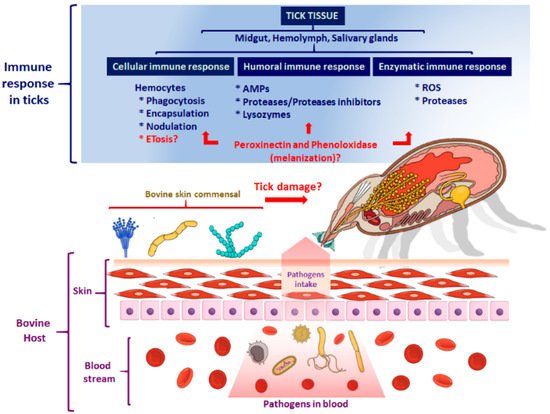
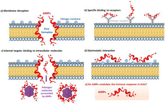
3. The Humoral Immune Response of Ticks

In ticks, this response consists of soluble effector molecules, called “humoral factors,” including AMPs, lysozymes, defensins, coagulation factors, proteases, and protease inhibitors (serpins, cystatins), enzymes involved in the oxidative burst and detoxification, as well as other recognition molecules (Figure 1) [33].

The Tick Repertoire of Humoral Factors

The AMPs are considered ancient evolutionary systems of immune defenses based on humoral components widely distributed in nature [38]. The size of AMPs is usually small (below 10 kDa) and has cationic character. However, some anionic peptides have been reported, and they also differ in their amino acid sequence and mode of action (Figure 2) [39].
Figure 2. Mechanisms of action of the AMPs. The mechanisms of action of the AMPs vary depending on the interaction with the components of the cytoplasmic membrane. (a) Membrane disruption. The AMPs disrupt the integrity of the pathogen’s membrane by forming pores that eventually destabilize the structure and leak the internal content. (b) Specific binding to receptors. Some AMPs bind specifically to pathogen receptors, causing a steric hindrance and avoiding the correct binding to the ligands. (c) Internal targets: binding to intracellular molecules. The AMPs also aim at targets inside the pathogen, where they reach the intracellular molecules blocking their function. (d) Electrostatic interaction. The electrostatic interactions between the AMPs and the phospholipids on the pathogen’s surface change the membrane potential, resulting in deficient activity of the membrane components with the external environment. (e) Do AMPs modulate the immune response in ticks? The AMPs may be involved either in signaling pathways or activating processes of the immune response to pathogens. They may also be acting as main effectors in specific immunological mechanisms. Figure created in the Mind the Graph platform (www.mindthegraph.com, accessed on 9 September 2021).
AMPs are expressed mainly in hemocytes, fat body, gut, ovaries, and salivary glands. They respond to either blood-feeding or microbial challenge, and their release to the hemolymph is essential in the immune system response [40][22][41]. The antibacterial mechanism of AMPs targets Gram-positive bacteria, yeast, and fungi [42]. Interestingly, ticks use some proteins from the blood meal as a source for the production of AMPs, as in hemoglobin-derived AMPs (hemocidines) produced by the proteolytic activity of proteinases present in the tick gut [43].
Lysozymes are another type of molecule with a broad microbicidal spectrum. These digestive enzymes hydrolyze N-acetyl-muramic bonds and N-acetyl-D-glucosamine residues of the bacterial peptidoglycan. In D. variabilis, lysozymes are more abundant in hemolymph than in other organs and synergize with some defensins, breaking the bacterial cell wall and accelerating some AMPs actions [33].
Several immune responses involved in the recognition and control of pathogens depend on the activity of specific proteases or peptidases. Serine proteases are key regulating molecules for several immune responses, including coagulation, AMPs synthesis, and protein degradation [44]. In addition, the activation of this pathway is controlled by three serine proteases: factor C, factor B, and pro-clotting enzymes, which are activated in the presence of lipopolysaccharide (LPS) and released to the hemolymph that in turn, cause the immobilization of binding pathogen [45]. Protease inhibitors control various proteolytic pathways that play an essential role in tick immunity [46]. Their functions include antimicrobial activity, egg production regulation, development, thrombin, trypsin, and elastase inhibition [47]. Interestingly, the pan protease inhibitors of alpha-macroglobulin type are in the tick hemolymph, where they protect the ticks against invading microbes’ proteases [48].


This entry is adapted from the peer-reviewed paper 10.3390/pathogens10111511

References

  1. Ghosh, S.; Azhahianambi, P.; Yadav, M.P. Upcoming and future strategies of tick control: A review. J. Vector Borne Dis. 2007, 44, 79–89.
  2. Maritz, C.; Louw, A.; Gothe, R.; Neitz, A. Neuropathogenic properties of Argas (Persicargas) walkerae larval homogenates. Comp. Biochem. Physiol. Part A Mol. Integr. Physiol. 2001, 128, 233–239.
  3. Hajdušek, O.; Šíma, R.; Ayllón, N.; Jalovecká, M.; Perner, J.; De La Fuente, J.; Kopáček, P. Interaction of the tick immune system with transmitted pathogens. Front. Cell. Infect. Microbiol. 2013, 3, 26.
  4. Jongejan, F.; Uilenberg, G. The global importance of ticks. Parasitology 2004, 129, S3–S14.
  5. Kunz, S.; Kemp, D. Insecticides and acaricides: Resistance and environmental impact. Rev. Sci. Tech. 1994, 13, 1249–1286.
  6. Abbas, R.Z.; Zaman, M.A.; Colwell, D.; Gilleard, J.; Iqbal, Z. Acaricide resistance in cattle ticks and approaches to its management: The state of play. Vet. Parasitol. 2014, 203, 6–20.
  7. George, J.E.; Pound, J.M.; Davey, R.B. Chemical control of ticks on cattle and the resistance of these parasites to acaricides. Parasitology 2004, 129, S353–S366.
  8. Kvell, K.; Cooper, E.; Engelmann, P.; Bovari, J.; Németh, P. Blurring borders: Innate immunity with adaptive features. Clin. Dev. Immunol. 2007, 83671.
  9. Fogaça, A.C.; Sousa, G.; Pavanelo, D.B.; Esteves, E.; Martins, L.A.; Urbanova, V.; Kopacek, P.; Daffre, S. Tick immune system: What is known, the interconnections, the gaps, and the challenges. Front. Immunol. 2021, 12, 119.
  10. Guerrero, F.; Miller, R.; Rousseau, M.-E.; Sunkara, S.; Quackenbush, J.; Lee, Y.; Nene, V. BmiGI: A database of cDNAs expressed in Boophilus microplus, the tropical/southern cattle tick. Insect Biochem. Mol. Biol. 2005, 35, 585–595.
  11. Megy, K.; Emrich, S.J.; Lawson, D.; Campbell, D.; Dialynas, E.; Hughes, D.S.; Koscielny, G.; Louis, C.; MacCallum, R.; Redmond, S.; et al. VectorBase: Improvements to a bioinformatics resource for invertebrate vector genomics. Nucleic Acids Res. 2012, 40, D729–D734.
  12. Gulia-Nuss, M.; Nuss, A.; Meyer, J.M.; Sonenshine, D.E.; Roe, R.M.; Waterhouse, R.M.; Sattelle, D.B.; De La Fuente, J.; Ribeiro, J.; Megy, K.; et al. Genomic insights into the Ixodes scapularis tick vector of Lyme disease. Nat. Commun. 2016, 7, 10507.
  13. Rosa, R.D.; Peixoto, J.; Mesquita, R.D.; Kalil, S.P.; Pohl, P.C.; Braz, G.R.; Fogaça, A.; Daffre, S. Exploring the immune signalling pathway-related genes of the cattle tick Rhipicephalus microplus: From molecular characterization to transcriptional profile upon microbial challenge. Dev. Comp. Immunol. 2016, 59, 1–14.
  14. Xi, Z.; Ramirez, J.L.; Dimopoulos, G. The Aedes aegypti Toll pathway controls dengue virus infection. PLoS Pathog. 2008, 4, e1000098.
  15. Lemaitre, B. The road to Toll. Nat. Rev. Immunol. 2004, 4, 521–527.
  16. Kleino, A.; Silverman, N. The Drosophila IMD pathway in the activation of the humoral immune response. Dev. Comp. Immunol. 2014, 42, 25–35.
  17. Palmer, W.J.; Jiggins, F.M. Comparative genomics reveals the origins and diversity of arthropod immune systems. Mol. Biol. Evol. 2015, 32, 2111–2129.
  18. Narasimhan, S.; Schuijt, T.J.; Abraham, N.M.; Rajeevan, N.; Coumou, J.; Graham, M.; Robson, A.; Wu, M.-J.; Daffre, S.; Hovius, J.W.; et al. Modulation of the tick gut milieu by a secreted tick protein favors Borrelia burgdorferi colonization. Nat. Commun. 2017, 8, 184.
  19. Tafesh-Edwards, G.; Eleftherianos, I. JNK signaling in Drosophila immunity and homeostasis. Immunol. Lett. 2020, 226, 7–11.
  20. Shaw, D.K.; Wang, X.; Brown, L.J.; Chávez, A.S.O.; Reif, K.E.; Smith, A.A.; Scott, A.J.; McClure, E.E.; Boradia, V.M.; Hammond, H.L.; et al. Infection-derived lipids elicit an immune deficiency circuit in arthropods. Nat. Commun. 2017, 8, 14401.
  21. Dostert, C.; Jouanguy, E.; Irving, P.; Troxler, L.; Galiana-Arnoux, D.; Hetru, C.; Hoffmann, J.A.; Imler, J.-L. The Jak-STAT signaling pathway is required but not sufficient for the antiviral response of Drosophila. Nat. Immunol. 2005, 6, 946–953.
  22. Liu, L.; Dai, J.; Zhao, Y.O.; Narasimhan, S.; Yang, Y.; Zhang, L.; Fikrig, E. Ixodes scapularis JAK-STAT pathway regulates tick antimicrobial peptides, thereby controlling the agent of human granulocytic anaplasmosis. J. Infect. Dis. 2012, 206, 1233–1241.
  23. Shuai, K.; Ziemiecki, A.; Wilks, A.F.; Harpur, A.G.; Sadowski, H.B.; Gilman, M.Z.; Darnell, J.E. Polypeptide signaling to the nucleus through tyrosine phosphorylation of Jak and Stat proteins. Nat. Cell Biol. 1993, 366, 580–583.
  24. Capelli-Peixoto, J.; Carvalho, D.D.; Johnson, W.C.; Scoles, G.A.; Fogaça, A.C.; Daffre, S.; Ueti, M.W. The transcription factor Relish controls Anaplasma marginale infection in the bovine tick Rhipicephalus microplus. Dev. Comp. Immunol. 2017, 74, 32–39.
  25. Karlikow, M.; Goic, B.; Saleh, M.-C. RNAi and antiviral defense in Drosophila: Setting up a systemic immune response. Dev. Comp. Immunol. 2014, 42, 85–92.
  26. Schnettler, E.; Tykalová, H.; Watson, M.; Sharma, M.; Sterken, M.; Obbard, D.; Lewis, S.H.; McFarlane, M.; Bell-Sakyi, L.; Barry, G.; et al. Induction and suppression of tick cell antiviral RNAi responses by tick-borne flaviviruses. Nucleic Acids Res. 2014, 42, 9436–9446.
  27. Blair, C.D. Mosquito RNAi is the major innate immune pathway controlling arbovirus infection and transmission. Future Microbiol. 2011, 6, 265–277.
  28. Asgari, S. Role of microRNAs in arbovirus/vector interactions. Viruses 2014, 6, 3514–3534.
  29. Budachetri, K.; Karim, S. An insight into the functional role of thioredoxin reductase, a selenoprotein, in maintaining normal native microbiota in the Gulf Coast tick (Amblyomma maculatum). Insect Mol. Biol. 2015, 24, 570–581.
  30. Narasimhan, S.; Sukumaran, B.; Bozdogan, U.; Thomas, V.; Liang, X.; DePonte, K.; Marcantonio, N.; Koski, R.A.; Anderson, J.F.; Kantor, F.; et al. A tick antioxidant facilitates the Lyme disease agent’s successful migration from the mammalian host to the arthropod vector. Cell Host Microbe 2007, 2, 7–18.
  31. Paradkar, P.N.; Duchemin, J.-B.; Voysey, R.; Walker, P.J. Dicer-2-dependent activation of Culex vago occurs via the TRAF-Rel2 signaling pathway. PLoS Negl. Trop. Dis. 2014, 8, e2823.
  32. Lässer, C. Exosomes in diagnostic and therapeutic applications: Biomarker, vaccine and RNA interference delivery vehicle. Expert Opin. Biol. Ther. 2015, 15, 103–117.
  33. Sonenshine, D.E.; Hynes, W.L. Molecular characterization and related aspects of the innate immune response in ticks. Front. Biosci. 2008, 1, 7046–7063.
  34. Strand, M.R. The insect cellular immune response. Insect Sci. 2008, 15, 1–14.
  35. Urbanová, V.; Šíma, R.; Sauman, I.; Hajdusek, O.; Kopáček, P. Thioester-containing proteins of the tick Ixodes ricinus: Gene expression, response to microbial challenge and their role in phagocytosis of the yeast Candida albicans. Dev. Comp. Immunol. 2015, 48, 55–64.
  36. Eggenberger, L.R.; Lamoreaux, W.J.; Coons, L.B. Hemocytic encapsulation of implants in the tick Dermacentor variabilis. Exp. Appl. Acarol. 1990, 9, 279–287.
  37. Ceraul, S.M.; Sonenshine, D.E.; Hynes, W. Resistance of the tick Dermacentor variabilis (Acari: Ixodidae) following challenge with the bacterium Escherichia coli (Enterobacteriales: Enterobacteriaceae). J. Med. Entomol. 2002, 39, 376–383.
  38. Zhang, L.; Gallo, R.L. Antimicrobial peptides. Curr. Biol. 2016, 26, R14–R19.
  39. Fogaça, A.C.; Lorenzini, D.M.; Kaku, L.M.; Esteves, E.; Bulet, P.; Daffre, S. Cysteine-rich antimicrobial peptides of the cattle tick Boophilus microplus: Isolation, structural characterization and tissue expression profile. Dev. Comp. Immunol. 2004, 28, 191–200.
  40. Kopácek, P.; Hajdusek, O.; Buresová, V.; Daffre, S. Tick innate immunity. Adv. Exp. Med. Biol. 2010, 708, 137–162.
  41. Taylor, D. Innate Immunity in Ticks: A review. J. Acarol. Soc. Jpn. 2006, 15, 109–127.
  42. Cabezas-Cruz, A.; Tonk, M.; Bleackley, M.R.; Valdés, J.J.; Barrero, R.A.; Hernández-Jarguín, A.; Moutailler, S.; Vilcinskas, A.; Richard-Forget, F.; Anderson, M.A.; et al. Antibacterial and antifungal activity of defensins from the Australian paralysis tick, Ixodes holocyclus. Ticks Tick-Borne Dis. 2019, 10, 101269.
  43. Nakajima, Y.; Naters-Yasui, A.V.D.G.V.; Taylor, D.; Yamakawa, M. Antibacterial peptide defensin is involved in midgut immunity of the soft tick, Ornithodoros moubata. Insect Mol. Biol. 2002, 11, 611–618.
  44. Janeway, C.A.J.; Medzhitov, R. Innate immune recognition. Annu. Rev. Immunol. 2002, 20, 197–216.
  45. Jiravanichpaisal, P.; Lee, B.L.; Söderhäll, K. Cell-mediated immunity in arthropods: Hematopoiesis, coagulation, melanization and opsonization. Immunobiology 2006, 211, 213–236.
  46. Valenzuela, J.G.; Francischetti, I.M.B.; Pham, V.M.; Garfield, M.K.; Mather, T.N.; Ribeiro, J.M.C. Exploring the sialome of the tick Ixodes scapularis. J. Exp. Biol. 2002, 205, 2843–2864.
  47. Blisnick, A.A.; Foulon, T.; Bonnet, S.I. Serine protease inhibitors in ticks: An overview of their role in tick biology and tick-borne pathogen transmission. Front. Cell. Infect. Microbiol. 2017, 7, 199.
  48. Saravanan, T.; Weise, C.; Sojka, D.; Kopáček, P. Molecular cloning, structure and bait region splice variants of α2-macroglobulin from the soft tick Ornithodoros moubata. Insect Biochem. Mol. Biol. 2003, 33, 841–851.
More
This entry is offline, you can click here to edit this entry!