Computed tomography (CT) proved to be a reliable, nondestructive, high-performance machine, enabling visualization and structure analysis at submicronic resolutions. CT allows both qualitative and quantitative data of the 3D model, offering an overall image of its specific architectural features and reliable numerical data for rigorous analyses. The precise engineering of scaffolds consists in the fabrication of objects with well-defined morphometric parameters (e.g., shape, porosity, wall thickness) and in their performance validation through thorough control over their behavior (in situ visualization, degradation, new tissue formation, wear, etc.).
1. Introduction
Basing their work on Röntgen’s X-rays [1] and W.H. Oldendorf’s findings regarding the radiodensity discontinuities of two different materials [2], G.H. Hounsfield and A.M. Cormack developed the first functional medical CT scanner [3]. As a medical device, the use of CT scanners raises great concerns regarding the damage caused by the absorbed X-ray. Therefore, the device is equipped with a low tube voltage (approximately 75 KeV [4]) so that low scanning times and adequate slice thickness (usually between 0.4 and 10 mm [5]) result in images with good resolution and sufficient contrast to permit the visualization of different forms of tissue. CT scanners are used for the purposes of diagnosis and control in oncology [6,7], dentistry [8,9] or orthopedics [10].
Although initially exclusively used in the medical field [11], nowadays, CT scanners are being exploited in a variety of nondestructive measurements. However, given the difference between the areas of application, the characteristics of the equipment are also different. Since no live subjects are imaged, the X-ray tube has a much higher voltage (up to 800 kV [12]), the scanning chamber is smaller, and the resolution and accuracy of the generated images can be easily adjusted by moving the object closer to the source [13]. As a result, the field of application for CT scanners has enlarged, comprising metrology [13,14], quality control [15,16], and even forensic studies [17,18,19,20,21,22] or paleontology [23,24,25,26].
Natural materials have been scanned using CT with the aim of using the obtained information for the fabrication of implantable scaffolds with similar structural features [27,28,29]. CT is often coupled with additive manufacturing techniques in order to obtain scaffolds that would ensure the best possible outcome in terms of architecture, mechanical properties, tissue ingrowth, and so on [29,30].
2. CT Imaging of Scaffolds with Biomedical Applications
Most scaffold-based strategies tackled in the regeneration or repair of a damaged organ require a precise control over the microarchitecture of the implanted object. Not only the geometry should perfectly fit the defect, but also the internal structure of the scaffold must be carefully evaluated. The porosity, phase distribution in hybrid or composite materials, and presence of potential defects are of main importance and exhibit a great influence not only on the scaffolds’ mechanical properties and integrity or stability but also on cells’ interactions, nutrients’ diffusion, or the angiogenesis process.
A precise imaging of the architectural features is of paramount importance in assessing the behavior of a scaffold in all phases of its evaluation—from fabrication to in vivo testing. Scanning a scaffold to visualize its architectural features is performed in all stages, and the obtained images and data sets may be used in various assessments. As an example, following fabrication, the pores’ distribution or walls’ thickness might be important to assess the homogeneity of a scaffold, while post-in vitro or in vivo testing, the data might offer valuable information regarding the scaffolds’ degradation or integration in the host tissue. Moreover, in vitro tests might be performed in either acellular or cellular conditions, a case in which several parameters might be of interest (e.g., mineralization potential, cells distribution). Regarding the imaging possibilities of visualizing scaffolds without explanting them, to evaluate either their degradation/wear or the tissue ingrowth, in situ imaging is also possible through CT scanning. However, these types of tests are performed using a scanner that resembles the medical CT equipment but is designed to accommodate small animals, such as laboratory rats or rabbits. The X-ray source and detector move around the scanned subject, and the beam’s intensity varies so that it would not hurt the animal, while rendering good-quality images [
54]. Considering the significantly different setup of the scanner, these tests are not part of the current review and will not be detailed here. An overview of the types of investigations performed using CT imaging is presented in
Figure 2.
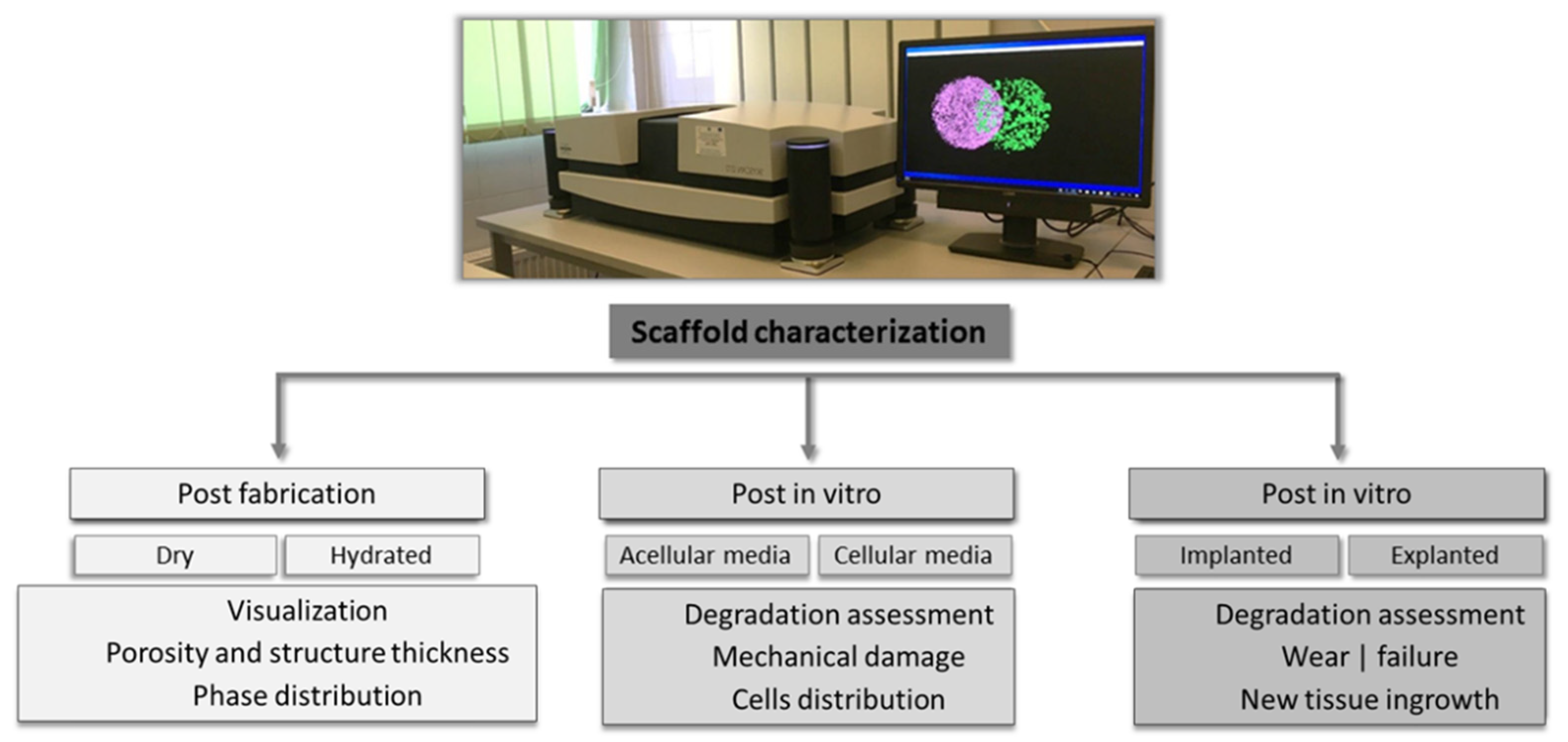 Figure 2.
 Types of scaffolds’ characterizations and corresponding assessments performed using CT scanners.
Other imaging techniques, such as scanning electron microscopy (SEM) associated with EDAX spectroscopy, atomic force microscopy (AFM), or confocal microscopy (CFM), provide important information regarding surface morphology, topography, and chemical composition. Nonetheless, their use is associated with several drawbacks, such as the need to destroy the sample to obtain suitable geometries that can be further analyzed, and the registered data provide information only with respect to the surface of the sample (SEM, AFM) or thin 3D sections (CFM). For instance, the limitation in terms of imaging depth for SEM is around 200 nm, while CFM, although may penetrate at higher depths (around 100 µm), cannot be applied for opaque scaffolds [
55]. CT is used as a complementary technique to obtain both qualitative and quantitative insights regarding the overall internal microarchitecture of the scaffold without causing any alteration to the sample. Its versatility is demonstrated by the possibility to scan all types of materials in hydrated or dried state (i.e., polymers, ceramics, metals, and composites) obtained through various fabrication methods (e.g., membranes, fibers, porous scaffolds, particles). A proper scanning offers not only high-quality images but also a relevant set of data, which will be further used in quantitative determination. Three-dimensional images of samples can reveal information regarding the overall microarchitecture of the scanned object, such as porosity [
56,
57,
58,
59], fiber orientation [
60], phase distribution [
61,
62,
63,
64], the presence and distribution of mineral clusters [
65], cell spreading on and within scaffolds [
66,
67], and effect of degradation evolution on scaffolds’ microstructure [
68].
3. Limitations and Perspectives of the CT Technique
Since its release to the public in the 1970s, the CT—both the technique and the equipment—has suffered significant changes. What seemed to be unreachable 50 years ago is now common practice: scanning of a 3D object within a reasonable period of time, obtaining high-resolution images that allow the observation of details at less than 5 µm, and even more, using the registered data sets for quantitative measurements [
203]. In addition, the fast progress of additive manufacturing technologies and the current focus on personalized medicine are also factors that put pressure on CT scanner producers to constantly improve their products, making them more accurate and user friendly. The obvious benefits of using CT as an advanced characterization equipment seem to be limited only by the relatively high cost of the apparatus and the need to be operated by trained professionals.
The development of new protocols as solutions for various issues encountered when scanning polymer-based scaffolds, hydrated materials, or soft tissue has also been established. To enable an accurate analysis, staining agents have been proposed [
69,
71,
96,
97,
98]. Still, a universal protocol is unlikely to emerge since the type of sample and its composition dictates the specimen preparation and the parameters for scanning and reconstruction. Furthermore, the image data processing depends a great deal on the users’ ability to perform a proper separation of the object from the surrounding background (binarization) [
204]. As a result, the necessity of using complementary techniques, such as SEM, AFM, or histology, to validate the obtained results has yet to be overcome. Due to the lack of generally accepted protocols and standards, the use of additional procedures is required to undoubtedly confirm the results of a study, leading to increased consumption of time and reagents and the use of complicated equipment and protocols. As far as the authors of this review are aware, using solely CT to discuss, for example, the architectural features or mineral composition of a scaffold has not been reported yet.
With respect to in vivo testing, CT proved to be a dependable technique. Not only it offers information about the degradation or integration of a scaffold into the native tissue, but it also allows scanning of the same sample at different time points, thus decreasing the number of sacrificed animals in preclinical trials.
The advances in the field of CT corroborated with the limitations of animal models for the validation of implantable scaffolds will soon render this technique indispensable in the field of tissue engineering. Time-lapse scanning can be successfully used to observe the behavior of a sample in various processes, such as degradation or integration [
155]. Moreover, the great number of high-quality research studies that prove the adequacy of CT in establishing phase distribution, scaffold degradation, or implant wear performed in either in vitro or in vivo conditions demonstrates the potential of this technique to replace the current gold standard for these analyses. The number of studies in which CT is employed as a characterization method for scaffolds with biomedical applications is increasing yearly. A simple search on sciencedirect.com using as the keywords “CT and tissue engineering” generated over three times more articles in 2021 when compared with 2011. Since research topics no longer subscribe to a single domain and emphasis is put on interdisciplinarity, it would be safe to assume that in the following years, CT will become a popular analysis for the in-depth qualitative and quantitative characterization of scaffolds with biomedical applications.
This entry is adapted from the peer-reviewed paper 10.3390/ma14226763