Alkaloids are natural products that possess numerous pharmacological activities and have been exploited effectively to treat cancer. However, the clinically approved anticancer alkaloids are generally limited by serious side effects due to their lack of specificity to cancer cells, indiscriminate tissue distribution and toxic formulation excipients. Lipid-based nanoparticles represent the most effective drug delivery system concerning clinical translation owing to their unique appealing characteristics for drug delivery.
1. Introduction
Cancer ranks as the leading cause of morbidity and mortality in the world with an estimated 19.3 million new cases and 9.9 million deaths reported in 2020 [
1]. Although stupendous advances have been made in understanding the molecular underpinnings and genomic landscape of cancers, the oncologic outcomes remain poor. Current treatments of cancer include surgery, radiation therapy and chemotherapy [
2,
3,
4]. However, the administration of anti-cancer drugs, including chemotherapeutic drugs, biologic agents and immunotherapeutic drugs using the conventional methods, has been hindered by various pharmacological issues, including toxicities, unsatisfactory therapeutic efficacy and drug resistance [
5,
6]. These unsatisfactory oncologic outcomes have revitalized the interest in natural product-derived anticancer agents.
Bioactive natural products have been serving as the primary source of medicines by numerous cultures around the world over the past millennia [
7]. With the rise of modern scientific approaches, the past century has witnessed a surge of highly active compounds derived from natural products and their derivatives with a precise mode of actions for the treatment of a myriad of diseases. Natural products have gained great interest due to their vast scaffold diversity and structural complexity unrivaled by current synthetic drugs [
8]. An analysis of all FDA-approved small-molecule drugs from 1981 to 2014 revealed that approximately 51% were natural products and their derivatives, and about 80% of anti-cancer small-molecule drugs were natural products and their derivatives [
9]. Several classes of natural products have been identified, including terpenoid, polyketide, phenylpropanoid and alkaloid [
10].
Alkaloid is a class of naturally occurring nitrogen containing heterocyclic organic compounds with a wide range of pharmacological activities, often considered privileged structures in drug discovery [
10,
11]. Since the commercialization of the first alkaloid morphine in 1826, numerous alkaloids have been isolated and exploited effectively for the betterment of mankind. Today, alkaloid drugs have been approved by the FDA for the treatment of cancer, Alzheimer’s disease, Parkinson’s disease, migraine, pain control, erectile dysfunction, heart failure and many more [
12,
13]. Alkaloids have demonstrated wide-spectrum anticancer activity by inhibiting topoisomerase I and suppressing microtubule dynamics [
14,
15]. The most notable anticancer alkaloid drugs that continue to maintain the palpable significance in clinical practice include paclitaxel, docetaxel, vincristine, vinblastine, irinotecan and topotecan. However, the administration of anticancer alkaloids has generally been limited by serious side effects due to their lack of specificity to cancer cells, indiscriminate tissue distribution and toxic formulation excipients [
16,
17,
18]. These limitations prompted unceasing investigational efforts to develop effective and safe nanoformulations and improve oncologic outcomes.
Cancer is a disease where the adequacy of delivery of extremely potent yet toxic chemotherapeutic drugs can result in either efficacious responses or serious morbidity [
19]. To mitigate these limitations, tailor-designed nanomedicines have emerged as a promising strategy for cancer treatment owing to their improved pharmacokinetic properties, therapeutic efficacy, specific targeting of tissues and minimized adverse effects [
20]. Furthermore, the use of nanotechnology allows drugs to traverse biological barriers such as the blood-brain barrier [
19,
21]. Among all the different classes of nanocarriers, lipid-based nanoparticles represent the most established and effective drug delivery system concerning clinical translation, with multiple formulations having already obtained U.S. Food and Drug Administration (FDA) approval for clinical use [
20]. Lipid-based nanoparticles have received great attention due to their unique, appealing characteristics for drug delivery, including (1) excellent biocompatibility and biodegradability; (2) improved solubility and stability of difficult-to-deliver drugs, including both hydrophobic and hydrophilic drugs; (3) enhanced therapeutic index by improving efficacy and reducing toxicity; (4) versatility which allows chemical modifications and surface coatings and (5) ability of co-deliver two different anticancer drugs to enable precise spatiotemporal multi-drug treatment [
22,
23,
24,
25]. These advantages of lipid-based nanoparticles have been exploited effectively in enhancing the efficacy and reducing the toxicity of anticancer alkaloids, the most exceptional of which are Onivyde (liposomal irinotecan), Marqibo (liposomal vincristine) and Lipusu (liposomal paclitaxel), which have received regulatory approval for clinical use [
26]. These uplifting translational successes have motivated an exponential increase in research investigating the potential and effectiveness of encapsulating anticancer alkaloids in different lipid-based nanoparticles. Nevertheless, despite the stupendous therapeutic potential demonstrated by nanomedicines in pre-clinical studies, there are still many shortcomings to be solved.
2. Nanotechnology
The prefix “nano” comes from ancient Greek which represents “dwarf” [
27]. One nanometer (nm) is an international System of Units that equals to one billionth of a meter. According to the National Nanotechnology Initiative (NNI), nanotechnology refers to the comprehension and manipulation of matter at atomic or molecular levels between 1 to 100 nm, where unique phenomena facilitate the novel applications [
28]. It involves various scientific disciplines (i.e., engineering, technology, medicine, chemistry, physics, biology or a combination of these disciplines) [
29]. Nanotechnology has been utilized in numerous medical-related fields including magnetic resonance imaging (MRI), hyperthermic destruction of tumor, proteins detection, diagnosis, pharmacological research, cells and biological molecules purification. Nowadays, numerous nanoparticles have been studied and developed for clinical use including liposomes, nanocapsules, nanorods, nanowires, nanospheres, nanoshells, nanotubes, nanopores and dendrimers [
27,
30,
31].
In general, nanomaterials can be divided into four different categories which are carbon-based, inorganic-based, organic-based and composite-based nanomaterials. Carbon-based nanomaterials can be found in morphologies such as sphere-shaped, ellipsoid-shaped, tube-shaped or horn-shaped [
32]. These nanomaterials can further be classified into graphene quantum dots (0-D), carbon nanotubes (1-D) and graphene (2-D) based on their dimensions, where 0-D refers to no dimension, 1-D refers to one dimension and 2-D refers to two dimensions at nanoscale [
33,
34]. Inorganic-based nanomaterials comprise metal-based and metal oxide-based nanoparticles. Metal-based nanoparticles are comprised of pure metal nanoparticles (i.e., iron, magnesium, zinc, platinum, titanium, copper, gold, silver and alginate nanoparticles). Metal-based nanoparticles can be bound to oxygen, becoming metal oxide nanomaterials (i.e., zinc oxide, silver oxide, etc.) [
35,
36]. Organic-based nanomaterials are mainly made of organic matter except inorganic and carbon-based nanomaterials. These organic-based nanomaterials can be transformed into liposomes, micelles, dendrimers and polymer nanoparticles that are very useful in drug delivery through noncovalent interactions [
36,
37]. Composite-based nanomaterials consist of several phases, with one of the phases on a nanoscale dimension that merges different nanoparticles together with enormous or complicated materials, for example hybrid nanofibers and metal-organic frameworks. A composite of these nanomaterials can be a mixture of any polymer, ceramic or metal materials with any organic-based, inorganic-based, metal-based or carbon based nanomaterials [
36]. These nanomaterials have potency to revolutionizes the manner at which diseases such as cancer are diagnosed and treated.
3. Alkaloid
Alkaloids are ubiquitous in nature. They are mostly found in plants, and can also be produced by terrestrial animals, marine organisms, microorganisms such as bacteria, fungi and insects. Approximately 20% of plant species contain alkaloids, most of which are biosynthetically derived from amino acids lysine (Lys), ornithine (Orn), tryptophan (Trp), tyrosine (Tyr) and phenylalanine (Phe) [
124].
Alkaloid is a class of naturally occurring heterocyclic organic compounds that contain a nitrogen atom. With over 20,000 structurally characterized members, alkaloids remain one of the most medicinally important classes of compounds with a wide range of pharmacological activities, often considered privileged structures in drug discovery [
10,
11]. In fact, the first naturally derived pure medicine was morphine, an alkaloid isolated from opium poppy in year 1805 and it was commercialized by Merck in 1826 [
13]. Since then, numerous alkaloids have been isolated and exploited effectively for the betterment of mankind.
Due to its vast structural diversity and widespread distribution in nature, several classification systems have been used to classify alkaloids, including chemical classification, taxonomic classification, pharmacological classification and biosynthetic classification, each with their own strengths and limitations. In this review, we have adopted the chemical classification to classify the alkaloids based on their chemical structures as this is the most established classification scheme for alkaloids. On this classification basis, the main classes of alkaloids in our review are indole, quinoline, isoquinoline, pyrrolidine, pyridine, piperidine, tropane, indolizidine, terpenoid, purine, imidazole and steroidal alkaloids. Table 1 summarizes different types of medicinally significant alkaloids according to the chemical classification.
Table 1. Medicinally Significant Alkaloids According to the Chemical Classification.
|
Class
|
Drugs
|
Molecular Formula
|
Origin
|
Indication/Uses |
|

Indole
|
Vincristine
|
C46H56N4O10
|
Catharanthus roseus
|
Anticancer
|
|
Vinblastine
|
C46H58N4O9
|
|
Vinorelbine

|
C45H54N4O8
|
|
Vincamine

|
C21H26N2O3
|
Vinca minor
|
Primary degenerative and vascular dementia
|
|
Physostigmine
|
C15H21N3O2
|
Physostigma venenosum
|
Glaucoma
|
|
Ajmaline

|
C20H26N2O2
|
Rauvolfia serpentina
|
Anti-arrhythmic
|
|
Ajmalicine

|
C21H24N2O3
|
Anti-hypertensive
|
|
Reserpine

|
C33H40N2O9
|
Rauvolfia serpentina
|
Anti-hypertensive
|
|
Yohimbine
|
C21H26N2O3
|
Erectile dysfunction
|
|
Strychnine

|
C21H22N2O2
|
Strychnos nux-vomica
|
Convulsant
|
|
Mitragynine
|
C23H30N2O4
|
Mitragyna speciosa
|
Stimulant, analgesic
|
|
Psilocin

|
C12H16N2O
|
Psilocybe cubensis
|
Hallucinogen
|
|
Psilocybin
|
C12H17N2O4P
|
|
Ephedrine
|
C6H5CH(OH)CH (CH3)NHCH3
|
Ephedra sinica
|
Bronchial asthma
|
|

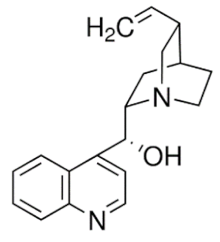
Quinoline
|
Irinotecan
|
C33H38N4O6
|
Catharanthus roseus
|
Anticancer
|
|
Topotecan
|
C23H23N3O5
|
|
Colchicine
|
C22H25NO6
|
Colchicum autumnale
|
Gout
|
|
Quinidine

|
C20H24N2O2
|
Cinchona officinalis
|
Anti-arrhythmic
|
|
Quinine

|
C20H24N2O2
|
Anti-malarial
|
|
Cinchonine

|
C19H22N2O
|
|
Cinchonidine
|
C19H22N2O
|
|
Isoquinolines
|
Morphine
|
C34H40N2O10S
|
Papaver somniferum
|
Analgesic
|
|
Codeine
|
C18H21NO3
|
|
Heroine
|
C21H23NO5
|
|
Apomorphine

|
C17H17NO2
|
Parkinson’s disease
|
|
Noscapine
|
C22H23NO7
|
Anti-tussive
|
|
Trabectedin
|
C39H43N3O11S
|
Ecteinascidia turbinata
|
Anticancer
|
|
Berberine
|
C20H18ClNO4
|
Coptis chinensis
|
Antimicrobial
|
|
Tubocurarine

|
C37H41ClN2O6
|
Chondrodendron tomentosum
|
Skeletal muscle relaxant
|
|
Atracurium

|
C53H72N2O12
|
Leontice leontopetalum
|
|
Tetrandrine

|
C38H42N2O6
|
Stephania tetrandra
|
Anti-arrhythmic
|
|
Galantamine

|
C17H21NO3
|
Galanthus nivalis
|
Alzheimer’s disease
|
|
Sanguinarine
|
C20H14NO4
|
Sanguinaria canadenis
|
Antibacterial, antiplaque
|
|
Papaverine

|
C20H21NO4
|
Papaver somniferum
|
Vasodilator
|
|

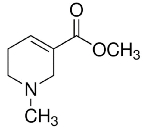
Pyrrolidines
|
Hygrine

|
C8H15NO
|
Erythroxylon coca
|
Laxative, diuretic
|
|
Cuscohygrine
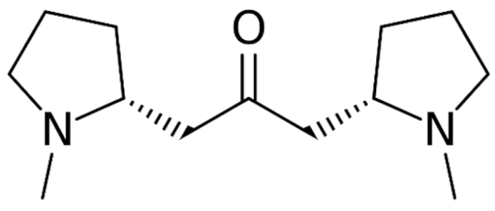
|
C13H24N2O
|
|
Stachydrine

|
C7H14NO2
|
Stachys tuberifera
|
Neuroprotectant
|
|

Pyridines
|
Arecoline
|
C8H13NO2
|
Areca catechu
|
Muscarinic agonist
|
|
Ricinine
|
C8H8N2O2
|
Ricinus communis
|
Insecticide
|
|
Trigonelline
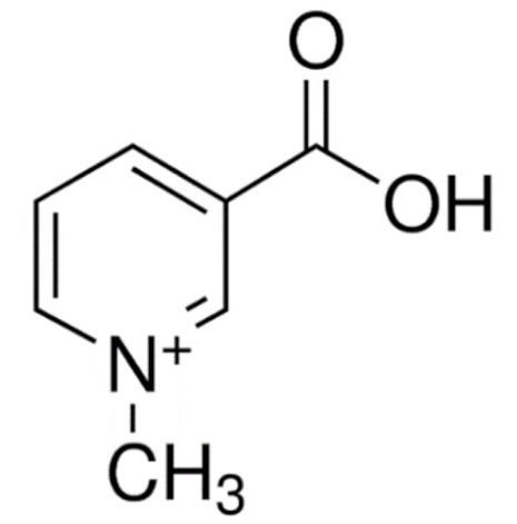
|
C7H7NO2
|
Trigonella foenum-graecum
|
Antidiabetic
|
|
Nicotine

|
C10H14N2
|
Nicotiana tabacum
|
Smoking cessation
|
|

Piperidine
|
Piperine

|
C17H19NO3
|
Piper nigrum
|
Anticancer
|
|
Piperlongumine

|
C17H19NO5
|
Piper longum
|
|
Pipernonaline

|
C21H27NO3
|
Antifungal
|
|
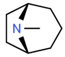
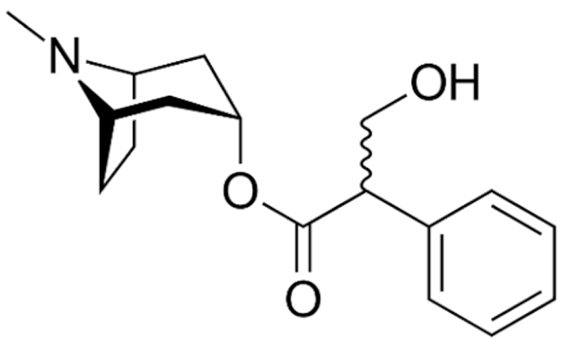
Tropanes
|
Atropine
|
C17H23NO3
|
Atropa belladonna
|
Anticholinergic
|
|
Cocaine
|
C17H21NO4
|
Erythroxylum coca
|
Local anaesthetic
|
|
Hyoscyamine
|
C17H23NO3
|
Atropa belladonna, Hyoscyamus niger
|
Anticholinergic
|
|
Hyoscine
|
C17H21NO4
|
Atropa belladonna
|
Motion sickness
|
|

Indolizidine
|
Swainsonine
|
C8H15NO3
|
Swainsona canescens
|
Anticancer
|
|
Castanospermine
|
C8H15NO4
|
Castanospermum australe
|
Antiviral
|
|
Securinine

|
C13H15NO2
|
Securinega suffruticosa
|
Neuroprotection
|
|
Tylophorine

|
C24H27NO4
|
Tylophora indica
|
Anticancer
|
|
Lycorine
|
C16H17NO4
|
Clivia miniata
|
|

Terpenoids
|
Paclitaxel
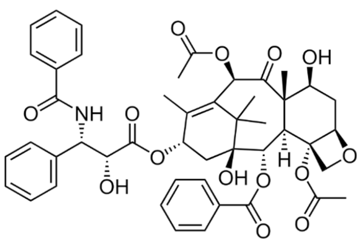
|
C47H51NO14
|
Taxus brevifolia
|
|
Docetaxel
|
C43H53NO14
|
Taxus baccata
|
|
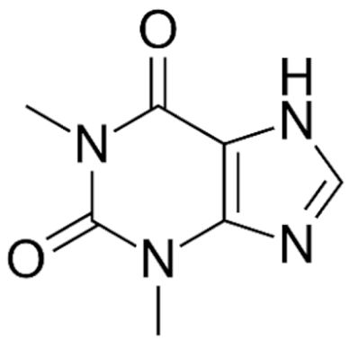
Purine
|
Caffeine
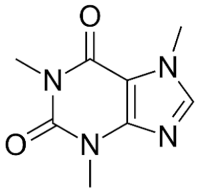
|
C8H10N4O2
|
Coffee arabica
|
CNS stimulant
|
|
Theobromine

|
C7H8N4O2
|
Theobroma cacao
|
Cardioprotectant
|
|
Theophylline
|
C7H8N4O2
|
COPD and asthma
|
|

Imidazole
|
Pilocarpine
|
C11H16N2O2
|
Pilocarpus microphyllus
|
Glaucoma
|
|
Epiisopiloturine
|
C₁₆H₁₈N₂O₃
|
Anthelmintic
|
|
Steroidal
|
Pancuronium

|
C35H60N2O4
|
Malouetia bequaertiana
|
Skeletal muscle relaxant
|
|
Conessine
|
C24H40N2
|
Holarrhena antidysenterica
|
Antidysenteric
|
|
Solanidine
|
C27H43NO
|
Solanum tuberosum
|
Anticancer
|
|
Tomatidine
|
C27H45NO2
|
Lycopersicon esculentum
|
Today, alkaloid drugs have been approved by the FDA for the treatment of cancer, Alzheimer’s disease, Parkinson’s disease, migraine, pain control, erectile dysfunction, heart failure and many more [
12]. Alkaloids have been widely utilized in various solid tumors and hematological malignancies as a monotherapy or in combination with other chemotherapeutic drugs [
125,
126,
127,
128,
129]. Thus, much effort has been devoted to elucidate and decipher the mechanism of action of these anticancer alkaloids.
4. Lipid-Based Nanoparticles for Encapsulation of Anticancer Alkaloids
Considering all the foregoing hints, nanocarriers could be a potential strategy to overcome the limitations of alkaloids. Nanocarriers are around 5 to 200 nm in size and can be used in a wide range of applications [
196]. They can be categorized into various types (e.g., organic, inorganic, polymeric, biological, lipid-based nanocarriers) according to their physical properties, chemical properties, morphology and size. Among the carriers, lipid-based nanocarriers offer an alternative to solubilize, encapsulate and deliver alkaloids in a programmed manner to enhance their water solubility, bioavailability and anticancer efficacy [
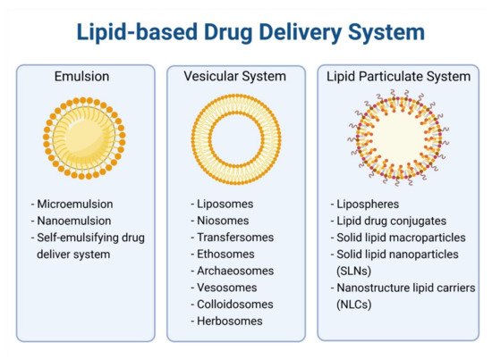
25]. Examples of lipid-based nanocarriers are liposomes, solid lipid nanoparticles (SLN) and nanostructured lipid-carriers (NLC) (
Figure 2). These drug carriers are made up of biocompatible lipids triglycerides, cholesterol and phospholipids in which most of them are derivatized based on or extracted from natural sources, resulting in their excellent biodegradability and biocompatibility [
197]. Excipients use in lipid carriers such as cholesterol, PEG and phosphatidylcholine have established toxicology data and safety profiles for their use in pharmaceutical products, further strengthening their potential as the ideal drug delivery system [
198]. Lipid-based nanoparticles with an average size of 100 nm and have longer circulation half-lives which enhance their propensity to extravasate through vascular fenestrations of tumors’ vasculature, thus enhancing the potency of anticancer agents [
199]. However, it is relatively difficult to prepare such small-sized lipid-based nanoparticles. In this regard, a “top down” size reduction approach that requires high energy input (e.g., sonication) and a “bottom up” method that produces nanoparticles by lipid condensation from solution can be used to solve this problem [
200]. As far as we know, clinically approved cancer nanomedicines that utilize lipid-based nanocarriers as drug delivery agents have particle sizes larger than 80 nm, for example, Doxil80–100 nm), Marqibo (100 nm) and Abraxane (130 nm) [
190,
201,
202]. Numerous studies have been conducted to encapsulate alkaloids into lipid nanocarriers.
Figure 2. Classification of Lipid-based Nanocarriers.
4.1. Liposome
Liposomes are one of the extensively studied lipid vesicles, which are made up of phospholipids and an aqueous medium. Lipid vesicles are formed when lipids interact with the aqueous medium, where the lipid hydrophilic head group envelops the aqueous core along with exposure of the hydrophilic tail group to the external medium. Owing to this distinct structural property, drugs can be entrapped in the lipid bilayer or loaded in the internal aqueous core of liposomes depending on their hydrophilicity, for example, Doxil and Onivyde [
203]. The first nano-based formulations approved by the FDA for cancer treatment are liposomal anticancer drugs. Doxil was the first doxorubicin-loaded liposomal drug which received FDA approval in 1995 to be used in the treatment of AIDS-related multiple myeloma and Kaposi sarcoma due to its lower cardiotoxicity and higher efficacy as compared to free doxorubicin alone. The clinical success of Doxil further established the potential of liposomes in drug delivery, and many promising liposomal formulations are currently being scrutinized intensively in clinical studies [
203,
204]. Reduced dose-limiting toxicities, improved undesirable pharmacokinetics and drug solubility are the primary objectives in liposomal development to deliver alkaloids. Liposomes’ stability is one of the major issues in the development of liposomal alkaloids [
197]. The drug to lipid ratio and lipid composition have to be taken into consideration in preparing physically stable liposomal alkaloids. Neutral zwitterionic lipids are the most commonly used lipids in preparing liposomes such as phosphatidylcholine. Phospholipids and cholesterol can be added to enhance stability, reduce aggregation and improve permeability of drugs [
205,
206]. Introduction of the PEGylation approach in the early nineties was shown to prolong the circulation time of liposome significantly due to steric stabilization of vesicles [
207]. Likewise, incorporation of PEG-modified lipids was found to enhance delivery and cytotoxicity of paclitaxel liposome to human cancer cells [
208]. However, PEG, which is hydrophobic in nature, was later found to lower the polarity of the aqueous matrix and destabilize the liposomes, leading to rapid drug leakage. To overcome the issue, poly(zwitterionic) polymers such as poly[2-(methacryloyloxy)ethyl phosphorylcholine) (PMPC) [
209] and poly(carboxybetaine) (PCB) [
210] were introduced to replace PEG. That said, liposomes have disadvantages including limited drug loading capacity due to space available in liposomal lipid membranes, inadequate control of drug release, reproducibility issues and stability issues [
211]. The drug-loading capacity of liposome can be solved by a remote loading approach where the drug will be added to preformed liposomes via pH gradient or ion gradient competent to create a pH gradient [
212].
In 2014, liposomes were used to co-encapsulate doxorubicin and a contrast agent (Magnevist) or hyperthermic agent (Fe
3O
4) [
213,
214]. Doxorubicin has also been encapsulated into ultrasound-sensitive liposomes in which the drug release can be facilitated by sonication that degrades liposomes, co-encapsulated with curcumin in liposomes to enhance anti-tumor efficacy of doxorubicin in the C26 colon cancer cell line, encapsulated in arginine-penetrating peptides/PEG modified liposomes to decrease in vitro cytotoxicity and enhance delivery of doxorubicin for the treatment of ovarian cancer [
215,
216,
217]. In addition, incorporation of docetaxel in anacardic acid and PEG-modified liposomes have proven to stabilize docetaxel [
218]. Many different liposomes and modified liposomes were designed to carry a variety of anticancer agents, such as paclitaxel and 5-fluorouracil [
219].
Liposomal preparation requires a cryogenic atmosphere brought on to the introduction of vesicular drug delivery systems using non-ionic surfactants. It was named niosome consisting of either uni- or multi- lamellar vesicles. Niosome was first introduced in the cosmetic industry and its potential application in drug delivery was only discovered thenceforth [
220]. The unique structure of niosome allows it to encapsulate both hydrophilic materials in vesicular aqueous core and lipophilic materials in the bilayer domain. It is composed of non-ionic surfactants with cholesterol and can increase the size and provide charge to vesicles, therefore, enhancing the entrapment efficiency of noisome. Niosome has a similar structure as liposomes and it is expected to be a better delivery system than liposomes due to its stability, cost and entrapment efficiency [
221,
222]. Nevertheless, only few niosome formulations are tested in clinical trials, and no formulations are commercially marketed heretofore owing to their low efficacy [
223]. Ethosome is also a modified version of classical liposomes and is mainly composed of phospholipids, ethanol and water in which the concentration of ethanol is relatively high, differentiating it from other vesicular carriers. High ethanol constituents of ethosome improve skin permeability by releasing the encapsulated materials into deeper layers and systemic circulation [
224,
225]. For transdermal delivery of drugs, ethosome is superior over classical liposomes due to its higher entrapment efficiency, more negative zeta potential and smaller size [
226,
227]. Despite having these superiorities, only few ethosome formulations enter clinical trials due to limitations such as low yield and suitability to carry potent drugs only but not drugs which require a high concentration of blood [
224].
4.2. Micelles
Micelles are colloidal systems formed through self-assembly of amphiphilic molecules. They can be further classified into polymeric micelles, lipid micelles and lipid polymeric hybrid micelles based on types of amphiphilic molecules. Amphiphilic molecules in lipid micelles are normally small-molecule surfactants. Unlike liposomes having a lipid bilayer, lipid micelles have a monolayer with an outer hydrophilic corona enclosing an inner hydrophobic core form by hydrophobic acyl chains [
228]. Critical micelle concentration (CMC) refers to concentration of surfactants above which micelles form. It is an important surfactants parameter to consider in designing micelles [
197]. Enhancing solubility of drugs is the primary objective of designing micelles as drug nano-carriers. Hydrophobic drugs such as docetaxel and paclitaxel are carried in the lipophilic core of micelles [
228]. Nevertheless, lipid micelles possess major limitations: unstable in bloodstream as dissociation occurred upon dilution below the micelle forming concentration (CMC) and limited interior hydrophobic space influencing the loading capacity of drugs. In order to stabilize the micelles by reinforcing weak intermolecular interactions, several strategies such as the formation of covalent crosslinking (for i.e., shell crosslinking) and non-covalent crosslinking (for i.e., diblock copolymers) can be used [
229]. Lipid micelle has been used to deliver various drugs including paclitaxel, doxorubicin and camptothecin in the preclinical stage [
230,
231,
232]. Cabral and co-workers identified that small polymeric micelles are suitable in delivering drug to the tumor site due to their favorable size range between 30 to 100 nm which penetrates well in highly permeable tumors [
200]. However, they are not up to the market until today due to insufficient cellular interaction with tumor cells for cellular uptake and poor physical stability in vivo [
233]. Even though both micelles and vesicles were formulated in the same principle whereby the lipid molecules reorganized and clustered together in an aqueous solution, the lipid layers were formed differently depending on their shapes. Lipid micelles were formed by wedge-shaped lipid molecules with the hydrophobic tails facing inwards, whereas the vesicles were formed by the cylinder-shaped phospholipid molecules with the hydrophobic tails sandwiched between the hydrophilic head groups [
234].
4.3. Solid Lipid Nanoparticles
Comparing the lipid-based nanocarriers discussed, solid lipid nanoparticles (SLN) represent a colloidal drug delivery system with an external aqueous phase and internal lipid phase introduced in the early 1990s. They are composed of a combination of various solid lipids such as waxes and fatty acids, as well as mono-, di- and triacylglycerols that form a lipid matrix entrapping drugs or other hydrophobic materials. Beneficial properties of SLN, such as cost effectiveness, non-toxicity, ease of preparation, controlled drug release, good system stability and provision of target-specific effects, made them outstrip other carriers when they were first introduced [
235,
236]. On top of that, use of IV acceptable solid lipids (e.g., phospholipids, glycerides) and surfactants (e.g., poloxamer 188, lecithin, tween-80) makes SLN a versatile platform for drug delivery readily to translate into clinical application [
235]. SLN-based formulations have shown a substantial enhancement in the anti-tumor efficacy of hydrophobic drugs. For example, resveratrol-SLN have greater inhibitory effects against the proliferation, invasion and migration of breast cancer cells than the free drug alone; talazoparib-SLN improve the therapeutic index against triple-negative breast cancer by overcoming homologous recombination-mediated resistance and talazoparib toxicity [
237]. However, the highly organized crystalline structure of solid lipids leaves a limited room for drug incorporation which contributes to low drug capacity and drug expulsion during storage [
238]. However, the highly organized crystalline structure of solid lipids leaves a limited room for drug incorporation which contributes to a low drug-loading capacity and drug expulsion during storage [
238]. To overcome this problem, Muller and colleagues have come out with a novel lipid delivery system called nanostructured lipid carriers (NLC) [
239].
4.4. Nanostructured Lipid Carriers
NLC are a modified version of conventional SLN by incorporating liquid lipids with a solid lipid. The room between crystal imperfections and fatty acid chains of NLC allows more drug accommodation. Moreover, certain drugs are more soluble in liquid lipids than solid lipids [
240]. Hence, NLC as the second generation lipid carriers after SLN reduce drug expulsion during storage and enhance the drug-loading capacity [
238]. NLC can be prepared using various surfactants and co-surfactants, allowing them to be formulated for various administration routes (e.g., parenteral, oral, topical, ocular, nasal) to deliver drugs and active substances for biochemical, cosmetic and pharmaceutical purposes [
240]. NLC possess favorable properties of low toxicity and controlled drug release, and provide target-specific effects and a high drug load for both hydrophilic and hydrophobic agents, making them outstrip SLN when they were introduced [
235,
241]. NLC-based formulations have been studied extensively for the delivery of anti-tumor agents. For example, 6-gingerol-NLC has a greater water solubility and oral bioavailability than the free drug alone, penetrating peptides, and hyaluronic-acid-modified artesunate-NLC has a better cell-membrane-penetrating ability against hepatic cancer; thymoquinone-NLC and fluvastatin-NLC have an improved anti-tumor efficacy against hepatic cancer and prostate cancer, respectively [
242].
Lipid-based delivery systems attracted a great deal of attention in the past decades as a strategy to enhance anticancer efficacy and overcome delivery barriers and the therapeutic index of various drugs, especially alkaloids. Myriads of preclinical studies have been focused on the use of lipid carriers to deliver alkaloids due to their potential in cancer treatment. Table 2 summarizes alkaloids that have been encapsulated in lipid-based nanocarriers for cancer treatment. Some of them utilized vesicular systems while others used lipid-particulate systems.
Table 2. Alkaloids that have been Successfully Encapsulated in Lipid-based Nanoparticles.
|
Class
|
Compound
|
Type of Lipid Carrier
|
Type of Cancer
|
In Vitro/In Vivo
|
Cell Type/Animal Model
|
Ref.
|
|
Terpenoids
|
Docetaxel
|
Liposome
|
Breast
|
in vitro; in vivo
|
MCF-7/4T1 xenograft mice
|
[243]
|
|
in vivo
|
MDA-MB-435/LCC6 xenograft mice
|
[244]
|
|
in vitro
|
MCF-7
|
[245]
|
|
Lung
|
in vivo
|
A549 xenograft rat
|
[246]
|
|
in vitro
|
A549
|
[247]
|
|
in vitro
|
A549
|
[245]
|
|
Liver
|
in vitro
|
HepG2
|
[245]
|
|
Melanoma
|
in vitro; in vivo
|
B16F10/B16-F10 xenograft mice
|
[248]
|
|
Micelle
|
Breast
|
in vitro
|
MCF-7
|
[249]
|
|
in vitro
|
MCF-7
|
[250]
|
|
Lung
|
in vitro
|
A549
|
[250]
|
|
Niosome
|
Breast
|
in vitro; in vivo
|
MDA-MB-s31 and MCF-7
|
[251]
|
|
NLC
|
Liver, Ovarian, Lung, Melanoma
|
in vitro; in vivo
|
HepG2, SKOV3, A549, B16 cells
|
[252]
|
|
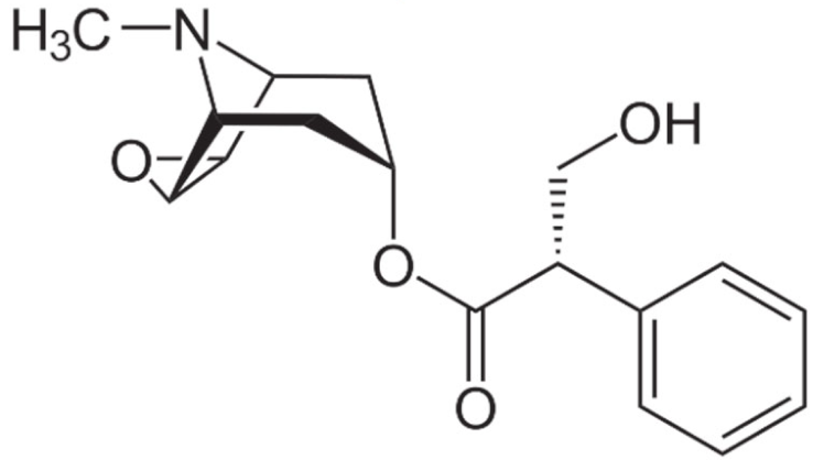
Paclitaxel

|
Liposome
|
Breast
|
in vitro; in vivo
|
4T1 xenograft mice
|
[253]
|
|
in vivo
|
ICR male mice
|
[254]
|
|
Lung
|
in vivo
|
A549 xenograft mice
|
[255]
|
|
in vitro; in vivo
|
A549
|
[69]
|
|
NLC
|
Breast, Ovarian
|
in vitro
|
MCF-7, SKOV3
|
[256]
|
|
Ethosome
|
Squamous Cell Carcinoma
|
in vitro
|
DJM-1
|
[257]
|
|
Micelle
|
Glioma
|
in vitro; in vivo
|
C6/C6 xenograft rat
|
[258]
|
|
Indole
|
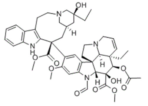
Vincristine

|
Liposome
|
Acute Lymphoblastic Leukemia
|
in vivo
|
Namwala xenograft mice
|
[190]
|
|
Brain Glioma
|
in vitro, in vivo
|
Glioma bearing mice
|
[259]
|
|
Nasopharyngeal cancer
|
in vitro, in vivo
|
KB/KBv200 xenograft mice
|
[260]
|
|
SLN
|
Breast
|
in vitro; in vivo
|
MDA-MB-231
|
[261]
|
|
NLC
|
Breast
|
in vitro
|
MCF-7
|
[262]
|
|
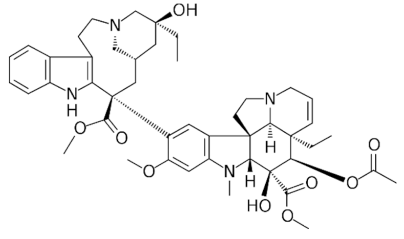
Vinblastine
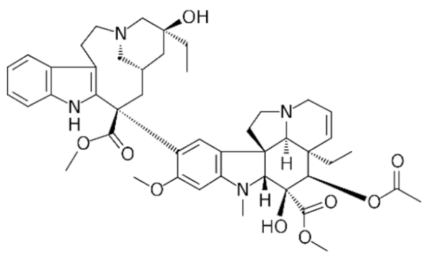
|
Liposome
|
Non-Small Cell Lung
|
in vitro; in vivo
|
LLT/LLT xenograft mice
|
[263]
|
|
Niosome
|
Lung
|
in vitro; in vivo
|
TC-1/TC-1 xenograft mice
|
[264]
|
|
Vinorelbine

|
Micelle
|
Breast
|
in vitro
|
MCF-7
|
[265]
|
|
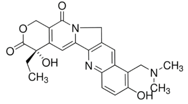
Quinoline
|
Topotecan
|
Liposome
|
Lung and Adenocarcinoma
|
in vitro
|
LLC
|
[266]
|
|
Breast
|
in vitro; in vivo
|
MCF-7/MCF-7 xenograft mice
|
[267]
|
|
SLN/NLC
|
Leukemia
|
in vitro
|
K-562
|
[268]
|
|
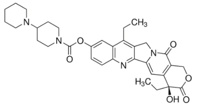
Irinotecan

|
Liposome
|
Colon
|
in vivo
|
Colo 320DM and Colon 26 xenograft mice
|
[269]
|
|
SLN
|
Rectal, Colon
|
in vivo
|
SCC7 xenograft mice
|
[270]
|
|
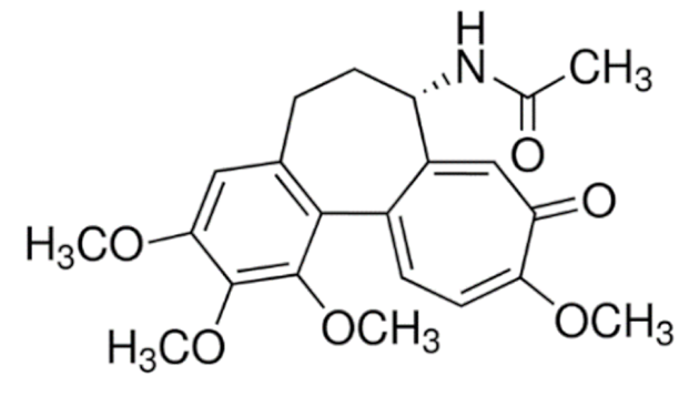
Isoquinoline
|
Berberine
|
Liposome
|
Hepatic
|
in vitro; in vivo
|
HepG2/HepG2 xenograft mice
|
[271]
|
|
Breast
|
in vitro; in vivo
|
MCF-7/MCF-7 CSC xenograft mice
|
[272]
|
|
SLN
|
Breast, Hepatic, Lung
|
in vitro
|
MCF-7, HepG2, A549
|
[273]
|
This entry is adapted from the peer-reviewed paper 10.3390/cancers13215346