Dittrichia viscosa (L.) Greuter: History
Please note this is an old version of this entry, which may differ significantly from the current revision.
Subjects: Plant Sciences
Contributor:

With the increasing interest in obtaining biologically active compounds from natural sources, Dittrichia viscosa (L.) Greuter (Asteraceae) came into our focus as a readily available and aromatic wild shrub widely distributed in the Mediterranean region. This work provides a phytochemical profile of D. viscosa in terms of parallel chemical composition in the lipophilic fraction (essential oil) and the water fraction (hydrosol). GC-MS analysis identified 1,8-cineole, caryophyllene oxide, α-terpenyl acetate, and α-muurolol as the major components of the essential oil, while in the hydrosol p-menth-1-en-9-ol, 1,8-cineole, linalool, cis-sabinene hydrate, and α-muurolol were the major volatile components. 3,4-Dihydroxybenzoic acid was found to be the predominant compound in the hydrosol composition by HPLC analysis. The antimicrobial potential of both extracts was evaluated against thirteen opportunistic pathogens associated with common skin and wound infections and emerging food spoilage microorganisms. The antimicrobial activity of the essential oil suggests that the volatiles of D. viscosa could be used as novel antimicrobial agents. 

  • Dittrichia viscosa
  • essential oil
  • hydrosol
  • GSH
  • antimicrobial
  • antiproliferative
  • antiphytoviral activity

1. Introduction

Plants are one of the most important sources of a variety of bioactive compounds that make them useful in daily life. Accordingly, a large number of plant species have recently become the focus of phytochemical and pharmacological studies. Considering that only 1–10% of plant species have been studied chemically and pharmacologically for their potential medicinal value [1], it is clear that plants are an under-researched natural source of bioactive compounds. Therefore, the study of their metabolites and biological effects will continue to be the focus of scientific interest with the aim of finding bioactive natural compounds and further development of alternative green and sustainable technologies that reduce or eliminate the use of hazardous substances in everyday life.
The Mediterranean climate favors the growth of a large number of plant species, many of which are aromatic plants used in folk medicine and nutrition. Dittrichia viscosa (L.) Greuter (syn. Inula viscosa L. (Aiton)) is a weed plant with numerous biological activities. It is a perennial herbaceous plant of the Asteraceae family. The plant is erect, 40–140 cm tall, and branched, with a prominent central shoot. The leaves are stalkless, alternate on the stem, and have a serrated margin directed towards the leaf tip. The yellow flower heads are 20–22 mm in size. The whole plant, especially the leaves, is covered with glandular hairs that secrete a sticky, aromatic-smelling resin [2][3][4][5]. Among the Mediterranean wild species, D. viscosa has been shown to be a remarkable source of potential bioactive metabolites that could find application in agriculture and other fields [6]. Folk medicine describes the use of this plant for the treatment of various diseases such as bronchitis and diabetes [1], as well as for its antipyretic, anti-inflammatory, and anthelmintic properties [7].
As an aromatic species with intense odor, this plant has been the subject of phytochemical profiling of volatile components performed by steam distillation, solvent extraction, and extraction by ultrasonic distillation [8][9][10]. The composition of the essential oil of D. viscosa is described in a large number of scientific papers and the main components of the essential oil obtained from different countries and regions are listed in the work of Zouaghi et al. [8]. Moreover, the phytochemical diversity of this plant has been described in detail by Grauso et al. [11], where all the compounds identified by different authors have been listed in view of explaining the antimicrobial, nematicidal and insecticidal activity of this versatile plant. Although D. viscosa is widely distributed along the Adriatic coast, to our knowledge, there are no data on the composition of volatile compounds in the essential oil and hydrosol of Croatian D. viscosa. Therefore, the first objective of this work is to determine the phytochemical composition, especially since we have not found any data on the hydrosol composition of D. viscosa from other regions either. Compared to essential oils, hydrosols contain more polar volatile compounds that are soluble in water [12], and we assume that these aqueous solutions are underestimated as mixtures of biologically active compounds that can be used as harmless natural products. Moreover, plant-derived natural products are environmentally friendly and often have a new mechanism of action that can overcome developed resistance [6]. Among the prominent biological effects described for this wild species [7][13][14][15], an interesting finding is the use of compounds from D. viscosa as a natural additive of polylactic acid, a biodegradable thermoplastic polymer, with the aim of modulating its physicochemical properties and achieving a bio-based packaging system [6]. Therefore, the present results on the analysis of the chemical composition of the essential oil and hydrosol of D. viscosa may be of great value for future studies and potential applications.
Recently, with the outbreak of a pandemic that we are still facing, the use of disinfectants in daily life has greatly increased, highlighting the need for natural disinfectants and antimicrobial products. An overview of the scientific literature revealed diverse reports of antimicrobial effects of D. viscosa, based on the variation in the chemical composition of the distillates, oils, and extracts tested, as well as the antimicrobial susceptibility assays chosen [11]. Ethanolic extract of D. viscosa leaves and flower buds showed antimicrobial activity against ATCC and foodborne isolates, with Candida albicans ATCC 10231 being the most sensitive strain [16]. We investigated the antimicrobial potential of the essential oil and, for the first time, hydrosol of D. viscosa by targeting thirteen opportunistic pathogens associated with common skin and wound infections and emerging food spoilage microorganisms.
Natural products with antiproliferative effects on tumor cells are the focus of modern medicine, with the goal of reducing the harmful effects of synthetic agents on healthy cells. Many studies have shown that D. viscosa is a plant with anti-cancer potential. Ozkan et al. [17] tested D. viscosa extracts on MCF-7 and T98-G cancer cells. The methanol extract showed a significantly stronger antiproliferative effect on both cell lines compared to the aqueous extract. Messaoudi et al. [18] also investigated the cytotoxic effect of ethanol and ethyl acetate extract of D. viscosa on two breast cancer cell lines. Both extracts inhibited the division of the tested cell lines after 72 h of treatment. The ethyl acetate extract showed higher cytotoxic activity, which the authors attributed to the synergistic effect of three dominant compounds: tomentosine, inuviscolide, and isocostic acid. Benbacer et al. [19] demonstrated a cytotoxic effect of D. viscosa extracts on two cervical cancer cell lines, in a manner that promoted apoptosis. Similar to the previous study, the key compound responsible for the cytotoxic activity is tomentosine, a sesquiterpene lactone that has been shown to be an extremely good anticancer agent [19][20]. We investigated the antiproliferative potential of D. viscosa volatiles on tree cancer cell lines: HeLa, HCT116, and U2OS. The possible mechanism of the antiproliferative activity of hydrosol was evaluated in relation to changes in intracellular GSH level since glutathione plays one of the most important roles in endogenous cell defense against oxidative stress.
Essential oils and other plant extracts have been used against a range of plant diseases caused by phytopathogenic bacteria, fungi, plant-parasitic nematodes, and parasitic and non-parasitic weeds [6]. Previous studies describe the activity of plant volatiles as natural antiphytoviral compounds [21][22][23][24][25][26][27][28][29]. The antiphytoviral activity of D. viscosa extracts has not been tested so far. Based on the chemical composition of essential oil and hydrosol, we assumed that this weed plant could be a readily available natural antiphytoviral agent. Indeed, the antiphytoviral activity of essential oils and hydrosols is of particular interest today from a green chemistry perspective. Low or no toxicity to non-target organisms and the possibility of obtaining them from renewable sources are just some of the advantages associated with the use of natural-based products in crop protection.

2. Current Results and Discussion

2.1. Gas Chromatography and Mass Spectrometry (GC-MS) Analysis of the Free Volatile Compounds from Essential Oil and Hydrosol

In this work, free volatile compounds were isolated from dried aerial parts of D. viscosa by water distillation in a Clevenger-type apparatus. The lipophilic (essential oil) and hydrofracture (hydrosol) collected from the inner tube of the Clevenger apparatus differ in chemical composition and biological activity due to the difference in solubility of the volatile compounds. In addition to water distillation, some authors have performed steam distillation, solvent extraction, and extraction by ultrasonic distillation from plant material of the genus Inula [8][9][10]. They obtained significant differences in the yield and composition of the essential oil [8]. In our study, the volatile compounds in the pentane layer (essential oil) and volatile compounds in the aqueous phase (hydrosol) were analyzed. The total oil yield was 0.09%, based on the dry weight of the samples. The composition and relative amounts of the compounds in both layers are shown in Table 1. Lipophilic compounds dissolved in pentane were analyzed by GC-MS. GC-MS analysis of the aqueous layer identified the more hydrophilic volatile components. Components that are soluble in both water and organic solvents were detected in the water and pentane layers (Table 1). Thus, GC-MS analysis of both phases, coupled with HPLC analysis of the hydrosol, provides us with a more complete phytochemical composition of the volatiles of this plant species. Haoui et al. [10] found that monoterpenes were the major chemical class of the essential oil of D. viscosa from Turkey and Algeria, while the class of oxygenated sesquiterpenes predominated in the plants from Spain, Italy, France, and Jordan [10]. In our study, twenty compounds, divided into six classes, and seventeen compounds, divided into three classes, were identified in the essential oil (EO) and hydrosol, accounting for 96.74% and 96.90% of the total oil and hydrosol composition, respectively. In terms of compound classes, the oxygenated monoterpenes dominate in the EO and hydrosol samples, accounting for 53.41% and 81.85% of the total composition, respectively. In addition to the oxygenated monoterpenes 1,8-cineole identified as the dominant compound in the oil (16.41%) and α-terpinyl acetate (13.92%), the oxygenated sesquiterpenes caryophyllene oxide (15.14%) and α-muurolol (13.75%) stand out in the overall composition of the oil of D. viscosa (Table 1). 1,8-Cineole (18.55%) represents the second most abundant component in the total hydrosol composition, and the compounds α-muurolol and caryophyllene oxide were also identified as frequent compounds in the hydrosol composition with proportions of 10.25% and 3.24%, respectively (Table 1). The oxygenated monoterpene p-menth-1-en-9-ol dominated the overall hydrosol composition (29.93%), while this compound was not detected in the oil composition. Linalool (11.67%) and cis-sabinene hydrate (10.97%) were also identified at a high percentage in the hydrosol and are also among the abundant components in the essential oil composition with proportions of 6.62% and 4.23%, respectively (Table 1). The oil contains a total of 30.11% oxygenated sesquiterpenes, with caryophyllene oxide (15.14%) and α-muurolol (13.75%) being the dominant compounds in this class and cyperotundone (1.22%) being less abundant. Madani et al. [30] compiled a table of the main components of the essential oils of D. viscosa from Algeria, Jordan, Italy, Turkey, Spain, and France. The composition of the oil of D. viscosa from Sardinia is most similar to the composition of the oil of Croatian D. viscosa in terms of caryophyllene oxide content. Caryophyllene oxide is also the main component obtained by water distillation from the leaves of this species from Algeria and Tunisia [30][31]. The fatty acid and hydrocarbon groups represent less than 6% of the total oil (Table 1). Differences in the composition of free volatiles of the species D. viscosa are influenced by population diversity, the time of collection of the plant material, and isolation techniques. We identified volatiles from two phases and, as shown in Table 1, some compounds were detected in both the lipophilic and aqueous phases, but some were identified in only one phase. This approach has given us a more complete insight into the chemical composition and potential application of specialized metabolites of the species D. viscosa.
Table 1. Phytochemical composition (% ± SD) of the essential oil (EO) and hydrosol (H) of Dittrichia viscosa.
Component RI* RI** EO
(Yield in %)
H
(Yield in %)
Monoterpene hydrocarbons     0.71 1.56
α-Pinene * 938 1036 0.71 ± 0.01 a 0.23 ± 0.01 b
Sabinene 971 1115 - 0.28 ± 0.01
Myrcene 992 1145 - 0.62 ± 0.03
Limonene 1032 1204 - 0.43 ± 0.03
Oxygenated monoterpenes     53.41 81.85
1,8-Cineole 1030 1211 16.41 ± 0.01 b 18.55 ± 0.01 a
cis-Sabinene hydrate 1065 1561 4.23 ± 0.01 b 10.97 ± 0.01 a
Linalool 1099 1548 6.62 ± 0.01 b 11.67 ± 0.01 a
Borneol * 1176 1699 0.32 ± 0.01 a 0.12 ± 0.05 b
α-Terpineol 1186 1646 2.65 ± 0.01 a 2.62 ± 0.01 b
β-Cyclocitral 1223 1629 4.81 ± 0.01 a 2.23 ± 0.01 b
Bornyl acetate 1287 1591 2.71 ± 0.01 a 0.67 ± 0.01 b
p-Menth-1-en-9-ol 1294 1915 - 29.93 ± 0.01
α-Terpinyl acetate 1349 1685 13.92 ± 0.01 a 1.31 ± 0.01 b
Cyclohexene, 1,5,5-trimethyl-6-methylene 1364 - - 2.02 ± 0.01
(E)-Isoeugenol 1446 2314 1.74 ± 0.01 1.76 ± 0.01
Sesquiterpene hydrocarbons     7.26 -
allo-Aromadendrene 1465 1662 2.34 ± 0.01 -
β-Bisabolene 1494 1729 4.31 ± 0.01 -
δ-Cadinene 1517 1754 0.61 ± 0.07 -
Oxygenated sesquiterpenes     30.11 13.49
Caryophyllene-oxide * 1581 1955 15.14 ± 0.01 a 3.24 ± 0.01 b
α-Muurolol 1645 2181 13.75 ± 0.01 a 10.25 ± 0.01 b
Cyperotundone 1696 - 1.22 ± 0.01 -
Fatty acids     2.58 -
Hexadecanoic acid 1959 2913 2.58 ± 0.01 -
Hydrocarbons     2.67 -
Heneicosane * 2100 2100 0.42 ± 0.03 -
Docosane * 2200 2200 1.73 ± 0.01 -
Tricosane * 2300 2300 0.52 ± 0.01 -
Total identification (%)     96.74 96.90
Retention indices (RI) were determined relative to a series of n-alkanes (C8–C40) on capillary columns VF5-ms (RI*) and CP Wax 52 (RI**); RI, identification by comparison of RIs with samples listed in a homemade library, reported in the literature [32] and/or authentic samples; comparison of mass spectra with those in the NIST02 Wiley 9 mass spectral libraries; * co-injection with reference compounds; -not identified; SD standard deviation of triplicate analyzes; significant differences were determined using multiple t-tests. a,b Mean values with different superscripts indicate a statistically significant difference between the data from EO and the H sample (p < 0.05).

2.2. HPLC Analysis of Hydrosol

Although hydrosols are important by-products of essential oil distillation, their chemical composition is generally analyzed relatively rarely. To detect more polar components of D. viscosa dissolved in the hydrosol, we subjected it to high-performance liquid chromatography (HPLC) in addition to GC-MS analysis. HPLC analysis of the hydrosol of Veronica saturejoides detected vanillin, cinnamic acid, and protocatechuic acid (synonim 3,4-dihydroxybenzoic acid), the latter being the most abundant compound with an average concentration of 7.33 mg/L [33]. Beara et al. [34] also found significant amounts of protocatechuic acid in 70% aqueous acetone extracts of Veronica teucrium and V. jacquinii. Stojković et al. [35] described protocatechuic acid as the main compound in water extracts of V. montana, while rosmarinic and caffeic acids were detected in hydrosol of Poliomintha longiflora, Mexican oregano [36]. Based on the retention times of standards and spiking samples, four phenolic acids and the flavonoid luteolin were identified in hydrosol of D. viscosa (Table 2). Together, these compounds account for more than 80% of the chromatogram area. The dominant phenolic acid with a concentration of 62.24 mg/L is 3,4-dihydroxybenzoic acid (protocatechuic acid). This benzoic acid derivative is characterized by its antioxidant, anti-inflammatory [37], and antitumor activities [38]. Other phenolic compounds, cinnamic acid and its derivatives caffeic acid and o-coumaric acid are represented with much lower concentrations in the hydrosol composition (Table 2).
Table 2. HPLC analysis of phenolic compounds.
Phenolic Compound mg/L ± SD
3,4-dihydroxybenzoic acid 62.24 ± 2.72
caffeic acid 0.90 ± 0.06
trans-o-coumaric acid 0.39 ± 0.02
cinnamic acid 1.16 ± 0.01
luteolin 1.75 ± 0.13

2.3. Wide-Spectrum Antimicrobial Activity

The antimicrobial activity of the hydrosol and essential oil was evaluated using a microdilution assay for a variety of human opportunistic pathogens associated with skin and wound infections, as well as foodborne pathogens. Bacterial and fungal growth was not affected by a 25% dilution of D. viscosa hydrosol. The essential oil was found to be effective against all the microorganisms tested as shown in Table 3. The essential oil inhibited bacterial growth at minimal inhibitory concentrations (MICs) ranging from as low as 0.09 mg/mL to 5.62 mg/mL, and fungi at MIC50 of 0.09 mg/mL and 2.81 mg/mL. Although the MIC50 values recorded for fungi were species-specific, the MIC90 of 5.62 mg/mL of essential oil was consistent for both yeast and mold strains. Of note, C. albicans is a commensal on human skin and mucoses but can cause infections ranging from superficial infections of the skin to life-threatening systemic infections [39]. Food-spoilage and food-poisoning mold Aspergillus niger is ubiquitous in the environment but has been implicated in serious opportunistic infections of humans, particularly pulmonary and cutaneous aspergillosis [40].
Table 3. Antimicrobial activity of Dittrichia viscosa essential oil by microdilution assay.
Species Strain Origin Essential Oil (mg/mL) a
MIC MBC
Gram-positive bacteria      
Staphylococcus aureus ATCC 29213 2.8 2.8
Staphylococcus aureus Clinical/MRSA 5.6 5.6
Staphylococcus epidermidis Human 1.4 1.4
Streptococcus pyogenes ATCC 19615 0.09 0.09
Streptococcus agalactiae Clinical 0.09 0.09
Enterococcus faecalis ATCC 29212 1.4 2.8
Listeria monocytogenes ATCC 19111 (1/2a) 2.8 2.8
Bacillus cereus Food 0.7 0.7
Clostridium perfringens Food 0.09 0.09
Gram-negative bacteria      
Escherichia coli ATCC 25922 2.8 2.8
Acinetobacter baumannii ATCC 19606 5.6 5.6
Yeast   MIC50 MIC90
Candida albicans ATCC 90029 2.8 5.6
Molds   MIC50 MIC90
Aspergillus niger Food 0.09 5.6
a Two-fold dilutions of essential oil were tested in a range from 22.5 to 0.02 mg/mL by the microdilution method.
The essential oil strongly inhibited the growth of all tested bacteria, regardless of their Gram discrimination, and showed a strong and concentration-dependent bactericidal effect mostly at dosages of MICs. The most profound effect was recorded against S. pyogenes ATCC 19615, the clinical isolate of Streptococcus agalactiae, and the foodborne isolate of Clostridium perfringens, killing them at a dose of only 0.09 mg/mL of essential oil. All three bacterial species contribute significantly to skin and soft tissue infections in adults [41]. Moreover, S. aureus ATCC 29213 and methicillin-resistant S. aureus (MRSA) clinical strain was killed by dilutions of 2.81 and 5.62 mg/mL of essential oil, respectively (Table 3). Of note, S. aureus is one of the most common pathogens of nosocomial and community-associated infections worldwide [41]. This bacterium commonly causes skin, soft tissue, and bloodstream infections, 70% of which are due to MRSA strains that are resistant to almost all commercially available β-lactam and other classes of antibiotics [42]. Moreover, the MIC and MBC value of essential oil against Acinetobacter baumannii was 5.62 mg/mL. Notably, this pathogen deserves particular attention as one of the most common agents of various nosocomial infections, such as ventilator-associated pneumonia, urinary tract infections, bacteremia, and complicated skin and soft tissue, abdominal and central nervous system infections [43]. These infections are particularly difficult to treat due to their intrinsic and acquired resistance mechanisms, which places them in the group of multidrug-resistant ESKAPE pathogens (Enterococcus faeciumS. aureusKlebsiella pneumoniaeA. baumanniiPseudomonas aeruginosa, and Enterobacter spp.) with high medical priority [44].
Ali-Shtayeh et al. [45] reported that the water extract of D. viscosa was active against C. albicans by disc-diffusion method, while Al-Masri et al. [46] demonstrated antifungal activity against Botryis cinerea in terms of reduction of mycelium growth and germination when the hydrodistillation of D. viscosa was applied in combination with a low dose of the fungicide iprodione. Moreover, a number of studies reported stronger activity of alcohol extracts of D. viscosa, mainly methanol and ethanol, on both fungal and bacterial strains [11]. On the other hand, the antimicrobial activity of the essential oil of D. viscosa has been sparsely studied. Only Blanc et al. [47] demonstrated an antifungal effect of the essential oil (obtained by hydrodistillation as in our study) against the pathogenic and food-poisoning fungi Aspergillus fumigatusA. nigerC. albicansCladosporidium cladosporioides, and Cryptococcus neoformans. Other authors used steam distillation to obtain the essential oil, which showed different antimicrobial activities ranging from no observed antifungal activity [48] to inhibition up to 84.11% [49].
Overall, the antimicrobial activity of the essential oil found in our study, in contrast to the hydrosol tested, is probably related to the higher concentration of oxygenated sesquiterpenes found in the oil (Table 1), compared to the hydrosol. In this context, the content of caryophyllene oxide, which was very abundant in the oil compared to the hydrosol, is of particular importance. This component has already been described as a potent antimicrobial substance when present in higher concentrations in D. viscosa hydrodistillation [8] and essential oil [31]. It should also be noted that the synergy between essential oil compounds may also contribute to the broad-spectrum antimicrobial activity shown in our study, as previously suggested by other authors [8].

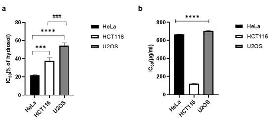
2.4. Antiproliferative Activity

Recently, much research has been done with the aim of finding natural chemotherapeutic agents. Numerous studies have shown that extracts from different parts of D. visocosa have promising cytotoxic activity [18][19][50][51][52][53][54][55]. In this study, we tested for the first time the antiproliferative activity of the volatile compounds in the essential oil and a hydrosol of D. viscosa. The essential oil showed potent antiproliferative activity on all three cancer cell lines used: HeLa, HCT116 and U2OS (IC50 0.66 mg/mL, 0.12 mg/mL and 0.7 mg/mL, respectively). The hydrosol significantly inhibited the division of cancer cells with IC50 values, 21.70% for HeLa cells, 37.73% for HCT116, and 54.51% for U2OS (Figure 1).
Figure 1. Antiproliferative activity of Dittrichia viscosa hydrosol (a) and essential oil (b) on HeLa, HCT116, and U2OS cancer cell lines assessed by MTS-based cell proliferation assay. Statistical significance was determined by one-way ANOVA, followed by Turkey’s multiple comparisons test. IC50 values are the means of three independent experiments. SD values are indicated with error bars. Statistically significant difference between HeLa and HCT116 cells is marked with *** p < 0.001, between HeLa and U2OS is marked with **** p < 0.0001 and between HCT116 and U2OS is marked with ### p < 0.001.
Ozkan et al. [17] showed that the methanol extract of areal parts of D. viscosa had a stronger antiproliferative effect than the aqueous extract on two tested cell lines, MCF-7 (human breast adenocarcinoma) and T98-G (human brain tumor). In the study by Benbacer et al. [19], methanol extract of Dittrichia also showed significant inhibition of proliferation of human cervical cancer cells, HeLa and SiHa. The main mechanism of action involves the induction of programmed cell death. The ethanol extract of D. viscosa flowers inhibited the growth and proliferation of Vero cells, with an IC50 value of 202.43 µg/mL. The methanol fraction had excellent antiproliferative activity on MCF-7 cells with an IC50 value of 15.78 µg/mL [50][54].
Numerous compounds present in D. viscosa extracts have shown significant biological activities [56][57][58]. These mainly include flavonoids such as nepetin, hispidulin, and methylquercetin. In the present study, GC-MS analysis revealed the presence of monoterpenoids p-menth-1-en-9-ol and 1,8-cineole as the dominant compounds in the hydrosol (Table 1). Oxygenated monoterpenes have been previously described as compounds with anticancer activity [59]. Moteki et al. [60] reported the cytotoxic activity of 1,8-cineole in leukemia cancer cells. Although the mechanism of cytotoxic activity is not fully elucidated, the authors showed that the suppression of leukemia cell growth was associated with the induction of apoptosis. Murata et al. [61] also showed that the main mechanism of inhibition of colorectal cell proliferation was apoptosis. Treatment with 1,8-cineole activated p38 and dephosphorylated Akt, leading to activation of caspase-3 and induction of apoptosis. A study conducted on three cancer cell lines (MCF7, A2780, and HT29) and one normal fibroblast cell line (MRC5) showed that 1,8-cineole acts selectively and causes a remarkable dose-dependent inhibition of the growth of cancer cells but not of healthy fibroblasts [62]. Thus, 1,8-cineole emerges as a promising, safe, and potent chemotherapeutic agent for the treatment of various cancers. In addition, 3,4-dihydroxybenzoic acid or protocatechuic acid (PCA), which is present in a variety of fruits, vegetables, and a number of medicinal plants [63] as well as in the hydrosol of D. viscosa (Table 2), possesses a wide range of biological activities such as antioxidant activity, antiviral, anti-inflammatory, anticancer, and many others [64][65][66][67][68][69]. Due to its chemical structure, it acts as an excellent antioxidant. It also has the possibility of prooxidant activity, which probably plays an important role in inhibiting the proliferation of cancer cells. Thus, the activity of hydrosol on cancer cells could be related to the high content of this phenolic compound in addition to 1,8-cineole. Previous studies have shown a significant antiproliferative effect of 3,4-dihydroxybenzoic acid on immortalized breast cells HBL 100, breast cancer cells PC14 and promyelocytic leukemia cells HL-60 [65][66][67][68] and liver cancer cell line HepG2 [69], possibly by generating oxygen free radicals that act as signaling molecules and affect genes involved in cell cycle regulation and apoptosis [70].
The results of our study confirmed that in addition to the previously demonstrated antiproliferative activity of various extracts of D. viscosa, hydrosol tested for the first time exhibited very significant antiproliferative activity on cancer cell lines. The anti-cancer potential of D. viscosa volatiles should be investigated on other cell lines, focusing on extracts from different parts of the plant, with the aim of finding new active molecules that could be used in the treatment of different types of cancer.

2.5. Glutathione (GSH) Assay

The effect of D. viscosa hydrosol on intracellular GSH level was measured using Ellman’s reagent. A change in GSH level is important for assessing toxicological responses and is an indicator of oxidative stress, possibly leading to apoptosis and cell death. The significant amount of 3,4-dihydroxybenzoic acid in hydrosol composition (Table 2) and its dual role of acting as both antioxidant and prooxidant, as well as its important role in the proliferation of cancer cells and prevention of carcinogenesis [71], prompted us to investigate the effect of hydrosol treatment on GSH level in HeLa cells. Glutathione is considered to be a very important factor in regulating carcinogenic mechanisms in cancer cells [72]. In contrast to its protective role in healthy cells, where it is crucial for neutralizing carcinogens, elevated GSH levels in tumor cells are associated with tumor progression and increased resistance to chemotherapeutic agents [73]. In recent years, several novel therapies targeting the antioxidant GSH system in tumor cells have been developed to achieve better response and reduced drug resistance. HeLa cells treated with the hydrosol for 1 h (IC50 from MTT measurements) showed a significant reduction in GSH level compared to untreated control cells (Table 4). The reduced GSH level indicates the direction of the cellular response in oxidative homeostasis, suggesting oxidative stress as a possible mechanism of the antiproliferative effect of hydrosol on tumor cells.
Table 4. GSH level (mmol/L) ±SD in hydrosol (H) treated HeLa cells compared to untreated cells.
  Control H Treated
GSH level 0.287 ± 0.007 a 0.187 ± 0.025 b
a, b Mean values with different superscripts indicate a statistically significant difference between the data (p < 0.05).

2.6. Antiphytoviral Activity
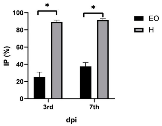
The search for substances of natural origin with antiphytoviral activity is particularly important today to support biological production and the replacement of synthetic chemicals with natural agents. Scientific literature reports that the application of water extracts of D. viscosa in combination with a low dose of the effective fungicide iprodione may be a viable way to reduce the severity of gray mold disease [46]. A mixture of acetone and n-hexane extract of D. viscosa emulsified in water effectively controlled downy mildew of cucumber, late blight of potato or tomato, powdery mildew of wheat, and rust of sunflower [74]. Based on the phytochemical composition of the essential oil and hydrosol (Table 1 and Table 2), we hypothesized that the biological activities of D. viscosa extracts could be extended in terms of antiphytoviral activity. The activity of both lipophilic and hydrophilic extracts of D. viscosa on the defense response of local host plants to tobacco mosaic virus (TMV) infection was investigated. TMV is a model virus in plant virology and a very important pathogen of agricultural crops causing significant yield losses. In addition to the antiphytoviral activity of essential oils of aromatic plants [21][22][23][24][25][26][27][28][29], the activity of hydrosol of Hypericum perforatum ssp. veronense was recently demonstrated [26], showing that hydrosols are a readily available natural source of bioactive compounds that can be used for plant protection against viral pathogens. Moreover, considering that hydrosols are a by-product of the essential oil distillation process, it is clear that the use of all products of this process is environmentally and biologically desirable. Although both essential oil and hydrosol-treated plants significantly reduced the number of local lesions compared to control plants (Table 5), the percentage inhibition of local lesions was more pronounced in hydrosol-treated plants (Figure 2). On the third day post-inoculation, the inhibition of lesions on the leaves of the essential oil- and hydrosol treated plants was 25.1% and 89.3%, respectively, and on the seventh day after inoculation, this inhibition was 37.5% and a promising 91.5%, respectively (Figure 2). Based on the results (Table 5Figure 2) and the fact that our preliminary study showed that simultaneous inoculation of hydrosol and virus did not reduce the number of local symptoms, we concluded that pretreatment with hydrosol activates the plant defense response and increases resistance to viral pathogens. Considering that salicylic acid (2-hydroxybenzoic acid) is one of the most important endogenous signals in the activation of plant defense response [75], we suggest that the antiviral activity of the hydrosol of D. viscosa may be related to the high content of benzoic acid derivative, namely 3,4-diydroxybenzoic acid (Table 2). In addition, it is also possible that other components contained in the hydrosol (Table 1 and Table 2) have synergic effects and activate plant signaling pathways leading to increased resistance to viral infections. The reported antiphytoviral activity of both the essential oil and hydrosol deserves more detailed analysis in the future and opens new areas of research regarding this unexplored bioactivity of D. viscosa. Further studies are required to evaluate the efficacy against viral diseases under field conditions.
Figure 2. Percentage of inhibition (IP) of lesions on leaves of treated Datura stramonium plants inoculated with tobacco mosaic virus compared to control plants on days three and seven postinoculation (dpi). Prior to inoculation, D. stramonium plants were treated with essential oil (EO) or hydrosol (H) of Dittrichia viscosa for two consecutive days. Error bars show the standard deviation of triplicate analyses; significant differences were marked with *.
Table 5. Local lesion number (LLN) on leaves of treated Datura stramonium plants inoculated with tobacco mosaic virus and on leaves of control plants (C) on days three and seven post-inoculation (dpi). Prior to inoculation, D. stramonium plants were treated with essential oil (EO) or hydrosol (H) of Dittrichia viscosa for two consecutive days.
dpi LLN ± SD
3rd C 6.2 ± 0.2 a
EO 4.6 ± 0.2 b
H 0.7 ± 0.2 c
7th C 14.2 ± 2.4 a
EO 8.9 ± 2.0 b
H 1.2 ± 0.4 c
SD, the standard deviation of triplicate analysis; significant differences were determined by one-way ANOVA. a, b, c Mean values with different superscripts indicate statistically significant differences between control and essential oil/hydrosol treatment data (p ˂ 0.05).

This entry is adapted from the peer-reviewed paper 10.3390/plants10091837

References

  1. Zaki, M.; Loubidi, M.; Oukhrib, A.; Mallouk, S. Natural products from Dittrichia Viscosa (Mini-Review). RHAZES Green Appl. Chem. 2019, 9, 30–46.
  2. Maleš, Ž.; Šarić, F. Kvantitativna analiza fenolnih spojeva ljepljivog omana-Inula viscosa (L.) Ait. Farm. Glas. 2009, 65, 143–148.
  3. Tutin, T.G.; Heywood, V.H.; Burges, N.A.; Valentine, D.H.; Walters, S.M.; Webb, D.A. Flora Europaea, 2nd ed.; Cambridge University Press: London, UK, 1964–1980; Volume 1–5.
  4. Pignatti, S. Flora Ditalia; Edagricole: Bologna, Italy, 1982; Volume I–III.
  5. Flora Croatica Database: 26865. Available online: https://hirc.botanic.hr/fcd/DetaljiFrame.aspx?IdVrste=26865&taxon=Dittrichia+viscosa+(L.)+Greuter (accessed on 20 July 2021).
  6. Moeini, A.; van Reenen, A.; Van Otterlo, W.; Cimmino, A.; Masi, M.; Lavermicocca, P.; Valerio, F.; Immirzi, B.; Santagata, G.; Malinconico, M.; et al. α-costic acid, a plant sesquiterpenoid from Dittrichia viscosa, as modifier of Poly (lactic acid) properties: A novel exploitation of the autochthone biomass metabolite for a wholly biodegradable system. Ind. Crops Prod. 2020, 146, 112134.
  7. Barrero, A.F.; Herrador, M.M.; Arteaga, P.; Catalán, J.V. Dittrichia viscosa L. Greuter: Phytochemistry and biological activity. Nat. Prod. Commun. 2008, 3, 1799–1804.
  8. Zouaghi, N.; El, N.; Bensiradj, H.; Cavaleiro, C.; Nadjemi, B.; Telfah, A. Antimicrobial activities of natural volatiles organic compounds extracted from Dittrichia viscosa (L.) by hydrodistillation. Jordan J. Biol. Sci 2021, 14, 41–49.
  9. Eljazi, J.S.; Selmi, S.; Zarroug, Y.; Wesleti, I.; Aouini, B.; Jallouli, S.; Limam, F. Essential oil composition, phenolic compound, and antioxidant potential of Inulaviscosa as affected by extraction process. Int. J. Food Prop. 2018, 21, 2309–2319.
  10. Haoui, I.E.; Derriche, R.; Madani, L.; Oukali, Z. Analysis of the chemical composition of essential oil from Algerian Inula viscosa (L.) Aiton. Arab. J. Chem. 2015, 8, 587–590.
  11. Grauso, L.; Cesarano, G.; Zotti, M.; Ranesi, M.; Sun, W.; Bonanomi, G.; Lanzotti, V. Exploring Dittrichia viscosa (L.) Greuter phytochemical diversity to explain its antimicrobial, nematicidal and insecticidal activity. Phytochem. Rev. 2020, 19, 659–689.
  12. Hamedi, A.; Pasdaran, A.; Zebarjad, Z.; Moein, M. A survey on chemical constituents and indications of aromatic waters soft drinks (Hydrosols) used in Persian nutrition culture and folk medicine for neurological disorders and mental health. J. Evid. Based Complementary Altern. Med. 2017, 22, 744–752.
  13. Rhimi, W.; Ben Salem, I.; Iatta, R.; Chaabane, H.; Saidi, M.; Boulila, A.; Cafarchia, C. Dittrichia viscosa L. leaves lipid extract: An unexploited source of essential fatty acids and tocopherols with antifungal and anti-inflammatory properties. Ind. Crops Prod. 2018, 113, 196–201.
  14. Bedoya, L.M.; Sanchez Palomino, S.; Abad, M.J.; Bermejo, P.; Alcami, J. Screening of selected plant extracts for in vitro inhibitory activity on human immunodeficiency virus. Phyther. Res. 2002, 16, 550–554.
  15. Yaniv, Z.; Dafni, A.; Friedman, J.; Palevitch, D. Plants used for the treatment of diabetes in Israel. J. Ethnopharmacol. 1987, 19, 145–151.
  16. Mohti, H.; Taviano, M.F.; Cacciola, F.; Dugo, P.; Mondello, L.; Marino, A.; Crisafi, G.; Benameur, Q.; Zaid, A.; Miceli, N. Inula viscosa (L.) Aiton leaves and flower buds: Effect of extraction solvent/technique on their antioxidant ability, antimicrobial properties and phenolic profile. Nat. Prod. Res. 2020, 34, 46–52.
  17. Ozkan, E.; Karakas, F.P.; Yildirim, A.B.B.; Tas, I.; Eker, I.; Yavuz, M.Z.; Turker, A.U. Promising medicinal plant Inula viscosa L.: Antiproliferative, antioxidant, antibacterial and phenolic profiles. Prog. Nutr. 2019, 21, 652–661.
  18. Messaoudi, M.; Chahmi, N.; El Mzibri, M.; Gmouh, S.; Amzazi, S.; Benbacer, L.; El Hassouni, M. Cytotoxic effect and chemical composition of Inula viscosa from three different regions of morocco. Eur. J. Med. Plants 2016, 16, 1–9.
  19. Benbacer, L.; Merghoub, N.; El Btaouri, H.; Gmouh, S.; Attaleb, M.; Morjani, H.; Amzazi, S.; Mzibri, M. Antiproliferative effect and induction of apoptosis by Inula viscosa L. and Retama monosperma L. Extracts in human cervical cancer cells. In Topics on Cervical Cancer with an Advocacy for Prevention; IntechOpen: London, UK, 2012.
  20. Rozenblat, S.; Grossman, S.; Bergman, M.; Gottlieb, H.; Cohen, Y.; Dovrat, S. Induction of G2/M arrest and apoptosis by sesquiterpene lactones in human melanoma cell lines. Biochem. Pharmacol. 2007, 75, 369–382.
  21. Dunkic, V.; Bezic, N.; Vuko, E.; Cukrov, D. Antiphytoviral activity of Satureja montana L. ssp. variegata (host) P.W. Ball essential oil and phenol compounds on CMV and TMV. Molecules 2010, 15, 6713–6721.
  22. Dunkić, V.; Bezić, N.; Vuko, E. Antiphytoviral activity of essential oil from endemic species Teucrium arduini. Nat. Prod. Commun. 2011, 6, 1385–1388.
  23. Dunkić, V.; Vuko, E.; Bezić, N.; Kremer, D.; Ruščić, M. Composition and antiviral activity of the essential oils of Eryngium alpinum and E. amethystinum. Chem. Biodivers. 2013, 10, 1894–1902.
  24. Bezić, N.; Vuko, E.; Dunkić, V.; Ruščić, M.; Blažević, I.; Burčul, F. Antiphytoviral activity of sesquiterpene-rich essential oils from four croatian teucrium species. Molecules 2011, 16, 8119–8129.
  25. Vuko, E.; Rusak, G.; Dunkić, V.; Kremer, D.; Kosalec, I.; Rađa, B.; Bezić, N. Inhibition of Satellite RNA Associated Cucumber Mosaic Virus Infection by Essential Oil of Micromeria croatica (Pers.) Schott. Molecules 2019, 24, 1342.
  26. Vuko, E.; Dunkić, V.; Ruščić, M.; Nazlić, M.; Mandić, N.; Soldo, B.; Šprung, M.; Fredotović, Ž. Chemical composition and new biological activities of essential oil and hydrosol of Hypericum perforatum L. ssp. veronense (Schrank) H. Lindb. Plants 2021, 10, 1014.
  27. Vuko, E.; Dunkić, V.; Bezić, N.; Ruščić, M.; Kremer, D. Chemical composition and antiphytoviral activity of essential oil of Micromeria graeca. Nat. Prod. Commun. 2012, 7, 1227–1230.
  28. Lu, M.; Han, Z.; Xu, Y.; Yao, L. In vitro and in vivo anti-tobacco mosaic virus activities of essential oils and individual compounds. J. Microbiol. Biotechnol. 2013, 23, 771–778.
  29. Bishop, C.D. Antiviral activity of the essential oil of melaleuca alternifolia (Maiden amp; Betche) cheel (tea tree) against tobacco mosaic virus. J. Essent. Oil Res. 1995, 7, 641–644.
  30. Madani, L.; Derriche, R.; Haoui, I.E. Essential oil of Algerian Inulaa viscosa leaves. J. Essent. Oil Bear. Plants 2014, 17, 164–168.
  31. Gharred, N.; Dbeibia, A.; Falconieri, D.; Hammami, S.; Piras, A.; Dridi-Dhaouadi, S. Chemical composition, antibacterial and antioxidant activities of essential oils from flowers, leaves and aerial parts of Tunisian Dittrichia Viscosa. J. Essent. Oil Res. 2019, 31, 582–589.
  32. Adams, R.P. Identification of Essential Oil Components by Gas Chromatography/Mass Spectrometry, 4.1 ed.; Allured Publishing: Carol Stream, IL, USA, 2017.
  33. Nazlić, M.; Kremer, D.; Grubešić, R.J.; Soldo, B.; Vuko, E.; Stabentheiner, E.; Ballian, D.; Bogunić, F.; Dunkić, V. Endemic Veronica saturejoides vis. ssp. saturejoides–chemical composition and antioxidant activity of free volatile compounds. Plants 2020, 9, 1646.
  34. Beara, I.; Živković, J.; Lesjak, M.; Ristić, J.; Šavikin, K.; Maksimović, Z.; Janković, T. Phenolic profile and anti-inflammatory activity of three Veronica species. Ind. Crops Prod. 2015, 63, 276–280.
  35. Stojković, D.S.; Živkovic, J.; Soković, M.; Glamočlija, J.; Ferreira, I.C.; Janković, T.; Maksimović, Z. Antibacterial activity of Veronica montana L. extract and of protocatechuic acid incorporated in a food system. Food Chem. Toxicol. 2013, 55, 209–213.
  36. Cid-Pérez, T.S.; Ávila-Sosa, R.; Ochoa-Velasco, C.E.; Rivera-Chavira, B.E.; Nevárez-Moorillón, G.V. Antioxidant and antimicrobial activity of mexican oregano (Poliomintha longiflora) essential oil, hydrosol and extracts fromwaste solid residues. Plants 2019, 8, 22.
  37. Li, X.; Wang, X.; Chen, D.; Chen, S. Antioxidant activity and mechanism of protocatechuic acid in vitro. Funct. Foods Health Dis. 2011, 1, 232–244.
  38. Abotaleb, M.; Liskova, A.; Kubatka, P.; Büsselberg, D. Therapeutic potential of plant phenolic acids in the treatment of cancer. Biomolecules 2020, 10, 221.
  39. Mayer, F.L.; Wilson, D.; Hube, B. Candida albicans pathogenicity mechanisms. Virulence 2013, 4, 119–128.
  40. Perfect, J.R.; Cox, G.M.; Lee, J.Y.; Kauffman, C.A.; de Repentigny, L.; Chapman, S.W.; Morrison, V.A.; Pappas, P.; Hiemenz, J.W.; Stevens, D.A.; et al. The impact of culture isolation of aspergillus species: A hospital-based survey of aspergillosis. Clin. Infect. Dis. 2001, 33, 1824–1833.
  41. Ki, V.; Rotstein, C. Bacterial skin and soft tissue infections in adults: A review of their epidemiology, pathogenesis, diagnosis, treatment and site of care. Can. J. Infect. Dis. Med Microbiol. 2008, 19, 173–184.
  42. Chambers, H.F.; Deleo, F.R. Waves of resistance: Staphylococcus aureus in the antibiotic era. Nat. Rev. Microbiol. 2009, 7, 629–641.
  43. Peleg, A.Y.; Seifert, H.; Paterson, D.L. Acinetobacter baumannii: Emergence of a successful pathogen. Clin. Microbiol. Rev. 2008, 21, 538–582.
  44. Pendleton, J.N.; Gorman, S.P.; Gilmore, B.F. Clinical relevance of the ESKAPE pathogens. Expert Rev. Anti. Infect. Ther. 2013, 11, 297–308.
  45. Ali-Shtayeh, M.S.; Yaghmour, R.M.; Faidi, Y.R.; Salem, K.; Al-Nuri, M.A. Antimicrobial activity of 20 plants used in folkloric medicine in the Palestinian area. J. Ethnopharmacol. 1998, 60, 265–271.
  46. Al-Masri, M.I.; Sharawi, S.M.; Barakat, R.M.; Al-Masri, M.I.; Sharawi, S.M.; Barakat, R.M. Effect of clammy inula (inula viscose) plant extract in combination with a low dose of the fungicide iprodione on botrytis cinerea in vitro and in vivo. Am. J. Plant Sci. 2015, 6, 1519–1526.
  47. Blanc, M.C.; Bradesi, P.; Gonçalves, M.J.; Salgueiro, L.; Casanova, J. Essential oil of Dittrichia viscosa ssp. viscosa: Analysis by 13C-NMR and antimicrobial activity. Flavour Fragr. J. 2006, 21, 324–332.
  48. Bonsignore, L.; Loy, G.; Secci, D.; Logu, A.; de Palmieri, G. A preliminary microbiological screening of Sardinian plants. Fitoterapia 1990, 61, 339–341.
  49. Bekkara, F.; Benhammou, N.; Panovska, T.K. Biological activities of the essential oil and ethanolic extract of Inula viscosa from the Tlemcen region of Algeria. Adv. Food Sci. 2008, 30, 132–139.
  50. Talib, W.H.; Mahasneh, A.M. Antiproliferative activity of plant extracts used against cancer in traditional medicine. Sci. Pharm. 2010, 78, 33.
  51. Çelik, T.A.; Aslantürk, Ö.S. Evaluation of cytotoxicity and genotoxicity of Inula viscosa leaf extracts with Allium test. J. Biomed. Biotechnol. 2010, 2010, 189252.
  52. Kaileh, M.; Vanden Berge, W.; Boone, E.; Essawi, T.; Haegman, G. Screening of indigenous Palestinian medicinal plants for potential anti-inflammatory and cytotoxic activity. J. Ethnopharmacol. 2007, 113, 510–516.
  53. Merghoub, N.; El Btaouri, H.; Benbacer, L.; Gmouth, S.; Trentesaux, C.; Brassart, B.; Terryn, C.; Attaleb, M.; Madoulet, C.; Benjouad, A.; et al. Inula viscosa extracts induces telomere shortening and apoptosis in cancer cells and overcome drug resistance. Nutr. Cancer 2016, 68, 131–143.
  54. Talib, W.H.; Mahasneh, A.M. Antimicrobial, cytotoxicity and phytochemical screening of Jordanian plants used in traditional medicine. Molecules 2010, 15, 1811–1824.
  55. Afifi-Yazar, F.U.; Kasabri, V.; Abu-Dahab, R. Medicinal plants from Jordan in the treatment of cancer: Traditional uses vs. in vitro and in vivo evaluations—Part 1. Planta Med. 2011, 77, 1203–1209.
  56. Talib, W.H.; Abu Zarga, M.H.; Mahasneh, A.M. Antiproliferative, antimicrobial and apoptosis inducing effects of compounds isolated from Inula viscosa. Molecules 2012, 17, 3291–3303.
  57. Seca, A.M.L.; Grigore, A.; Pinto, D.C.G.A.; Silva, A.M.S. The genus Inula and their metabolites: From ethnopharmacological to medicinal uses. J. Ethnopharmacol. 2014, 154, 286–310.
  58. Zeouk, I.; Sifaoui, I.; López-Arencibia, A.; Reyes-Batlle, M.; Bethencourt-Estrella, C.J.; Bazzocchi, I.L.; Bekhti, K.; Lorenzo-Morales, J.; Jiménez, I.A.; Piñero, J.E. Sesquiterpenoids and flavonoids from Inula viscosa induce programmed cell death in kinetoplastids. Biomed. Pharmacother. 2020, 130, 110518.
  59. Asakawa, Y. Dietary monoterpenoids. In Handbook of Dietary Phytochemicals; Springer: Singapore, 2021; pp. 607–731.
  60. Moteki, H.; Hibasami, H.; Yamada, Y.; Katsuzaki, H.; Imai, K.; Komiya, T. Specific induction of apoptosis by 1,8-cineole in two human leukemia cell lines, but not a in human stomach cancer cell line. Oncol. Rep. 2002, 9, 757–760.
  61. Murata, S.; Shiragami, R.; Kosugi, C.; Tezuka, T.; Yamazaki, M.; Hirano, A.; Yoshimura, Y.; Suzuki, M.; Shuto, K.; Ohkohchi, N.; et al. Antitumor effect of 1, 8-cineole against colon cancer. Oncol. Rep. 2013, 30, 2647–2652.
  62. Abdalla, A.N.; Shaheen, U.; Abdallah, Q.M.A.; Flamini, G.; Bkhaitan, M.M.; Abdelhady, M.I.S.; Ascrizzi, R.; Bader, A. Proapoptotic activity of achillea membranacea essential oil and its major constituent 1,8-cineole against a2780 ovarian cancer cells. Molecules 2020, 25, 1582.
  63. Anter, J.; Romero-Jiménez, M.; Fernández-Bedmar, Z.; Villatoro-Pulido, M.; Analla, M.; Alonso-Moraga, Á.; Muñoz-Serrano, A. Antigenotoxicity, cytotoxicity, and apoptosis induction by apigenin, bisabolol, and protocatechuic acid. J. Med. Food 2011, 14, 276–283.
  64. Kan, A.K.; Rehana, R.; Fatima, N.; Mahmood, S.; Mir, S.; Khan, S.; Jabeen, N.; Murtaza, G. Pharmacological activities of protocatechuic acid. Acta Pol. Pharm. 2015, 72, 643–650.
  65. Hudson, E.A.; Dinh, P.A.; Kokubun, T.; Simmonds, M.S.J.; Gescher, A. Characterization of potentially chemopreventive phenols in extracts of brown rice that inhibit the growth of human breast and colon cancer cells. Cancer Epidemiol. Prev. Biomark. 2000, 9, 1163–1170.
  66. Lee, I.R.; Yang, M.Y. Phenolic compounds from Duchesnea chrysantha and their cytotoxic activities in human cancer cell. Arch. Pharm. Res. 1994, 17, 476–479.
  67. Tseng, T.H.; Kao, T.W.; Chu, C.Y.; Chou, F.P.; Lin, W.L.; Wang, C.J. Induction of apoptosis by hibiscus protocatechuic acid in human leukemia cells via reduction of retinoblastoma (RB) phosphorylation and Bcl-2 expression. Biochem. Pharmacol. 2000, 60, 307–315.
  68. Kampa, M.; Alexaki, V.-I.; Notas, G.; Nifli, A.-P.; Nistikaki, A.; Hatzoglou, A.; Bakogeorgou, E.; Kouimtzoglou, E.; Blekas, G.; Boskou, D.; et al. Antiproliferative and apoptotic effects of selective phenolic acids on T47D human breast cancer cells: Potential mechanisms of action. Breast Cancer Res. 2004, 6, R63.
  69. Yip, E.C.; Chan, A.S.; Pang, H.; Tam, Y.K.; Wong, Y.H. Protocatechuic acid induces cell death in HepG2 hepatocellular carcinoma cells through a c-Jun N-terminal kinase-dependent mechanism. Cell Biol. Toxicol. 2006, 22, 293–302.
  70. Klauning, J.E.; Kamendulis, L.M. The role of oxidative stress in carcinogenesis. Annu. Rev. Pharmacol. Toxicol. 2004, 44, 239–267.
  71. Tanaka, T.; Tanaka, T.; Tanaka, M. Potential cancer chemopreventive activity of protocatechuic acid. J. Exp. Clin. Med. 2011, 3, 27–33.
  72. Ortega, A.L.; Mena, S.; Estrela, J.M. Glutathione in cancer cell death. Cancers 2011, 3, 1285–1310.
  73. Kennedy, L.; Sandhu, J.K.; Harper, M.E.; Cuperlovic-culf, M. Role of glutathione in cancer: From mechanisms to therapies. Biomolecules 2020, 10, 1–27.
  74. Wang, W.; Ben-Daniel, B.H.; Cohen, Y. Control of plant diseases by extracts of Inula viscosa. Phytopathology 2004, 94, 1042–1047.
  75. Widhalm, J.R.; Dudareva, N. A familiar ring to it: Biosynthesis of plant benzoic acids. Mol. Plant 2015, 8, 83–97.
More
This entry is offline, you can click here to edit this entry!
Video Production Service