1. Introduction
Phytochemicals are bioactive polyphenolic compounds naturally found in plants that have been studied extensively due to their potential medicinal and nutritional benefits to humans. They not only play a protective role for the plant but are responsible for its color, aroma, and flavor. These compounds have attracted the attention of scientists worldwide, owing to their potent bioactivity against different diseases, their low cytotoxicity and their ability to be utilized in the production of cosmetics and dietary supplements [1,2,3].
Nowadays, the skin is more likely to become infected as a result of changing environmental conditions and an increase in pollution levels. Consequently, there has been a significant increase in the demand for herbal medicine in both developed and developing countries owing to their potent biological efficacy, higher safety margins, and lower cost than synthetic agents [13]. Numerous herbal extracts originating from different plant species have been evaluated for their potential to treat skin conditions due to their medicinal benefits, which include antimicrobial and anti-inflammatory properties, their ability to promote blood clotting and wound healing, and to relieve burns and other skin diseases [14,15]. Several common skin conditions, including eczema, acne, urticaria, pruritus, psoriasis, and other bacterial and fungal skin diseases, can be treated efficiently using medicinal herbs [16,17]. The chemical structure of most plant extracts used in pharmaceutical and cosmeceutical applications is based on flavonoids or other polyphenol rings, which have high molecular weight and, hence, have poor solubility and are poorly absorbed through the skin [18,19].
The human skin is a remarkably complex and sophisticated organ that serves as a barrier to external exposure by enveloping the inner body components. It consists of three main layers—the epidermis, dermis and hypodermis—and each has different degrees of specialization. Along with its function as a waterproof barrier, the epidermis contains melanocytes, which determine the skin’s tone and pigmentation. The dermis is the skin layer underneath the epidermis that composed of connective tissue, sweat glands and hair follicles and helps to maintain the skin’s flexibility. In comparison to the other layers of the skin, the hypodermis is made up of fat and connective tissue and is responsible for shock absorption [20]. The stratum corneum (SC), or outer layer of the epidermis, is entirely made up of dead, keratinized epithelium cells and other metabolically inactive cells that cover the outer surface of the skin [21]. The SC and epidermis are regarded as the most effective barriers against hydrophilic and lipophilic compounds, respectively [22,23]. Furthermore, in terms of skin mechanical properties, elastic fibres are essential for skin elasticity and are directly responsible for skin performance and appearance [24]. Figure 1 shows a simplified structure of the skin and its barriers.
There are various types of nanotechnology-based materials that are currently used in the delivery of polyphenolic phytochemicals. Nanosized delivery systems can be classified into two main groups: Organic delivery systems (i.e., liposomes and polymeric nanoparticles) and inorganic delivery systems (i.e., silver, gold, and copper nanoparticles) [48]. Liposomes are one of the most commonly used nanoparticles that have been successfully used in the pharmaceutical and cosmetics fields [49]. The encapsulation of curcumin into liposomal nanoparticles showed potent activity against lung, pancreatic, and colorectal cancer at a lower dose in comparison to free curcumin [50]. Polymeric nanoparticles can also be used as an efficient nanocarrier of phytochemicals, and the encapsulation of curcumin extract into chitosan and polylactic-co-glycolic acid (PLGA) nanoparticles exhibited a significant improvement in its solubility profile compared to a conventional curcumin formulation [51].
2. Advantages of Phytosomes in Topical Applications
In topical applications, phytosomes have several potential advantages over the conventional topical formulations. Phytosomes increase skin absorption and bioavailability, and they induce the delivery of herbal active constituents to tissues [
111]. Moreover, phytosomes improve skin functions by enhancing hydration, the enzyme balance, and collagen structure [
30]. The high affinity of phytosomes to skin phospholipids intensified its effectiveness compared to conventional free compounds [
112]. As mentioned earlier, there are several barriers facing topical applications of phytosomes formulation. For example, one of the most important barriers of the transdermal application of phytochemicals is the SC, which is the thick outer layer of the epidermis [
113]. Bioactive molecules can cross the SC via different pathways, which are either intercellular or intracellular. Intercellular penetration can be achieved via sweat glands, sebaceous, or hair follicles, whereas the intercellular lipid matrix and corneocytes are the main pathways of intracellular penetration [
114,
115]. It has been reported that enhancing the diffusion coefficient of the drug can increase the concentration of biomolecules and enhance partitioning between the these molecules and the SC layer, and that all these factors can improve the permeability of biomolecules to the SC for transdermal application [
26]. The transdermal permeability of topical products can be increased when the active ingredients are lipophilic and have a low molecular weight.
Most of the widely studied phytocompounds are polyphenols, which have poor bioavailability and lipid solubility due to their hydrophilic nature, limiting their in vivo activity [
28]. The phospholipid moieties of phytosomes have a high affinity to bind several flavonoids compounds tightly [
116]. There are several herbal extracts, such as hawthorn, grape seed, green tea, milk thistle and ginseng, that are more effective when they are loaded into phytosomes, even more so than when they are carried in a liposomal formulation [
112]. The formulation of polyphenol-based phytochemicals phytosomes nanoparticles enhances the application of standard herbal materials as the phospholipid molecules of phytosomes interact with the active phytoconstituents, increasing their stability [
117]. Furthermore, the phytosomes–herbal complex has a higher affinity to the skin phospholipid moiety, which can improve the lipid solubility of the topical formulation [
112].
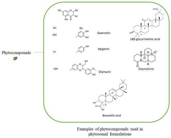
Figure 5 demonstrates some examples of the phytocompounds incorporated into a phytosomal delivery system.
Figure 5. Scheme demonstrating examples of phytocompounds incorporated into a phytosomal delivery system.
G. biloba extract has been used for different applications and can be applied topically for its antioxidant and antiaging agents [
118]. There are various reports that have compared standard
G. biloba extract and the extract complexed with phospholipids. It was reported that the topical application of
G. biloba improved peripheral circulation due to its topical anti-inflammatory activity, and it was more effective in a complex with phospholipid moieties of phytosomes [
119]. The bioavailability and pharmacokinetics profiles of the conventional
G. biloba extract and the phytosomal form were evaluated by Chen et al., who showed that the bioavailability of the herbal extract increased significantly in the phytosomal complex [
120]. In a clinical study by Kennedy et al., they compared the cognitive and mood effects of a low dose of
G. biloba extract and products complexing the extract with two types of phospholipids (PS or PC) in human subjects. Their findings demonstrated that all treatments were associated with improved calmness; however, there was a modest enhancement in the therapeutic benefit of secondary memory performance for
G. biloba extract complexed with PC [
121].
Quercetin is a phenolic phytochemical compound found in various vegetables, fruits, and leaves, and has soothing antioxidant and anti-itching effects [
122]. In a study by Maramaldi et al., the formulation of quercetin complexed with phytosomes nanoparticles exhibited potent dermal activity above that of standard quercetin and similar to the conventional anti-inflammatory drugs that are usually used [
122]. The quercetin-phytosomes complex had a significant impact by reducing redness, itching, and inflammation of damaged skin. Research also suggested that this complex may also support restoration of the skin barrier function, increasing hydration, and reducing water loss [
122]. In another report by El-Fattah et al., quercetin phytosomes demonstrated superiority in the dermal therapeutic benefit over free quercetin in an ovariectomized rat model [
123]. Lycopene is a terpene molecule found in fruits and vegetables that is known for its antioxidant, antiproliferative, and anti-inflammatory activities. Its incorporation into lipid nanocarriers enhanced the dermal absorption significantly to tackle skin aging and other skin conditions [
124,
125]. Moreover, it can also produce anti-proliferative effect against tumour [
126].
Molecular interaction of phytochemical-phospholipid complex has been studied using various analysis methods. Studies using 13C-NMR, 1H-NMR, 31P-NMR, IR, DSC, and X-ray revealed that complex formation resulted in chemical shifts and signal changes different from their original components [
33,
85,
127,
128,
129,
130,
131]. All these studies confirm the generation of chemical bonds between the phospholipid’s moieties and phytochemicals moieties. Chemical interactions include H-bonding, van der waals interaction, and hydrophobic effect, dipole-dipole interaction [
132]. The chemical structure of the phytochemicals can significantly affect the type of bonds formed during complex formation. For instance, lycopene, a type of hydrophobic β -carotenoids, consists of tetraterpene carbon chain interacts mainly via hydrophobic effect with the acyl chain of the phospholipid (i.e., fatty acid chain moiety and hydrophobic tail). This interaction has been confirmed by x-ray study where the signal of hydrophobic part of PC disappeared completely among the formation of the complex [
133]. Quercetin, a polyphenolic compound, on the other hand, has a cyclic hydrophobic part and hydroxyl groups which makes it an amphiphilic compound. Studies have shown that quercetin mainly interacts via H-bonding with the polar head group and with a lesser extent with the acyl chain moiety of the phospholipid [
127,
128]. Saponins, are glycoside compounds consists of a parent compound (such as terpene) and sugar derivative. A study of saponins formed from pentacyclic triterpenes and one or more sugar units showed that saponin interacts with the polar head group of the phospholipid during complex formation [
15]. Polyphenolic compounds extracted from olive oil fruit (i.e., tyrosol, verbascoside, hydroxytyrosol), interact mainly with the polar head group via H-bonding [
33]. 18-β-glycyrrhetinic acid, a steroid like structure, contains pentacyclic rings and an acid group. The polar group of 18-β-glycyrrhetinic acid interacts via H-bond with the polar head group of phospholipids and formed a complex [
132,
134]. In the DSC thermogram, the complex of 18-β-glycyrrhetinic acid-phospholipid showed a signal different than that of 18-β-glycyrrhetinic acid alone or the phospholipid alone. For example, 18-β-glycyrrhetinic Acid (18β-GA) showed a sharp endothermic peak at 294° which revealed its melting and thermogram of phospholipids showed mild and broad endothermic peak at 277°, 203° and 96°, while thermogram of 18β-GA phytosome revealed endothermic peak at 245°, 198° and 80° [
130].
Table 4 summarizes the main types of chemical interactions between phospholipids and phytochemicals.
Table 4. Examples of phytochemicals-phospholipids chemical interactions during phytosomes preparation.
| Phytochemical |
Type of Phytochemical |
Type of Phospholipid |
Chemical Interaction |
Analysis Method |
Reference |
| Quercetin |
Polyphenols |
PC |
H-bonds with the polar group of the phosphplipid |
1H-NMR, 31P-NMR, 13C-NMR |
[127] |
| Polyphenols |
DPPC 1
(PC) |
(1) Electrostatic interactions,
(2) H-bonds with the polar group of the phosphplipid,
(3) Hydrophobic
interaction with fatty acyl chains |
1H-NMR, 31P-NMR, 13C-NMR |
[128] |
| Lycopene |
Carotenoids (Terpenoid) |
DPPC
(PC) |
Hydrophobic interaction with the acyl fatty acid chain |
X-Ray |
[133] |
| β-carotene, Lycopene |
Carotenoids (Terpenoid) |
POPC 2
(PC) |
Hydrophobic interaction with the acyl fatty acid chain |
X-Ray |
[131] |
| Tyrosol, Verbascoside, Hydroxytyrosol |
Polyphenols |
PC |
H-bonds with the polar group of the phosphplipid |
1H-NMR, 31P-NMR, 13C-NMR |
[33] |
| Saponin |
Triterpene glycosides |
PC |
H-bonds with the polar group of the phosphplipid |
1H-NMR, 31P-NMR, 13C-NMR |
[15] |
| 18-β-glycyrrhetinic Acid |
Triterpenoids |
Soy lecithin (PC) |
H-bonds with the polar group of the phosphplipid |
DSC |
[132] |
1 DPPC: Dipalmitoylphosphatidylcholine. 2 POPC: 1-palmitoyl-2-oleoyl-sn-glycero-3-phosphocholine.
In vitro skin permeation studies have shown better permeation parameters and higher permeability rate into skin when phytochemicals complexed with PC. For example, oxymatrine (OXM) when complexed with PC (OXM-PC) in microemulsion formulation demonstrated better flux (
Jss) and permeability coefficient (
Kp) compared to control, which is free oxymatrine solution (
Jss was 253.63 ± 8.62 and 67.87 ± 8.03 µg/cm
2.h for complexed and free formula, respectively) when complexed with PC [
135]. Oxymatrine is a cyclic water-soluble compound found in a number of Chinese herbs, such as
Sophora flavescens,
Sophora macrocarpa,
Ammothamnus lehmannii,
Euchresta horsfieldii and
Leguminosae, and can be used as anti-inflammatory agent [
136].
Another example is boswellic acid (BA), which is the active phytoconstituent in
Boswellia serrata extract. The extract has been widely used in the treatment of inflammatory conditions and has some cosmetic applications [
137]. In vitro permeation studies on Caco-2 cell model of
B. serrata extract complexed with phospholipid showed superior mass flux (J) than the free extract alone (J was 24.02 ± 2.08 and 3.07 ± 0.09 ng/ cm
2.min, for complexed and free formula, respectively) [
138].
As mentioned earlier, SC is a barrier for drugs and chemicals to penetrate to the skin and deliver topically. In a study by F.-H. Cao et al., where in vivo skin permeation studies in mice have shown that percent of OXM-PC complex retention in destartum corneum skins is higher than that of free oxymatrine solution [
135]. The percent of retention ratio of OXM-PC reached a peak after 6 h (31.41%) and maintained a level of 22.37% after 24 h, while free (OXM) solution reached a peak after 9 h (17.23%) and decreased quickly to 4.56% after 24 h. This data confirmed that complexation with phospholipid can increase accumulation of OXM and enhance topical activity [
135].
Moreover, phytosomes was found to release the phytoconstituent in higher percent than other vesicular systems such as liposomes and niosomes. In a study by Sharma et al. investigated the effect of BA phytosomes in producing topical anti-inflammatory effect in induced paw edema in rats compared to BA liposomes and BA niosomes. BA phytosomes were found to be the most in reducing the inflammation and edema after 1, 3, 5 h of topical application (78.26 ± 3.67, 89.23 ± 3.11, and 88.89 ± 3.17%), while BA liposomes and BA niosomes could reduce the inflammation after topical application, but it was less than the effect of BA phytosomes (52.17 ± 2.14, 80.00 ± 3.19, 77.78 ± 3.02% for liposome and 60.87 ± 2.54, 81.54 ± 3.24, 79.63 ± 3.14% for niosome). The least inhibition of inflammation was by BA free formula (39.13 ± 1.97, 70.77 ± 2.71, and 68.52 ± 2.37% at 1, 3, and 5 h, respectively). This study indicate that vesicular system was more effective than free extract probably due to the encapsulation of the extract inside the vesicle and their small size [
139].
The bioavailability of phytochemicals has been greatly improved when formulated into phytosomes compared to free phytochemicals. Ju Ho et al. investigated the anti-inflammatory effect of
C. asiatica phytosomes in a mouse model of phthalic anhydride-induced atopic dermatitis. They found that
C. asiatica phytosomes successfully inhibited inflammatory activity by macrophage, which could be a promising tool for the management of atopic dermatitis [
140].
C. asiatica extract contains different groups of phytoconstituents, such as siaticoside, asiatic acid and madecassic acid, and is known for its anti-inflammatory activity [
141].
Phytosomes have been formulated in gel [
130] and cream [
122] for topical applications. Djekic et al. reported the formulation of a 18β-GA phytosomes loaded hydrogel by dispersion of 18β-GA phytosomes and carbomer with water. Then, formula was neutralized by 10% sodium hydroxide. Humectant was added later to form hydrogel. The study reported satisfactory physical stability during the first 30 days at room temperature and refrigerator with no organoleptic signs of change [
130]. 18β-GA is a triterpenoid derivative extracted from
Glycyrrhiza glabra which has anti-inflammatory, anti-irritant, and soothing effects [
132,
142,
143]. Quercetin phytosomes cream is available in a cream form in the market (Quercevita
®) which mainly contains lecithin, lecithin-quercetin, hydrogenated polydecene, glycerin and water [
122].
In terms of scale up production of phytosomal formulations, the manufacturing process and the obstacles that could be faced should be recognized to ensure the successful transfer of phytosome technology from the laboratory to the market. One of the main advantages of phytosomes scale up process is that the materials required for phytosomes preparation are mainly safe which make phytosomes as good target to be synthesized in large scale for industrial production [
144]. In addition, these materials were well evaluated in terms of toxicological effects which show low hazard report [
144]. Moreover, the process of phytosome preparation is simple, does not required complicated and expensive instruments, and does not interfere with the encapsulated herbal substances as high chemical binding between phytosome’s phospholipid and phytochemical is occurred [
144]. The easy process of scale up production of phytosomes from laboratory scale to industrial scale led to the successful reach of several phytosomal formulations to the market, and most of these products were developed by Indena [
144,
145]. For example, silybin phytosomes (Siliphos
®) and curcumin phytosomes (Meriva
®) are commercially available products currently used in cancer therapy [
129]. Despite the easy scale up production of phytosomes, the high pH sensitivity of some phytosome components could limit the large scale synthesis of such phytosme-based formulations and should be considered during the manufacturing [
146].
This entry is adapted from the peer-reviewed paper 10.3390/pharmaceutics13091475