Olfactory Optogenetics: History
Please note this is an old version of this entry, which may differ significantly from the current revision.
Contributor:

The mammalian olfactory system has an amazing ability to distinguish thousands of odorant molecules at the trace level. Scientists have made great achievements on revealing the olfactory sensing mechanisms in decades; even though many issues need addressing. Optogenetics provides a novel technical approach to solve this dilemma by utilizing light to illuminate specific part of the olfactory system; which can be used in all corners of the olfactory system for revealing the olfactory mechanism. 

  • optogenetics
  • olfactory
  • chemical sensing
  • neuronal
  • light

1. Introduction

As one of the oldest sensory systems, the mammalian olfactory system is capable of recognizing thousands of different odorant molecules, which can help creatures avoiding danger, looking for food, identifying spouses. The olfactory system has evolved a mature and perfect odor information processing mechanism. Odorant molecules are firstly sensed by olfactory sensory neurons (OSNs) of the olfactory epithelium (OE), where odorant receptors (ORs) are expressed in OSNs and interact specifically with odorant molecules. An OSN expresses only one type of OR protein, which belongs to the superfamily of G protein-coupled receptors (GPCRs). The specific interactions of ORs and odorant molecules cause OSNs to generate an electrical signal, which can be transmitted to the glomerular layer of the OB. The odor information is then projected by the mitral/cluster (M/T) cells of the OB through the lateral olfactory tract into the olfactory cortex (OC), including the anterior olfactory nucleus (AON), the piriform cortex (PC), the amygdaloid cortex (AOC), the olfactory tubercle (OT), and the lateral entorhinal cortex (LEC) [1]. At present, there is a general understanding of the structure of the olfactory system. However, the detailed mechanism of olfactory system requires further exploration. Much progress has been made in exploring the olfactory information processing mechanism, which mainly focuses on the structure and function of ORs [2][3][4], internal neural circuits of the OB [5][6][7], and feedback and centrifugal modulation of the OB [8][9][10]. The research progress of olfactory coding has been reviewed by many excellent articles [11][12]. OB transmits sensory input from OE to the OC and is modulated by intrabulbar circuits and centrifugal inputs. Therefore, it is a very necessary and difficult task to understand how different circuits mediate the various aspects of odor information encoding in OB.
How to deliver accurate odor stimuli to sensory cells has been a key technical challenge for investigating the olfactory system for decades. The emergence of optogenetic technology provides a novel and promising tool for this challenge [13][14]. The principle of optogenetics is to use light to control genetically engineered neuron populations with millisecond precision to activate or silence cells. The most commonly used optogenetics probes include depolarization and hyperpolarization genetics tools, such as channelrhodopsin-2 (ChR2) and Natromonas pharaonis halorhodopsin (NpHR) [15][16]. Some more flexible and refined tools have been expanded, such as ChETA [17] and ReaChR [18], which can activate target neurons at a higher frequency. Optogenetics can be used to elucidate neural circuit activity by controlling specific neuronal populations, which has ushered in important breakthroughs in the field of neuroscience. At the same time, optogenetic technology has also been used to reveal the mysteries of the olfactory system. Some optogenetic transgenic animal models have been developed for the research on the chemical sensing mechanisms of biological olfactory system, and are illustrated in Figure 1. Light–sensitive proteins have been expressed in a variety of neurons in the olfactory system, including OSNs in the OE, main output neurons and inhibitory interneurons in the OB, neurons in the OC, and neurons in the neuromodulation system dominating the OB. Thanks to its relationship between mammals and humans, as well as its technological maturity and expansion, the applications of optogenetics in non-human primates have also progressed, focusing on the primary motor cortex (M1) [19] or the frontal eye field (FEF) [20].
Interestingly, the precise light stimulation can replace the unstable odor delivery when necessary, making optogenetic technology extremely attractive in olfaction research. In recent years, optogenetics has been extensively applied in many fields, and some excellent reviews have summarized the progress of the application of optogenetics in the research of hippocampus related to memory [21], the prefrontal cortex associated with cognition [22], the amygdala associated with pain and anxiety [23], and neurological disorders such as depression and psychosis [24]. However, although optogenetics has been widely applied in the research of the olfactory sensing mechanism, the important applications of optogenetics in the olfactory system have rarely been outlined and discussed. Grimaud et al., reviewed the contributions of optogenetics in the study of olfactory learning and memory, but in recent years, scientists have made many outstanding advances in olfactory research using optogenetics [25].
Figure 1. Application of optogenetics in various parts of the olfactory system. Olfactory epithelium: ChR2 is expressed in all OSNs to test whether mice can perceive the time of olfactory stimulation [26]. Olfactory bulb: ChR2 was expressed in a single glomerulus to study its response to optogenetic stimulation [27]. Express ChrimsonR-tdTomato in mitral cells and granule cells of mice to explore the coding characteristics of its perception detection [28]. Olfactory cortex: explore whether light-activated piriform cortex neurons of different subtypes can induce different behaviors [29]. Archaerhodopsin is used to inhibit the hippocampus dominating the AON subregions, revealing the principle of odor memory [30]. Encephalic region: ChR2 was expressed in the dorsal raphe nucleus to study the olfactory regulation of serotonin [31]. Express ChR2 in HDB and explore the olfactory perceptual learning involving cholinergic neurons [32]. Reproduced with permission from [26], Copyright 2012 Society for Neuroscience [27], Copyright 2018 Springer Nature [28], Copyright 2020 Elsevier [29], Copyright 2011 Elsevier [30], Copyright 2018 Springer Nature [31], Copyright 2016 Society for Neuroscience [32], Copyright 2019 Elsevier.

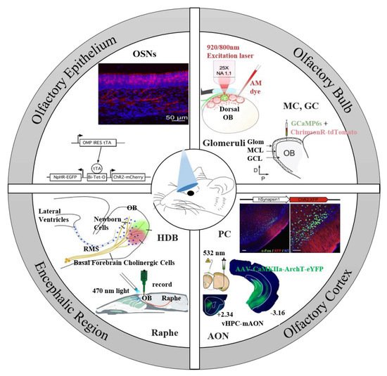
2. Optogenetic Tools for the Olfactory System

Optogenetic tools include dozens of light-sensitive proteins, which can be activated by different wavelengths of light with various operating speeds. Microbial rhodopsins undergo membrane depolarization or cellular signaling cascades caused by light-induced photochemical reactions [33], and are widely used in neuroscience, including the olfactory system. Channelrhodopsin-2 (ChR2) was the first optogenetic tool to activate neurons with light, and is the most commonly used one due to its rapid on-rate [16]. When ChR2-expressed neurons are illuminated by light with specific wavelength (450–490 nm), neurons will be depolarized by activating channels [14]. These light-sensitive proteins such as ChR2 could be introduced to olfactory neurons by a viral vector or transgenic animals. Under the driving of various promoters, adeno-associated viruses (AAVs) are used to target ChR2 in M/T cells, interneurons in OB, cells in AON, and PC [34][35][36][37]. Lentiviral vectors (LVs) are also used to target ChR2 in the rostral migratory stream (RMS) and PC [29][38]. Optogenetic tools can be guided to the nervous system through a viral expression system, and targeted neurons with the help of specific promoters are summarized in Table 1 and Table 2. Specific promoters drive light-sensitive proteins to target different neurons, such as Pcdh21 targeting M/T cells, TH targeting short-axon cells, CHR targeting interneurons in the EPL, and choline acetyltransferase promoter targeting choline acetyltransferase neurons [34][39][40][41]. This genetic technology enables specific types of neurons of the olfactory system to be activated or silenced by light in order to study the role and functional connection of specific neurons in the olfactory system.
Several transgenic animal lines have been created for the research of the olfactory system. Arenkiel et al., generated transgenic mice expressing ChR2-YFP from the Thy1 promoter for the precise and rapid activation of mitral cells in the OB [42]. Then, they used Thy1-ChR2-YFP mice for mitral-cell-specific light stimulation to map the functional connectivity between mitral cells and interneurons [40]. They also generated VGAT-ChR2 transgenic mice with ChR2-expressed GABAergic neurons and glycinergic inhibitory neurons to study the neuronal connectivity [43]. Additionally, Dhawale et al., generated transgenic mice by expressing ChR2-EYFP into OSNs and their axons with a promoter OMP [44]. In generated transgenic mice, the individual M/T cell can be activated by illuminating a single glomerulus. OMP-ChR2-YEP transgenic mice and M72-ChR2 transgenic mice lines were created to study olfactory perception (details in Section 4) [45][46]. In summary, these ChR2 transgenic animals allow scientists to selectively activate olfactory neurons and study the signal processing and perception in olfactory system [47].
Gunaydin and colleagues designed and verified the E123T mutation in ChR2 (ChETA) to address the precision limitations of ChR2 [48]. The Cre-dependent AAV-ChETA-EYFP vector was injected into the OT for cell-type-specific expression to study the roles of OT neurons in attractive and aversive behaviors [49]. Similarly, Aqrabawi and colleagues used AAV-ChETA-eYFP vectors to express ChETA-eYFP in AON neurons in FosCreER mice to manipulate populations of neurons in AON which constitute odor engrams [50]. Lin and colleagues engineered a variant of channelrhodopsins named denoted red-activatable ChR (ReaChR), which enables transcranial optical activation of neurons [51]. Using the novel optogenetic tool, Inagaki and colleague generated the UAS-ReaChR transgenic Drosophila for neural manipulation [52]. This transgenic model has helped to understand the neuronal functions of Drosophila mushroom body (a higher olfactory circuit) and the sensitivity regulation in insect primary olfactory neurons [53][54].
The proton pump archaerhodopsin (Arch) from Halorubrum sodomense or the archaerhodopsin from the Halorubrum strain TP009 (ArchT) were used as optogenetic neuronal silencing tools. To selectively silence GABAergic inhibitory neurons, scientists injected AAV-ArchT virus into granule cell layers in the OB of transgenic mice expressing Cre recombinase. This optogenetic technology, combined with intracellular recordings, examined the contribution of inhibition to rhythmic activity in the mouse olfactory bulb [55]. McCarthy and colleagues injected AAV-ArchT virus into the accessory OB of Pcdh21-Cre mice in which expression of Cre-recombinase is restricted to M/T cells to examine whether light inhibition of accessory OB neurons could affect lordosis in sexually mice [34]. In addition, another commonly used silencing tool is Natronomonas pharaonis halorhodopsin (NpHR). The modified NPHR is called eNpHR3.0, which has better targeting of cell membrane, a longer current, shorter response time, and more sensitive response. The pLenti-hSyn-eNpHR3.0-EYFP lentivirus [56] were injected in the OB to study the OB projection to the olfactory tubercle [57] and to examine the effect of anterior OB inhibition on odorant attraction [58].
Table 1. Optogenetics approach in interneurons in OB.

Level

Expression Target

Model Animal

Expression Approach

Light Delivery

Electrophysiology

Recordings

Behavior

Ref.

Tool

Wavelength (nm)

Duration

(ms)

Frequency (Hz)

Power

(mW)

GL

Glomeruli

OMP-ChR2-YFP transgenic mice

Transgenic animal model

A 470 nm LED coupled with an objective

470

10

-

-

M/T cells: patch-clamp

-

[6]

SACs

TH-Cre mice

Injection of AAV-ChR2 into the GL

A solid-state laser coupled with an optical fiber

473

-

-

100

M/T cells or ETCs: whole-cell patch-clamp or cell-attached and tungsten microelectrodes

-

[39]

EPL

EPL-INs

Crh-Cre mice

Injection of AAV-ChR2 into the OB

A BLM-Series 473 nm blue laser system coupled with an objective

473


20–40

EPL INs: whole-cell patch-clamp

Olfactory associative learning training

[40]

EPL-INs

CRH-Cre mice

Injection of AAV-ChR2 into the OB

A blue laser system guided by implanted fiber optics

473

10

-

30

MCs: whole-cell patch-clamp and extracellular recording electrodes

Olfactory associative learning training

[59]

IPL

dSACs

Chrna2-Cre mice

Injection of AAV-ChR2 into the IPL

A 75 W xenon arc lamp coupled with an objective

-

-

-

-

TCs: whole cell patch clamp

-

[35]

GCL

GCs

Dlx5/6-Cre mice

Injection of AAV-ChR2 into the OB

A BLM-Series 473 nm blue laser system coupled with an objective

473


20–40

GCs: whole-cell patch-clamp

Olfactory associative learning training

[40]

GCs

OMP-Cre mice

Injection of AAV-ChR2 into the GCL

An implanted LEDs driven with a high-power LED driver

470

5

40

23

M/T cells: a silicon-based recording electrode and 32 channels optrode

Habituation task; Olfactory discrimination task

[60]

Table 2. Optogenetics approach in neuromodulation projections to the OB.

Encephalic Region

Expression Target

Model Animal

Expression Approach

Light Delivery

Electrophysiology

Recordings

Ref.

Tool

Wavelength (nm)

Duration

(ms)

Frequency (Hz)

Power

(mW)

Basal forebrain

HDB cholinergic neurons

VGLUT3-Cre mice

Injection of AAV-ChR2 into the HDB

A 75W xenon arc lamp coupled with an objective

-

10–20

-

-

OB cells: patch clamp

[61]

ChAT-ChR2-EYFP transgenic mice

Transgenic animal model

A diode-pumped solid-state 473 nm laser coupled with an optical fiber target the HDB

473

15

5–50

-

M/T cells and brain slices: patch clamp

[41]

ChAT-ChR2-EYFP mice transgenic

Transgenic animal model

A blue light diode laser and a blue LED coupled with implanted fiber

473

15

5–50

-

-

[32]

HDB GABAergic neurons

DLX5/6-Cre mice

Injection of AAV-ChR2 into the HDB

A blueCoolLED pE 100 coupled with an objective

490

-

-

-

OB cells: whole-cell

[62]

ChAT/GAD2-Cre mice

Injection of AAV-ChR2/eNpHR into the HDB

A 470 or 565 nm LED coupled with an optical fiber positioned in the OB

470; 565

10000

-

10;3

M/T cells: sixteen channel electrodes

[63]

Raphe nuclei

5-HT axons

TPH2-ChR2-YFP transgenic mice

Transgenic animal model

A bright light-emitting diode (LED) array coupled with a microscope

473

10

10

15

M/T cells: tungsten electrodes and whole-cell

[64]

serotonergic cells

Slc6a4-Cre mice

Injection of AAV-ChR2 into DRN

A 470 nm LED coupled with a glass fiber positioned close to the OB

470

10000


1–10

OB cells: 16-channel electrode

[31]

serotonergic cells

SERT-Cre mice

Injection of AAV-ChR2 in the DRN

A 470 nm laser coupled with an optrode lowered into the DRN

470

10

1–30

-

APC neurons: microelectrodes;

[65]

locus coreuleus

noradrenergic neurons

DBH-Cre-NpHR transgenic mice

Transgenic animal model

A solid-state laser coupled with an optical fiber implanted in the OB

532

-

-

2–10

MCs: tetrodes

[66]

3. The Sensory Input from OSNs to the OB

The OE, as the first level to receive odor information, plays an important role in the processing of odor information. OSNs expressing the same type of ORs usually project their axons into two bilateral glomeruli located in the OB [67][68]. When OSNs are stimulated by odors, the M/T cells in the OB will generate dynamic and rapid electrical signals [69]. Therefore, it is crucial to reveal how OSNs transmit odor information to M/T cells. Researchers expressed the opsins in OSNs of the OE and activated these neurons by light for the study of the light-induced responses of neurons in combination with electrophysiological, pharmacological, or behavioral methods [70][71]. The use of optogenetics has proven to be feasible to deliver precise stimuli in order to simulate the transmission of odor information from OE to OB as well as the processing process in OB [72].
Gire et al., used the patch-clamp to record light-induced OB signals in transgenic mice that expressed the ChR2 selectively in OSNs to investigate mechanisms of OSN signaling into mitral cells (MCs) [26]. Under the control of the OMP olfactory receptor promoter, ChR2-mCherry mice were crossed with mouse strains encoding the TTA gene to produce offspring that specifically expressed ChR2 in the OE. The patch-clamp recording of OB slices showed that most MCs did not display significant fast signals in response to light stimulation of ChR-expressing OSNs, and the OSN signals were shunted in MC. Recording the signals of tufted cells in the same way indicated that MCs received strong multistep signals through the tufted cells, suggesting that the olfactory information in OSNs was processed considerably before reaching MC, i.e., the tufted cells only mediated processing. The input from OSNs arrived at the glomerulus first, and each glomerulus accepted the input convergence of many OSNs expressing the same OR [73][74]. In addition, sister cells received input from the same glomerulus when the odor information in the OE reached the M/T cells from the glomerulus. Therefore, whether the higher olfactory center received redundant information was worth pondering. Albeanu et al., determined the M/T sister cells via the light-activated electrophysiological signals of a single glomerulus [44]. Transgenic mice expressing ChR2-EYFP in OSNs were used in this study to enable the glomerular layer (GL) to be accurately activated by light, while extracellular recordings showed that M/T cells only responded to the light stimulation of the GL. Because the size of the light spot was close to the average size of a single glomerulus, a single glomerulus can be activated for recording the response of M/T cells, which allowed the correct identification of the parental glomerulus to each mitral/tufted unit, and was further used as a basis to divide M/T cells into sister cells and non-sister cells. Studying the odor responses of sister cells found that the changes in their firing rates were related, indicating that sister cells received a common excitatory input. Moreover, the non-redundancy was found in the temporal characteristics of sister cell activity, which was of great significance for the transmission of information from the OB to the cerebral cortex.
In short, in the process of transmitting odor information to the OB, OSNs, glomeruli, MCs, and TCs played different roles to complete this task, respectively. After the direct OSN-EPSCs arrived at the TC, the MCs received a strong multi-step signal through the TC. Meanwhile, although a pair of sister cells (M/T) receive a common glomerular input, there are non-redundant odor responses. With the help of optogenetics, the goal of precise control of OSNs or a single glomerulus can be achieved, and then the downstream neural response can be measured to analyze the projection relationship with M/T cells.

4. How Do Activities of the Olfactory Bulb Neurons Affect Perception?

An important issue in olfactory research is how the brain encodes and processes odor information, i.e., how to convert chemical perception signals stimulated by odors into complex brain activities and behaviors. Imaging, electrophysiology, pharmacology, genetics, and other methods were used to reveal the basic principles of olfactory codes, including phase coding [75], combined coding [76], sparse coding [27], and concentration coding [77]. More studies on olfactory coding have been carefully discussed in some excellent reviews [11][78][79], and this review focuses more on the role of optogenetics in it. Light can selectively activate glomeruli instead of odor stimuli, and the perceptual responses can be explored by adjusting different characteristics of light stimulation, including the start and end time, the duration, the population, and number of glomeruli of light stimulation, etc. (Figure 2). As a result, odor perception is associated with complex neural activity patterns, and optogenetics is used as a powerful weapon to unlock the mystery of olfactory encoding.
Figure 2. A general strategy for exploring the link between brain activity and olfactory perception using optogenetics. Optogenetics is used to manipulate the spatiotemporal patterns of glomerular or M/T cell activity (such as activation population, latency, duration, etc.), followed by behavioral measurement of animal olfactory discrimination and perceptual limits.
The timing of odor stimulation is a key parameter and may represent important information of the olfactory system. In order to better understand the role of stimulus timing in the olfactory system, and to explore whether and how animals read the time of olfactory activation relative to the sniff cycle (‘sniff phase’), Rinberg et al., from the New York University Neuroscience Institute used optogenetics to generate time-controllable light stimulation to activate olfactory sensory neurons [45]. The results of behavioral and electrophysiological recordings indicated that the mouse’s olfactory system can accurately detect the beginning of the stimulus relative to the olfactory cycle and read out the time pattern. However, another group of researchers from the John B. Pierce Laboratory used the same method to prove that mice can successfully distinguish the onset time and delay of virtual odor stimulation, regardless of sniffing [80]. The opposite result did not negate the role of the sniffing cycle but indicated that the internal glomerular timing (the activation time relative to other neurons in the sequence) also participated in the encoding of time information, thereby independently affecting the perception of smell. Animals can not only perceive the beginning and end of light stimulus, but also distinguish the duration of the stimulus. Li et al., stimulated ChR2-expressed OSNs of mice at different durations [81]. Animal behavior results showed that mice could distinguish stimuli with a duration of 10 milliseconds, demonstrating that the mammalian olfactory system could accurately sense the difference in odor input duration. They also explored the thorny issue of neuron processing of input duration. The results of electrophysiological records showed that, when the glomeruli were activated by light of different durations, M/T cells responded strongly, and the frequency of their stimulation spikes carried the information of the stimulation duration.
Using time-controllable precise light stimuli instead of odor stimuli, scientists discovered that the characteristics of the time activity can impact on olfactory perception, including the start time, end time, and duration of the stimulus. In addition, the neural activity that induces perception should also include some spatial characteristics, such as what cells are responding to and the impact of the response of a single cell or multiple cells on cognition. Rinberg’s group have made many striking breakthroughs on this subject using optogenetics technology in recent years. They first explored whether the mouse can perceive the activation of a single glomerulus [46]. The coding sequence of ChR2-YFP was inserted into the gene encoding the olfactory receptor M72 to activate individual glomeruli with light. By changing the intensity and duration of light stimulation and other parameters, it was found that a single glomerulus can convey odor information using intensity and time coding prompts. However, odor stimuli usually induced a group of glomerular responses, which not only varied with odor, but also varied with the concentration of a single odor. Their team proposed an odor coding scheme called “primary coding”, which meant that some of the earliest activated glomeruli were very crucial for identifying odors [82]. In order to test this hypothesis, they delivered light stimuli to the ChR2 expressing OSN to generate masking stimuli. Combined with the electrophysiological recording and behavioral tests, they found that the earliest induced neural activity can be used to make olfactory judgments, confirming this olfactory primary code scheme.
Using optogenetic technology to explore the sense of smell, researchers can measure the limit of behavior discrimination by precisely manipulating a single feature (a single time or space feature), which confirmed the importance of the above single feature for olfactory perception. Thus, how will the combined characteristics of neuron spatiotemporal activities affect olfactory perception? Researchers performed optogenetic operations on mice to link the complex spatiotemporal activity patterns of neurons with olfactory perception. Chong et al., studied the importance of the combined characteristics of the spatial identity of glomerular activation and the temporal latency for perceptual meaning [83]. They used OMP-ChR2-YFP mice to manipulate the activated neuron groups or the activation latency with precise light stimulation, and then measured the changes in mouse cognition under different activity patterns. The results showed that activating different cell groups or changing their activation latency relative to other cells would cause varying degrees of changes in cognition. In this study, they did not explore the animal’s perception limit, and a more advanced optogenetic technology would provide the possibility to solve this problem. The holographic two-photon optogenetic technology can selectively activate neuron collections with the single cell resolution [84], achieve the spike–scale stimulation with millisecond resolution via the latest soma-covering light spots technology [85], and reproduce the temporal and spatial resolution of olfactory neuron activity patterns. In the latest study, Rinberg et al., used a modified light stimulation to activate neurons expressing red-shifted opsin ChrimsonR, and explored the three stimulus characteristic dimensions of mice (i.e., number of response cells, synchrony between neurons, and latency from inhalation onset) [28]. Their latest results showed that mice can detect a single action potential synchronously evoked by less than 20 M/T cells. At the same time, the synchronization of activation between neurons affected the olfactory perception of mice more than the inhalation period.
Odor stimulation induces neuronal activity, and the spatio-temporal combination of glomerular activity corresponds to different odors and even their concentrations [86], so the temporal and spatial features of odor perception have extraordinary significance for odor decoding. Recent research has explored the influence of multi-dimensional features such as number, synchronization (relative to the inhalation phase or not), latency and duration of glomeruli, and M/T cells activation on olfactory perception. Dr. Rinberg’s group has made outstanding contributions to the research on this topic. Although it may not completely replace natural odor stimulation, optogenetics provides precise parameterization and good control of causal operation with minimal damage, combines behavioral means to explore the contribution of these features to olfactory perception, and helps us better understand the basic principle of olfactory coding.

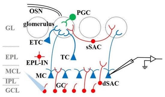
5. The Function of OB Interneurons in Odor Information Processing

The OB is the first relay station for odor information processing, connecting the sensory input of the OE with the olfactory area of the brain. The M/T cells in the OB receive odor information from the glomerulus, and the information is then transmitted to higher brain regions for further processing. M/T cells are modulated by various local interneurons in the OB during odor information processing. As shown in Figure 3, modulation involved neurons mainly include external tufted cells (ETCs), periglomerular cells (PGCs), superficial short-axon cells (sSACs) in the glomerular layer (GL), external plexiform layer interneurons (EPL-INs), deep short-axon cells (dSACs) in the internal plexiform layer (IPL), and granule cells (GCs) in the granule cells layer (GCL). Understanding the basic function of neuronal circuits in the OB may help decrypt the complex odor coding principles. Recently, optogenetics has been employed to reveal the complex modulation mechanism in OB. Table 1 summarizes the application of optogenetics approaches in investing the function of OB interneurons. Similar to the section on the functional connection between OE and OB, the general strategy of optogenetics in OB interneurons is to manipulate the characteristics of neuronal activation (in this case, the type of cell) and then measure the response of the downstream neuron to explore the projection from one area to another, where it is mainly the regulation of various types of interneurons on the M/T cells of main cells in OB.
Figure 3. Schematic diagram of the structure of the OB. The method of using optogenetics to study interglomerular interactions is to activate specific interneurons, and then record the electrophysiological signals of M/T cells.

This entry is adapted from the peer-reviewed paper 10.3390/bios11090309

References

  1. Giessel, A.J.; Datta, S.R. Olfactory maps, circuits and computations. Curr. Opin. Neurobiol. 2014, 24, 120–132.
  2. Genva, M.; Kenne Kemene, T.; Deleu, M.; Lins, L.; Fauconnier, M.-L. Is It Possible to Predict the Odor of a Molecule on the Basis of its Structure? Int. J. Mol. Sci. 2019, 20, 3018.
  3. Mainland, J.D.; Keller, A.; Li, Y.R.; Zhou, T.; Trimmer, C.; Snyder, L.L.; Moberly, A.H.; Adipietro, K.A.; Liu, W.L.L.; Zhuang, H.; et al. The missense of smell: Functional variability in the human odorant receptor repertoire. Nat. Neurosci. 2014, 17, 114–120.
  4. Xu, L.; Li, W.; Voleti, V.; Zou, D.-J.; Hillman, E.M.C.; Firestein, S. Widespread receptor-driven modulation in peripheral olfactory coding. Science 2020, 368, eaaz5390.
  5. Cavarretta, F.; Burton, S.D.; Igarashi, K.M.; Shepherd, G.M.; Hines, M.L.; Migliore, M. Parallel odor processing by mitral and middle tufted cells in the olfactory bulb. Sci. Rep. 2018, 8, 7625.
  6. Geramita, M.; Urban, N.N. Differences in Glomerular-Layer-Mediated Feedforward Inhibition onto Mitral and Tufted Cells Lead to Distinct Modes of Intensity Coding. J. Neurosci. 2017, 37, 1428–1438.
  7. Liu, G.; Froudarakis, E.; Patel, J.M.; Kochukov, M.Y.; Pekarek, B.; Hunt, P.J.; Patel, M.; Ung, K.; Fu, C.-H.; Jo, J.; et al. Target specific functions of EPL interneurons in olfactory circuits. Nat. Commun. 2019, 10, 3369.
  8. Bolding, K.A.; Franks, K.M. Recurrent cortical circuits implement concentration-invariant odor coding. Science 2018, 361.
  9. Otazu, G.H.; Chae, H.; Davis, M.B.; Albeanu, D.F. Cortical Feedback Decorrelates Olfactory Bulb Output in Awake Mice. Neuron 2015, 86, 1461–1477.
  10. Wang, C.Y.; Liu, Z.; Ng, Y.H.; Südhof, T.C. A Synaptic Circuit Required for Acquisition but Not Recall of Social Transmission of Food Preference. Neuron 2020, 107, 144–157.
  11. Uchida, N.; Poo, C.; Haddad, R. Coding and Transformations in the Olfactory System. Annu. Rev. Neurosci. 2014, 37, 363–385.
  12. Wilson, D.A.; Sullivan, R.M. Cortical processing of odor objects. Neuron 2011, 72, 506–519.
  13. Sohal, V.S.; Zhang, F.; Yizhar, O.; Deisseroth, K.J.N. Parvalbumin neurons and gamma rhythms enhance cortical circuit performance. Nature 2009, 459, 698–702.
  14. Boyden, E.S.; Zhang, F.; Bamberg, E.; Nagel, G.; Deisseroth, K. Millisecond-timescale, genetically targeted optical control of neural activity. Nat. Neurosci. 2005, 8, 1263–1268.
  15. Zhang, F.; Wang, L.; Brauner, M.; Liewald, J.F.; Kay, K.; Watzke, N.; Wood, P.G.; Bamberg, E.; Nagel, G.; Gottschalk, A.J.N. Multimodal fast optical interrogation of neural circuitry. Nature 2007, 446, 633–639.
  16. Nagel, G.; Szellas, T.; Huhn, W.; Kateriya, S.; Adeishvili, N.; Berthold, P.; Ollig, D.; Hegemann, P.; Bamberg, E. Channelrhodopsin-2, a directly light-gated cation-selective membrane channel. Proc. Natl. Acad. Sci. USA 2003, 100, 13940–13945.
  17. Klapoetke, N.C.; Murata, Y.; Kim, S.S.; Pulver, S.R.; Birdsey-Benson, A.; Cho, Y.K.; Morimoto, T.K.; Chuong, A.S.; Carpenter, E.J.; Tian, Z.; et al. Independent optical excitation of distinct neural populations. Nat. Methods 2014, 11, 338–346.
  18. Chaigneau, E.; Ronzitti, E.; Gajowa, M.A.; Soler-Llavina, G.J.; Tanese, D.; Brureau, A.Y.B.; Papagiakoumou, E.; Zeng, H.; Emiliani, V. Two-Photon Holographic Stimulation of ReaChR. Front. Cell. Neurosci. 2016, 10, 234.
  19. Watanabe, H.; Sano, H.; Chiken, S.; Kobayashi, K.; Fukata, Y.; Fukata, M.; Mushiake, H.; Nambu, A. Forelimb movements evoked by optogenetic stimulation of the macaque motor cortex. Nat. Commun 2020, 11, 3253.
  20. Tamura, K.; Takeda, M.; Setsuie, R.; Tsubota, T.; Hirabayashi, T.; Miyamoto, K.; Miyashita, Y. Conversion of object identity to object-general semantic value in the primate temporal cortex. Science 2017, 357, 687–692.
  21. Hardt, O.; Nadel, L. Systems consolidation revisited, but not revised: The promise and limits of optogenetics in the study of memory. Neurosci. Lett. 2018, 680, 54–59.
  22. Parnaudeau, S.; Bolkan, S.S.; Kellendonk, C.J.B.P. The Mediodorsal Thalamus: An Essential Partner of the Prefrontal Cortex for Cognition. Biol. Psychiatry 2017, 83, 648–656.
  23. Cheng, Z.; Cui, R.; Ge, T.; Yang, W.; Li, B. Optogenetics: What it has uncovered in potential pathways of depression. Pharmacol. Res. 2020, 152, 104596.
  24. Jarrin, S.; Finn, D.P.J.N.; Reviews, B. Optogenetics and its application in pain and anxiety research. Neurosci. Biobehav. Rev. 2019, 105, 200–211.
  25. Grimaud, J.; Lledo, P.M. Illuminating odors: When optogenetics brings to light unexpected olfactory abilities. Learn. Mem. 2016, 23, 249–254.
  26. Gire, D.H.; Franks, K.M.; Zak, J.D.; Tanaka, K.F.; Whitesell, J.D.; Mulligan, A.A.; Hen, R.; Schoppa, N.E. Mitral cells in the olfactory bulb are mainly excited through a multistep signaling path. J. Neurosci. 2012, 32, 2964–2975.
  27. Braubach, O.A.-O.; Tombaz, T.; Geiller, T.; Homma, R.; Bozza, T.A.-O.; Cohen, L.B.; Choi, Y. Sparsened neuronal activity in an optogenetically activated olfactory glomerulus. Sci. Rep. 2018, 8, 1–17.
  28. Gill, J.V.; Lerman, G.M.; Zhao, H.; Stetler, B.J.; Rinberg, D.; Shoham, S. Precise Holographic Manipulation of Olfactory Circuits Reveals Coding Features Determining Perceptual Detection. Neuron 2020, 108, 382–393.
  29. Choi, G.B.; Stettler, D.D.; Kallman, B.R.; Bhaskar, S.T.; Fleischmann, A.; Axel, R. Driving Opposing Behaviors with Ensembles of Piriform Neurons. Cell 2011, 146, 1004–1015.
  30. Aqrabawi, A.J.; Kim, J.C. Hippocampal projections to the anterior olfactory nucleus differentially convey spatiotemporal information during episodic odour memory. Nat. Commun. 2018, 9, 2735.
  31. Brunert, D.; Tsuno, Y.; Rothermel, M.; Shipley, M.T.; Wachowiak, M. Cell-Type-Specific Modulation of Sensory Responses in Olfactory Bulb Circuits by Serotonergic Projections from the Raphe Nuclei. J. Neurosci. 2016, 36, 6820.
  32. Nitenson, A.S.; Nieves, G.M.; Poeta, D.L.; Bahar, R.; Rachofsky, C.; Mandairon, N.; Bath, K.G. Acetylcholine Regulates Olfactory Perceptual Learning through Effects on Adult Neurogenesis. iScience 2019, 22, 544–556.
  33. Rost, B.R.; Schneider-Warme, F.; Schmitz, D.; Hegemann, P. Optogenetic Tools for Subcellular Applications in Neuroscience. Neuron 2017, 96, 572–603.
  34. McCarthy, E.A.; Kunkhyen, T.; Korzan, W.J.; Naik, A.; Maqsudlu, A.; Cherry, J.A.; Baum, M.J. A comparison of the effects of male pheromone priming and optogenetic inhibition of accessory olfactory bulb forebrain inputs on the sexual behavior of estrous female mice. Horm. Behav. 2017, 89, 104–112.
  35. Burton, S.D.; Larocca, G.; Liu, A.; Cheetham, C.E.J.; Urban, N.N. Olfactory Bulb Deep Short-Axon Cells Mediate Widespread Inhibition of Tufted Cell Apical Dendrites. J. Neurosci. 2017, 37, 1117–1138.
  36. Choy, J.M.C.; Suzuki, N.; Shima, Y.; Budisantoso, T.; Nelson, S.B.; Bekkers, J.M. Optogenetic Mapping of Intracortical Circuits Originating from Semilunar Cells in the Piriform Cortex. Cereb. Cortex 2015, 27, 589–601.
  37. Markopoulos, F.; Rokni, D.; Gire, D.H.; Murthy, V.N. Functional Properties of Cortical Feedback Projections to the Olfactory Bulb. Neuron 2012, 76, 1175–1188.
  38. Alonso, M.; Lepousez, G.; Wagner, S.; Bardy, C.; Gabellec, M.M.; Torquet, N.; Lledo, P.M. Activation of adult-born neurons facilitates learning and memory. Nat. Neurosci. 2012, 15, 897–904.
  39. Liu, S.; Puche, A.C.; Shipley, M.T. The Interglomerular Circuit Potently Inhibits Olfactory Bulb Output Neurons by Both Direct and Indirect Pathways. J. Neurosci. 2016, 36, 9604–9617.
  40. Huang, L.; Ung, K.; Garcia, I.; Quast, K.B.; Cordiner, K.; Saggau, P.; Arenkiel, B.R. Task Learning Promotes Plasticity of Interneuron Connectivity Maps in the Olfactory Bulb. J. Neurosci. 2016, 36, 8856–8871.
  41. Ma, M.; Luo, M. Optogenetic Activation of Basal Forebrain Cholinergic Neurons Modulates Neuronal Excitability and Sensory Responses in the Main Olfactory Bulb. J. Neurosci. 2012, 32, 10105–10116.
  42. Arenkiel, B.R.; Peca, J.; Davison, I.G.; Feliciano, C.; Deisseroth, K.; Augustine, G.J.; Ehlers, M.D.; Feng, G. In vivo light-induced activation of neural circuitry in transgenic mice expressing channelrhodopsin-2. Neuron 2007, 54, 205–218.
  43. Zhao, S.; Ting, J.T.; Atallah, H.E.; Qiu, L.; Tan, J.; Gloss, B.; Augustine, G.J.; Deisseroth, K.; Luo, M.; Graybiel, A.M.; et al. Cell type–specific channelrhodopsin-2 transgenic mice for optogenetic dissection of neural circuitry function. Nat. Methods 2011, 8, 745–752.
  44. Dhawale, A.K.; Hagiwara, A.; Bhalla, U.S.; Murthy, V.N.; Albeanu, D.F. Non-redundant odor coding by sister mitral cells revealed by light addressable glomeruli in the mouse. Nat. Neurosci. 2010, 13, 1404–1412.
  45. Smear, M.C.; Shusterman, R.; Oconnor, R.P.; Bozza, T.C.; Rinberg, D. Perception of sniff phase in mouse olfaction. Nature 2011, 479, 397–400.
  46. Smear, M.C.; Resulaj, A.; Zhang, J.; Bozza, T.C.; Rinberg, D. Multiple perceptible signals from a single olfactory glomerulus. Nat. Neurosci. 2013, 16, 1687–1691.
  47. Luna, V.M.; Morozov, A. Input-specific excitation of olfactory cortex microcircuits. Front. Neural Circuits 2012, 6, 69.
  48. Gunaydin, L.A.; Yizhar, O.; Berndt, A.; Sohal, V.S.; Deisseroth, K.; Hegemann, P. Ultrafast optogenetic control. Nat. Neurosci. 2010, 13, 387–392.
  49. Murata, K.; Kinoshita, T.; Fukazawa, Y.; Kobayashi, K.; Yamanaka, A.; Hikida, T.; Manabe, H.; Yamaguchi, M. Opposing Roles of Dopamine Receptor D1- and D2-Expressing Neurons in the Anteromedial Olfactory Tubercle in Acquisition of Place Preference in Mice. Front. Behav. Neurosci. 2019, 13, 50.
  50. Aqrabawi, A.J.; Kim, J.C. Olfactory memory representations are stored in the anterior olfactory nucleus. Nat. Commun. 2020, 11, 1246.
  51. Lin, J.Y.; Knutsen, P.M.; Muller, A.; Kleinfeld, D.; Tsien, R.Y. ReaChR: A red-shifted variant of channelrhodopsin enables deep transcranial optogenetic excitation. Nat. Neurosci. 2013, 16, 1499–1508.
  52. Inagaki, H.K.; Jung, Y.; Hoopfer, E.D.; Wong, A.M.; Mishra, N.; Lin, J.Y.; Tsien, R.Y.; Anderson, D.J. Optogenetic control of Drosophila using a red-shifted channelrhodopsin reveals experience-dependent influences on courtship. Nat. Methods 2014, 11, 325–332.
  53. Inada, K.; Tsuchimoto, Y.; Kazama, H. Origins of Cell-Type-Specific Olfactory Processing in the Drosophila Mushroom Body Circuit. Neuron 2017, 95, 357–367.e354.
  54. Guo, H.; Kunwar, K.; Smith, D. Odorant Receptor Sensitivity Modulation in Drosophila. J. Neurosci. 2017, 37, 9465.
  55. Fukunaga, I.; Herb, J.T.; Kollo, M.; Boyden, E.S.; Schaefer, A.T. Independent control of gamma and theta activity by distinct interneuron networks in the olfactory bulb. Nat. Neurosci. 2014, 17, 1208–1216.
  56. Gradinaru, V.; Zhang, F.; Ramakrishnan, C.; Mattis, J.; Prakash, R.; Diester, I.; Goshen, I.; Thompson, K.R.; Deisseroth, K. Molecular and Cellular Approaches for Diversifying and Extending Optogenetics. Cell 2010, 141, 154–165.
  57. Midroit, M.; Chalençon, L.; Renier, N.; Milton, A.; Thevenet, M.; Sacquet, J.; Breton, M.; Forest, J.; Noury, N.; Richard, M.; et al. Neural processing of the reward value of pleasant odorants. Curr. Biol. 2021, 31, 1592–1605.e1599.
  58. Kermen, F.; Midroit, M.; Kuczewski, N.; Forest, J.; Thévenet, M.; Sacquet, J.; Benetollo, C.; Richard, M.; Didier, A.; Mandairon, N. Topographical representation of odor hedonics in the olfactory bulb. Nat. Neurosci. 2016, 19, 876–878.
  59. Huang, L.; Garcia, I.; Jen, H.; Arenkiel, B.R. Reciprocal connectivity between mitral cells and external plexiform layer interneurons in the mouse olfactory bulb. Front. Neural Circuits 2013, 7, 32.
  60. Gschwend, O.; Abraham, N.M.; Lagier, S.; Begnaud, F.; Rodriguez, I.; Carleton, A. Neuronal pattern separation in the olfactory bulb improves odor discrimination learning. Nat. Neurosci. 2015, 18, 1474–1482.
  61. Case, D.T.; Burton, S.D.; Gedeon, J.Y.; Williams, S.P.G.; Urban, N.N.; Seal, R.P. Layer- and cell type-selective co-transmission by a basal forebrain cholinergic projection to the olfactory bulb. Nat. Commun. 2017, 8, 652.
  62. Diez, A.S.; Najac, M.; De Saint Jan, D. Basal forebrain GABAergic innervation of olfactory bulb periglomerular interneurons. J. Physiol. 2019, 597, 2547–2563.
  63. Böhm, E.; Brunert, D.; Rothermel, M. Input dependent modulation of olfactory bulb activity by HDB GABAergic projections. Sci. Rep. 2020, 10, 10696.
  64. Kapoor, V.; Provost, A.C.; Agarwal, P.; Murthy, V.N. Activation of raphe nuclei triggers rapid and distinct effects on parallel olfactory bulb output channels. Nat. Neurosci. 2016, 19, 271–282.
  65. Lottem, E.; Lorincz, M.L.; Mainen, Z.F. Optogenetic Activation of Dorsal Raphe Serotonin Neurons Rapidly Inhibits Spontaneous but Not Odor-Evoked Activity in Olfactory Cortex. J. Neurosci. 2016, 36, 7–18.
  66. Ramirezgordillo, D.; Ma, M.; Restrepo, D. Precision of Classification of Odorant Value by the Power of Olfactory Bulb Oscillations Is Altered by Optogenetic Silencing of Local Adrenergic Innervation. Front. Cell. Neurosci. 2018, 12, 48.
  67. Mori, K.; Sakano, H. How is the olfactory map formed and interpreted in the mammalian brain? Annu Rev Neurosci. 2011, 34, 467–499.
  68. Murthy, V.N. Olfactory maps in the brain. Annu Rev Neurosci. 2011, 34, 233–258.
  69. Najac, M.; De Saint Jan, D.; Reguero, L.; Grandes, P.; Charpak, S. Monosynaptic and polysynaptic feed-forward inputs to mitral cells from olfactory sensory neurons. J. Neurosci. 2011, 31, 8722–8729.
  70. Shmuel, R.; Secundo, L.; Haddad, R. Strong, weak and neuron type dependent lateral inhibition in the olfactory bulb. Sci Rep. 2019, 9, 1602.
  71. Vaaga, C.E.; Westbrook, G.L. Parallel processing of afferent olfactory sensory information. J. Physiol. 2016, 594, 6715–6732.
  72. Genovese, F.; Thews, M.; Mohrlen, F.; Frings, S. Properties of an optogenetic model for olfactory stimulation. J. Physiol. 2016, 594, 3501–3516.
  73. Mombaerts, P. Axonal wiring in the mouse olfactory system. Annu. Rev. Cell Dev. Biol. 2006, 22, 713–737.
  74. Vassar, R.; Chao, S.K.; Sitcheran, R.; Nuñez, J.M.; Vosshall, L.B.; Axel, R. Topographic organization of sensory projections to the olfactory bulb. Cell 1994, 79, 981–991.
  75. Iwata, R.; Kiyonari, H.; Imai, T. Mechanosensory-Based Phase Coding of Odor Identity in the Olfactory Bulb. Neuron 2017, 96, 1139–1152.e1137.
  76. Malnic, B.; Hirono, J.; Sato, T.; Buck, L.B. Combinatorial receptor codes for odors. Cell 1999, 96, 713–723.
  77. Zhou, Z.; Belluscio, L. Coding odorant concentration through activation timing between the medial and lateral olfactory bulb. Cell Rep. 2012, 2, 1143–1150.
  78. Grabe, V.; Sachse, S. Fundamental principles of the olfactory code. Biosystems 2018, 164, 94–101.
  79. Renou, M.; Party, V.; Rouyar, A.; Anton, S. Olfactory signal coding in an odor background. Biosystems 2015, 136, 35–45.
  80. Rebello, M.R.; McTavish, T.S.; Willhite, D.C.; Short, S.M.; Shepherd, G.M.; Verhagen, J.V. Perception of odors linked to precise timing in the olfactory system. PLoS Biol. 2014, 12, e1002021.
  81. Li, A.; Gire, D.H.; Bozza, T.C.; Restrepo, D. Precise Detection of Direct Glomerular Input Duration by the Olfactory Bulb. J. Neurosci. 2014, 34, 16058–16064.
  82. Wilson, C.D.; Serrano, G.O.; Koulakov, A.A.; Rinberg, D. A primacy code for odor identity. Nat. Commun. 2017, 8, 1477.
  83. Chong, E.; Moroni, M.; Wilson, C.; Shoham, S.; Panzeri, S.; Rinberg, D. Manipulating synthetic optogenetic odors reveals the coding logic of olfactory perception. Science 2020, 368.
  84. Mardinly, A.R.; Oldenburg, I.A.; Pégard, N.C.; Sridharan, S.; Lyall, E.H.; Chesnov, K.; Brohawn, S.G.; Waller, L.; Adesnik, H. Precise multimodal optical control of neural ensemble activity. Nat. Neurosci. 2018, 21, 881–893.
  85. Chen, I.W.; Ronzitti, E.; Lee, B.R.; Daigle, T.L.; Dalkara, D.; Zeng, H.; Emiliani, V.; Papagiakoumou, E. In Vivo Submillisecond Two-Photon Optogenetics with Temporally Focused Patterned Light. J. Neurosci. Off. J. Soc. Neurosci. 2019, 39, 3484–3497.
  86. Carey, R.M.; Verhagen, J.V.; Wesson, D.W.; Pírez, N.; Wachowiak, M. Temporal structure of receptor neuron input to the olfactory bulb imaged in behaving rats. J. Neurophysiol. 2009, 101, 1073–1088.
More
This entry is offline, you can click here to edit this entry!