Dental Plaque: History
Please note this is an old version of this entry, which may differ significantly from the current revision.

Dental plaque is a community of microorganisms found on the surface of teeth or other hard surfaces like dentures and embedded in a matrix of polymers of both host and bacterial origin.

  • dental plaque
  • dental caries
  • propolis
  • COVID-19 complications

1. Introduction

The microorganisms have the ability to attach to other microorganisms in a way that allows them to survive and resist host defense mechanisms or antibiotic treatments. Most of them attach to different surfaces to form some type of biofilm matrix that is highly structured and spatially organized.

If not removed regularly, the biofilm undergoes maturation, which is connected with a progressive shift from a Gram-positive to a Gram-negative anaerobic species, which results in formation under the gingival surface, where bacteria grow profusely [1]. The pathogenic bacterial complex can lead to dental caries, periodontitis and gingivitis. Tissue injury, flossing, dental treatment and even chewing and eating can induce blood vessel injury near spaces covered in dental plaque. It allows the leakage of Gram-negative anaerobic species into the systemic bloodstream followed by bacteremia.

In addition, dental biofilm, especially subgingival plaque in patients with periodontitis, has been associated with cardiovascular and respiratory disease and diabetes mellitus, and recently constituting a high risk for developing severe illness due to Severe Acute Respiratory Syndrome Coronavirus 2 (SARS-CoV-2) infection [2].

The Role of Dental Plaque in The Etiology of Dental Caries and Periodontitis

It was shown that 40–50% of plaque remains after tooth brushing, and the effects of biofilm retention are clearly evident in the prevalence of caries, gingivitis and periodontal disease. Sucrose fermentation causes pH to drop rapidly to 5.0 or less at the point where the plaque contacts the tooth enamel. The enzyme invertase splits sucrose into its component glucose and fructose molecules, which are then converted to lactic acid by the glycolytic pathway [3].

When the pH of the dental plaque drops below 5.0–5.2, the saliva buffer cannot compensate. Tooth mineral is solubilized, thereby buffering the plaque and maintaining an environment suitable for Streptococcus mutans growth. Then the lactic acid that comes from the bacterial fermentation of sucrose and other carbohydrates in the diet reach the enamel surface, which begins to dissolve, releasing Ca and PO 4 ions from the surfaces under the enamel [4].

Presence of plaque rich in bacteria (especially Streptococcus mutans ) directly destroys the enamel layer by dissolving tooth minerals (mainly hydroxyapatite, Ca 10(PO 4) 6(OH) 2) resulting in caries. Periodontal diseases include two main conditions: gingivitis and periodontitis. Periodontitis is a complex, chronic inflammatory disease caused by an abnormal host response to bacteria from dental plaque. It affects the supporting structures of the teeth (root cementum, periodontal ligament and alveolar bone), causing irreversible attachment and bone loss, which are observed histologically and clinically. It leads finally to tooth loss. It has been estimated that periodontitis affects about 40% of the adult population. The transition from a healthy periodontium to a diseased one depends on the microbial dysbiosis and the abnormal immune host response. While the more severe forms of periodontal disease associated with alveolar bone loss are less frequent, gingivitis is widespread at all ages and is the most common form of periodontal disease.

It was shown, that in patients with severe periodontitis, there was a significant reduction in serum albumin concentration and an increase in loss of attachment [5].

2. Possible Influence of Dental Plaque on Severe COVID-19 Complications
In late 2019, a new coronavirus SARS-CoV-2 was detected in Wuhan, China, that spread all over the world to cause a pandemic. The disease caused by SARS-CoV-2 infection was named Coronavirus Disease 2019 (COVID-19). Patients suffering from diabetes, hypertension, obesity, asthma, and kidney and liver disease are in the high-risk group for severe COVID-19 symptoms [6]. Dental plaque, which leads to oral diseases, may be a risk factor causing a severe course of COVID-19 similar to those in above mentioned chronic diseases. The argument for this hypothesis is based on data from the most recent literature. Recent studies underline the association between poor oral health and the severity of COVID-19 via direct and indirect mechanisms. The direct mechanism is connected with angiotensin-converting enzyme II (ACE-2) receptors, while the indirect mechanism is connected with inflammatory pathways and bacterial superinfections.
ACE-2 receptors play a key role in SARS-CoV-2 entry into host cells. Periodontal disease, which is caused by a long-term accumulation of a dental plaque biofilm, exhibits a relationship with COVID-19 infection mediated by ACE-2 receptors. An association between ACE-2 and SARS-CoV-2 spike protein has been found, so patients with greater ACE-2 expression may be more susceptible to SARS-CoV-2 infection. ACE-2 receptors are present not only in blood vessels, kidney and brain but also in the tongue, buccal mucosa and gingiva. Therefore, ACE-2 in the oral mucosa may have an impact on COVID-19 infection [7][8]. Takahashi et al. confirmed that bacteria and their pathogenic factors, such as endotoxins, increases the expression of ACE-2 receptors in the altered oral microbiome [9][10].
Hence, the greater expression of ACE-2 receptors promotes SARS-CoV-2 infection, which Gupta et al. discovered in gingival cervicular fluid [11]. It is possible that oral diseases caused by the long-term accumulation of bacteria in dental plaque may be a risk factor for SARS-CoV-2 infection transmitted by viral droplets spread while coughing or sneezing.
The indirect mechanism, which explains the link between dental plaque and severe COVID-19 complications, is connected with inflammatory pathways due to the overproduction of proinflammatory cytokines. The increased number of cells producing cytokines and chemokines appears in the gingiva, which leads to increased levels of proinflammatory cytokines in the patient’s serum [12].
In 2020, COVID-19 patients presenting with oral pain, desquamative gingivitis, ulcers and blisters were reported [13]; however, mild cases of COVID-19 may not lead to oral symptoms. Severe periodontitis may worsen the progression of COVID-19 because of cytokine infiltration from periodontally diseased tissues to saliva followed by cytokine aspiration into the lungs and subsequent inflammation. Cytokines may alter the respiratory epithelium and lead to infection by respiratory pathogens. Moreover, the increased serum levels of proinflammatory cytokines—IL1-β, IL1RA, IL2, IL7, IL8, IL9, IL10, MCP1, MIP1α, FGF2, GCSF, GMCSF, IFNγ, IP10 and TNF-α—influence the severity of diseases including COVID-19 [13]. When the virus enters host cells it activates an immune response causing cytokine release syndrome or a “cytokine storm”. Thus, hypercytokinemia constitutes a consequence of SARS-CoV-2 infection and is responsible for complications such as lung injury, hypercoagulation, multiorgan failure and shock.
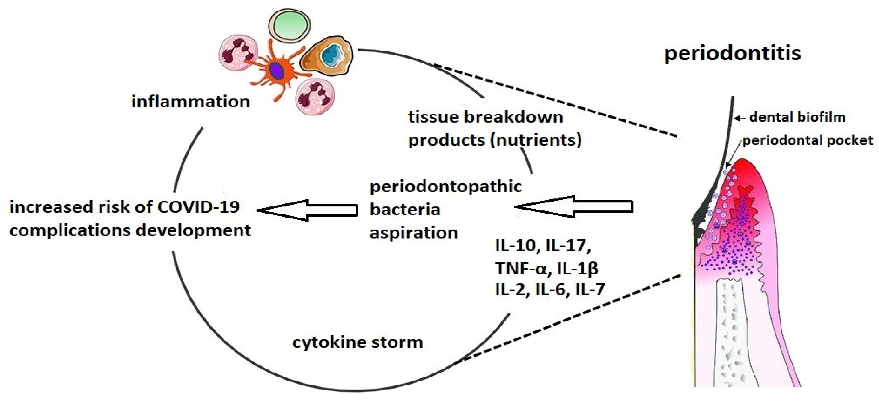
Hojyo et al. demonstrated that a high severity of COVID-19 is dependent on a cytokine storm [14]. IL-6 plays a key role in the cytokines release syndrome and a high blood level is highly correlated with the COVID-19 mortality [15]. Because the production of IL-6 and IL-17 is overexpressed in periodontitis, dental biofilm, which leads to periodontal diseases, has been recognized as a cause of a cytokine storm (Figure 1). Thus, we would like to highlight the association between altered biofilm and an increased risk of complications among patients with COVID-19.

Molecules 26 05516 g001

Figure 1. The connection between altered oral biofilm and complications of COVID-19.
Periodontal inflammation is shown by the elevated level of acute phase proteins (APPs), especially the C-reactive protein (CRP). One of proinflammatory proteins is Galectin-3, which takes part in T-cell mediated inflammation. It belongs to proteins from the group of lectins that bind to β-galactoside. Galectin-3 (Gal-3) takes part in the homeostasis of immune cells and inflammatory response. Gal-3 and CRP have been found among different predictors of COVID-19 [16][17]. In the case of Gal-3, its elevated level is connected with periodontitis [18], and this has been highlighted as a risk factor for a severe course of SARS-CoV-2 infection.
The relationship between COVID-19 infection and Gal-3 has been observed. Inhibition of Gal-3 decreases the viral activity because it exhibits a structure that is similar to an important area in the spike protein of SARS-CoV-2: increasing the immune response and viral attachment to host cells [18][19][20][21].
Moreover, during persistent inflammation accompanying SARS-CoV-2 infection, the coagulation cascade increases plasma levels of D-dimers, which are produced during fibrin degradation [22]. Some of the Streptococcus sanguis strains located in the oral cavity, produce platelet aggregation-associated protein (PAAP) which takes part in platelet activation and aggregation. Porphyromonas gingivalis may release proteins similar to PAAP [23]. Therefore, the periodontal therapy in patients with initial COVID-19 symptoms may reduce the plasma D-dimer level, which is, from our point of view, very important for preventing severe COVID-19 complications [24].
This bacterial potential, in addition to being detrimental to periodontitis, influences distant tissues and organs.
The oral cavity also constitutes a reservoir for Chlamydia pneumoniae, which is an obligate intracellular Gram-negative bacterium with a biphasic developmental cycle. C. pneumoniae is more frequent in subgingival plaques from patients with periodontal disease than in healthy controls [25]. It can cause persistent infections, such as airway infections, asthma and atherosclerosis. Some studies have shown that patients with severe periodontitis are often carriers of C. pneumoniae, which may increase the risk of a host inflammatory response with serious clinical consequences.
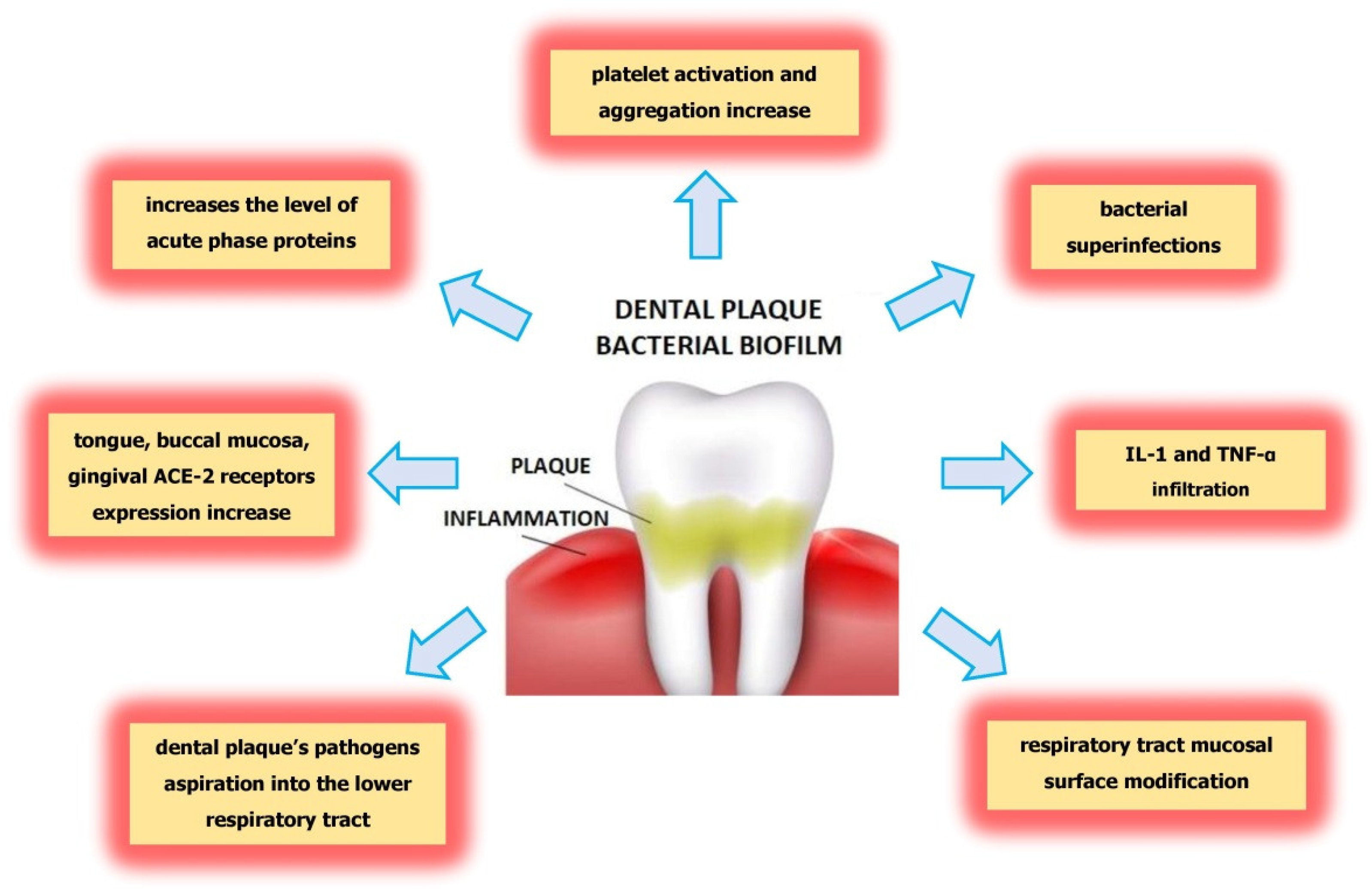
Several mechanisms indicate the ability of oral pathogens to lead to pneumonia during SARS-CoV-2 infection. Among the mechanisms that explain the association between oral pathogens and serious COVID-19 complications are the aspiration of oral pathogens into the lower respiratory tract, modification of mucosal surfaces by salivary enzymes in the respiratory tract that facilitate colonization by pathogens, and secretion of proinflammatory cytokines during periodontitis. (Figure 2). Moreover, proinflammatory cytokines increase the adhesion of pathogens to the lung epithelium. The aspiration of pathogens during coughing may lead to the development of pneumonia requiring hospitalization [26][27]. Therefore, there are many mechanisms which, due to the action of pathogens in the oral cavity, may worsen the course of COVID-19 and lead to superinfections and further complications. It is known that the cause of patient death is not the virus itself but bacterial superinfections. It is clear that poor oral health in the form of altered biofilm is associated with an increased bacterial exchange between the lungs and mouth, which leads to post-viral complications leading to respiratory infections [28][29].
Molecules 26 05516 g002
Figure 2. The mechanisms explaining the dental plaque influence on severity of COVID-19 complications.
High frequency of dental plaque occurrence and periodontitis development have brought about the need to investigate their possible correlation with the severity of COVID-19 complications to introduce appropriate preventive actions [30][31]. The recent study by Yang demonstrated that periodontal treatment reduced the risk of pneumonia in patient with COVID-19 [32].

3. Conclusions

Based on recent studies and the researchers’ suggestions, hypothesis and concepts, we can underline an association between poor oral health and the severity of the course of COVID-19, via direct and indirect mechanisms. The direct mechanism is connected with angiotensin-converting enzyme II (ACE-2) receptors, while the indirect mechanism is connected with inflammatory pathways and bacterial superinfections. Among the important mechanisms that explain the association between oral pathogens and the severity of SARS-CoV-2 infection are the aspiration of oral pathogens into the lower respiratory tract and the modification of mucosal surfaces in the respiratory tract that allows the adhesion and colonization of respiratory pathogens. The pro-inflammatory cytokine secretion promotes infection by means of respiratory pathogens, and bacterial infection is one of the most frequently occurring complications. Dental biofilm, especially covering subgingival plaque, has been associated with the development of severe complications from SARS-CoV-2 infection. Dental plaque leads to an imbalance in the pathological bacteria in the mouth, contributes to bacterial superinfections, and causes complications such as pneumonia or acute respiratory distress syndrome. As presented, there may be a strong connection between dental plaque occurrence and the severity of SARS-CoV-2 infection. A reduction in dental plaque by means of propolis may diminish the risk of COVID-19 complications. Therefore, propolis is a natural substance that can bring advantages in maintaining oral health and may reduce the risk of COVID-19 complications.

This entry is adapted from the peer-reviewed paper 10.3390/molecules26185516

References

  1. Yumoto, H.; Hirota, K.; Hirao, K.; Ninomiya, M.; Murakami, K.; Fujii, H.; Miyake, Y. The Pathogenic Factors from Oral Streptococci for Systemic Diseases. Int. J. Mol. Sci. 2019, 20, 4571.
  2. Kleinstein, S.; Nelson, K.; Freire, M. Inflammatory Networks Linking Oral Microbiome with Systemic Health and Disease. J. Dent. Res. 2020, 99, 1131–1139.
  3. Bowen, W.; Koo, H. Biology of Streptococcus mutans-Derived Glucosyltransferases: Role in Extracellular Matrix Formation of Cariogenic Biofilms. Caries Res. 2011, 45, 69–86.
  4. Dunne, W.M. Bacterial Adhesion: Seen Any Good Biofilms Lately? Clin. Microbiol. Rev. 2002, 15, 155–166.
  5. Kaur, N.; Kaur, N.; Sarangal, V. A study to evaluate the correlation of serum albumin levels with chronic periodontitis. Ind. J. Dent. Res. 2015, 26, 11–14.
  6. Richardson, S.; Hirsch, J.S.; Narasimhan, M.; Crawford, J.M.; McGinn, T.; Davidson, K.W.; Northwell COVID-19 Research Consortium. Presenting Characteristics, Comorbidities, and Outcomes Among 5700 Patients Hospitalized with COVID-19 in the New York City Area. JAMA 2020, 323, 2052–2059.
  7. Knypl, K. Pharmacotherapy of arterial hypertension: Antagonists of angiotensin receptors AT1. Med. Rodz. 2001, 2, 58–60.
  8. Xu, H.; Zhong, L.; Deng, J.; Peng, J.; Dan, H.; Zeng, X.; Li, T.; Chen, Q. High expression of ACE2 receptor of 2019-nCoV on the epithelial cells of oral mucosa. Int. J. Oral Sci. 2020, 12, 8.
  9. Takahashi, Y.; Watanabe, N.; Kamio, N.; Kobayashi, R.; Iinuma, T.; Imai, K. Aspiration of periodontopathic bacteria due to poor oral hygiene potentially contributes to the aggravation of COVID-19. J. Oral Sci. 2021, 63, 1–3.
  10. Takahashi, Y.; Watanabe, N.; Kamio, N.; Yokoe, S.; Suzuki, R.; Sato, S.; Iinuma, T.; Imai, K. Expression of the SARS-CoV-2 Receptor ACE2 and Proinflammatory Cytokines Induced by the Periodontopathic Bacterium Fusobacterium nucleatum in Human Respiratory Epithelial Cells. Int. J. Mol. Sci. 2021, 22, 1352.
  11. Gupta, S.; Mohindra, R.; Chauhan, P.; Singla, V.; Goyal, K.; Sahni, V.; Gaur, R.; Verma, D.; Ghosh, A.; Soni, R.; et al. SARS-CoV-2 Detection in Gingival Crevicular Fluid. J. Dent. Res. 2021, 100, 187–193.
  12. Liccardo, D.; Cannavo, A.; Spagnuolo, G.; Ferrara, N.; Cittadini, A.; Rengo, C.; Rengo, G. Periodontal Disease: A Risk Factor for Diabetes and Cardiovascular Disease. Int. J. Mol. Sci. 2019, 20, 1414.
  13. Huang, C.; Wang, Y.; Li, X.; Ren, L.; Zhao, J.; Hu, Y.; Zhang, L.; Fan, G.; Xu, J.; Gu, X.; et al. Clinical features of patients infected with 2019 novel coronavirus in Wuhan, China. Lancet 2020, 395, 497–506.
  14. Hojyo, S.; Uchida, M.; Tanaka, K.; Hasebe, R.; Tanaka, Y.; Murakami, M.; Hirano, T. How COVID-19 induces cytokine storm with high mortality. Inflamm. Regen. 2020, 40, 1–7.
  15. Sukumar, K.; Tadepalli, A. Nexus between COVID-19 and periodontal disease. J. Int. Med Res. 2021, 49, 3000605211002695.
  16. Praczyk, Ł.; Hoffmann, K.; Bryl, W. Galectin 3 as a Biomarker in Cardiovascular Diseases. HYGIEA Public Health 2019, 54, 75–79. Available online: http://www.h-ph.pl/pdf/hyg-2019/hyg-2019-2-075.pdf (accessed on 4 March 2021).
  17. Kazancioglu, S.; Yilmaz, F.M.; Bastug, A.; Ozbay, B.O.; Aydos, O.; Yücel, Ç.; Bodur, H.; Yilmaz, G. Assessment of Galectin-1, Galectin-3, and PGE2 Levels in Patients with COVID-19. Jpn. J. Infect. Dis. 2021, JJID.2021.020.
  18. Kara, C.; Çelen, K.; Dede, F.O.; Gökmenoğlu, C.; Kara, N.B. Is periodontal disease a risk factor for developing severe Covid-19 infection? The potential role of Galectin-3. Exp. Biol. Med. 2020, 245, 1425–1427.
  19. Liu, F.-T. Galectins: A New Family of Regulators of Inflammation. Clin. Immunol. 2000, 97, 79–88.
  20. De Oliveira, F.L.; Gatto, M.; Bassi, N.; Luisetto, R.; Ghirardello, A.; Punzi, L.; Doria, A. Galectin-3 in autoimmunity and auto-immune diseases. Exp. Biol. Med. 2015, 240, 1019–1028.
  21. Argüeso, P.; Panjwani, N. Focus on Molecules: Galectin-3. Exp. Eye Res. 2011, 92, 2–3.
  22. Cao, W.; Li, T. COVID-19: Towards understanding of pathogenesis. Cell Res. 2020, 30, 367–369.
  23. Herzberg, M.C.; Meyer, M.W. Effects of Oral Flora on Platelets: Possible Consequences in Cardiovascular Disease. J. Periodontol. 1996, 67, 1138–1142.
  24. Dikshit, S. Fibrinogen Degradation Products and Periodontitis: Deciphering the Connection. J. Clin. Diagn. Res. 2015, 9, ZC10–ZC12.
  25. Almeida-da-Silva, C.L.C.; Alpagot, T.; Zhu, Y.; Lee, S.S.; Roberts, B.P.; Hung, S.-C.; Tang, N.; Ojcius, D.M. Chlamydia pneu-moniae is present in the dental plaque of periodontitis patients and stimulates an inflammatory response in gingival epithelial cells. Microb. Cell 2019, 6, 197–208.
  26. Gomes-Filho, I.; Passos-Soares, J.; Da Cruz, S.S. Respiratory disease and the role of oral bacteria. J. Oral Microbiol. 2010, 2, 5811.
  27. Varanat, M.; Haase, E.; Kay, J.; Scannapieco, F. Activation of the TREM-1 pathway in human monocytes by periodontal pathogens and oral commensal bacteria. Mol. Oral Microbiol. 2017, 32, 275–287.
  28. Sampson, V.; Kamona, N.; Sampson, A. Could there be a link between oral hygiene and the severity of SARS-CoV-2 infections? Br. Dent. J. 2020, 228, 971–975.
  29. Paju, S.; A Scannapieco, F. Oral biofilms, periodontitis, and pulmonary infections. Oral Dis. 2007, 13, 508–512.
  30. Wojtkowska, A.A.; Wysokiński, A. Periodontitis and prevalence of cardiovascular diseases. Chor. Serca I Naczyń 2015, 12, 289–294.
  31. Botros, N.; Iyer, P.; Ojcius, D.M. Is there an association between oral health and severity of COVID-19 complications? Biomed. J. 2020, 43, 325–327.
  32. Yang, L.-C.; Suen, Y.-J.; Wang, Y.-H.; Lin, T.-C.; Yu, H.-C.; Chang, Y.-C. The Association of Periodontal Treatment and Decreased Pneumonia: A Nationwide Population-Based Cohort Study. Int. J. Environ. Res. Public Health 2020, 17, 356.
More
This entry is offline, you can click here to edit this entry!
ScholarVision Creations