Targeting SUMO Pathway in Cancer: History
Please note this is an old version of this entry, which may differ significantly from the current revision.
Subjects: Oncology
Contributor:

SUMOylation is a dynamic and reversible post-translational modification, characterized more than 20 years ago, that regulates protein function at multiple levels. Due to the reversible nature of this post-translational protein modification, the balance between SUMOylation and the removal of SUMO is critical. SUMO pathway regulates the hallmark properties of cancer cells. Moreover, alterations in activity and in levels of SUMO machinery components have been observed in human cancer. Many molecular mechanisms relevant to the pathogenesis of specific cancers involve SUMO, highlighting the potential relevance of SUMO machinery components as therapeutic targets. Early-phase clinical trials are currently evaluating the safety and efficacy of SUMO pathway inhibition in cancer patients.

  • cancer
  • post-translational modification (PTM)
  • small ubiquitin-like modifier (SUMO)
  • protein inhibitor of activated STAT (PIAS)
  • sentrin-specific protease (SENP)

Note: The entry will be online only after author check and submit it.

1. Introduction

Cancer is consistently ranked among the leading causes of death worldwide [1]. Hallmarks of cancer cells include uncontrolled proliferation, inhibition of apoptosis and differentiation, immune evasion, and the potential to metastasize [2]. Cellular homeostasis and responses to various stimuli are regulated by a wide array of post-translational modifications (PTM), including phosphorylation, acetylation, hydroxylation, glycosylation, and methylation as well as small protein modifications, such as ubiquitination [3,4]. Dysregulation of PTMs is frequent in cancer and plays a significant role in the pathogenesis of the disease [5,6]. Ubiquitination and its analogous modifications neddylation and SUMOylation are increasingly viewed as key regulators of tumorigenesis of various cancers [7,8,9]. In this review, we discuss SUMOylation’s contribution to the malignant phenotype and the pathway’s role as a potential therapeutic target in cancers originating from different cell types and organs.

2. Basic Principles of SUMOylation and Its Role in Physiology

Small ubiquitin-like modifiers (SUMOs) are a group of ubiquitin-like small proteins that are attached to substrate proteins in a reversible post-translational modification termed SUMOylation that is highly conserved in eukaryotes [9,10]. The SUMO pathway is involved in several central cellular processes. Modification by SUMO mediates the protein–protein interactions of target substrates, and influences their subcellular localization, stability, and enzymatic function [9,11]. SUMOylation can simultaneously target several members of a protein group to modulate the activity of a specific pathway [12]. The SUMO pathway shares some structural and functional similarities with the ubiquitin pathway [11]. The global molecular structure of SUMO is similar to ubiquitin, and both have a ββαββαβ fold structure and a conserved position of a diglycine (GG) motif required for isopeptide bond formation [13,14].
To date, five isoforms of SUMO (SUMO1, 2, 3, 4, and 5) have been identified in the human genome [9]. SUMO1, SUMO2, and SUMO3 are ubiquitously expressed in tissues, while the expression of SUMO4 and SUMO5 is restricted only to specific tissues [9,15,16,17,18]. SUMO1, SUMO2, and SUMO3 account for most of SUMO modifications, whereas the functional roles of SUMO4 and SUMO5 are more unclear [15,18]. SUMO2 and SUMO3 both share 97% sequence identity, but share only 50% sequence similarity with SUMO1 [19]. Expression levels of SUMOs are dynamic and fluctuate between different developmental stages [17]. SUMOs can be covalently attached to a single lysine residue (monoSUMOylation) or to multiple lysine residues (multiSUMOylation) of the substrate (Figure 1). SUMO is preferentially attached to SUMO consensus ΨKxE motifs of substrates, in which Ψ denotes a hydrophobic amino acid, K is lysine, x is any amino acid and E is glutamic acid, although some substrates are also SUMOylated at lysines in non-consensus sites [10,20,21]. SUMO itself may also be SUMOylated (polySUMOylation) to form polymeric SUMO chains, which are restricted to SUMO2 and SUMO3 [22]. SUMO1 does not form chains efficiently and is predominantly associated with monoSUMOylation [22]. However, SUMO1 can be attached to SUMO2/3 chains, resulting in the termination of chain elongation [23]. Non-covalent attachment of SUMO is regulated through the SUMO interaction motif (SIM) of binding partners [24,25].
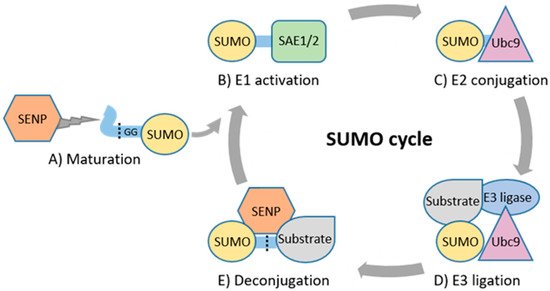
Figure 1. The enzymatic SUMO cascade. (A) SENPs cleave off amino acids from precursor SUMO to produce a mature SUMO. (B) SAE1/SAE2 forms of a thioester bond between SUMO’s GG and catalytic cysteine residue of SAE1/2. (C) Thioester bond is formed between SUMO’s GG and catalytic cysteine residue of Ubc9. (D) Ubc9 catalyzes formation of an isopeptide bond between GG and target substrate’s lysine residue often with assistance of an E3 ligase. (E) SENPs deconjugate SUMO from the substrate.
The enzymatic cascade regulating SUMOylation comprises SUMO E1-activating, E2-conjugating, E3 ligase and deconjugating enzymes (Figure 1) [9]. Maturation and deconjugation (deSUMOylation) of SUMO is regulated by the family of cysteine proteases, sentrin-specific proteases (SENP1, 2, 3, 5, 6, 7, and 8) [26]. SENP1–7 have specific, varying subcellular localizations and regulate SUMO processing, whereas SENP8 displays substrate specificity only for ubiquitin-like protein NEDD8 [26,27,28]. SUMO E1-activating enzyme (SAE1/2) activates mature SUMO and transfers it to the only known mammalian E2-conjugating enzyme, Ubc9 (encoded by UBE2I) [29,30,31]. Ubc9 alone is capable of conjugating SUMO to substrates in vitro. However, E3 ligase enzymes are required for specificity and efficiency of SUMOylation. The family of protein inhibitors of activated STAT (PIAS) is the major class of SUMO E3 ligases, and the human genome has four PIAS genes (PIAS1, 2, 3, and 4) that encode seven protein products, PIAS1, PIAS2a (PIASxα), PIAS2b (PIASxβ), PIAS3, PIAS3L, PIAS4 (PIASy), and PIASyE6− [32,33]. Other classes of SUMO E3 ligases include the well-characterized Ran-binding protein 2 (RanBP2), and few other E3 ligases, such as ZNF451, polycomb protein PC2 (also known as CBX4), and some members of the tripartite motif (TRIM) protein family [34,35,36,37,38].
The SUMO pathway undergoes constant cross talk with other post-translational modifications, including phosphorylation and methylation [39,40,41]. For instance, phosphorylation can influence SUMOylation of substrates containing the conserved phosphorylation-dependent SUMOylation motif (PDSM), which mediates phosphorylation-dependent SUMOylation of various transcriptional regulators [39]. In addition, lysine methylation of the KKxE motif can create a motif resembling the SUMO consensus site in substrates containing the KKxE motif [40]. HMGA2 SUMOylation is dependent on its methylation at K66 and K67 of KKAE motif by methyltransferase SET7/9. The KKxE motif is present in several other proteins, including the polyhomeotic complex 1 (PHC1), which also requires methylation for its complete SUMOylation.
The importance of SUMOylation in physiology is apparent from early development. A functional SUMO pathway is essential for embryogenesis. Loss of Ubc9 leads to severe defects of chromosome condensation and segregation, as well as disruption of nuclear organization, resulting in death at early embryonic development in mice [42]. SUMO1 knockout mice are viable, as SUMO2/3 can compensate for most functions of SUMO1 [17,43]. Interestingly, SUMO3−/− mice are also viable, but the knockout of SUMO2 leads to severe developmental defects and early death before adulthood, indicating that SUMO2 is essential for the development. Balance of SUMOylation and deSUMOylation is critical for embryonic development, as knockout of SENP1, SENP2, or SENP3 results in embryonic lethality in mice, suggesting non-redundancy and substrate specificity among SENPs [44,45,46]. SENP1−/− mice have severe fetal anemia and SENP2−/− mice display defects in trophoblast development, whereas conditional knockout Senp3+/− mice have impaired cytokine and inflammatory signaling. PIAS1−/− and PIAS4−/− knockout mice are produced at a lower frequency than the expected Mendelian ratio due to increased perinatal lethality, and both knockouts display defects in cytokine signaling [47,48]. Surviving PIAS1−/− mice are runts compared to their wild-type counterparts, whereas PIAS4−/− mice show no obvious phenotype. PIAS2−/− knockout mice have reduced testis weight and sperm count but remain viable and fertile [49]. PIAS3−/− mice are viable and show no overt phenotype but have an impaired visual response of medium wavelength cones [50].

3. Altered Expression and Prognostic Significance of SUMO Pathway in Cancer

Dysregulated mRNA and protein levels of the SUMO machinery components have been reported in tissue samples of cancer patients with potential prognostic associations (Table 1). In the majority of the published reports, the expression levels of SUMO pathway components are upregulated in cancer and are associated with a higher histological grade, higher stage, presence of metastases and poor prognosis. Interestingly, genome-wide analyses have identified concurrently increased levels of multiple SUMO machinery components in certain cancer types [51]. However, high expression has been associated with better prognosis in some cancer types. Downregulated expression levels in cancer tissues have consistently been reported for SENP2 [52,53,54,55,56,57] and PIAS2 [58,59,60]. The prognostic value of a single SUMO machinery component can also vary between cancer (sub)types, and, e.g., the value for PC2 [61,62] and PIAS4 [63,64] as prognostic biomarkers in breast cancer and for PIAS3 [65,66] in mesothelioma remains ambiguous (Table 1). Bioinformatic analyses of the SUMO machinery protein and mRNA levels, and their association with prognosis, utilizing datasets from various databases are generally well in line with the other published analyses of often smaller sample sizes [67,68,69]. Mass spectrometry-based methods might decrease variability in protein level analyses caused by the use of different antibodies, whereas IHC allows analysis of the subcellular localization of a protein, which might be crucial for the prognostic significance of the SUMO machinery components in particular [63,70,71].
Table 1. Prognostic value of SUMO pathway components.
High Expression Associates with
Poor Prognosis
High Expression Associates with
Good Prognosis
Cancer Type Protein(s) Cancer Type Protein(s)
adrenocortical PIAS3, PIAS4, SAE1, SAE2, SENP1, SENP3, SUMO1, SUMO2, SUMO4 bladder Ubc9
breast PC2, PIAS3, PIAS4, SAE1, SAE2, SENP5, SENP7L, SUMO1, SUMO2, SUMO3, Ubc9 breast PC2, PIAS1, PIAS4
colorectal SAE2, SENP1, SUMO1 cervical PIAS3
gastric PC2, PIAS2, SAE2, SUMO3, Ubc9 colorectal PC2
glioma SAE1, Ubc9 gastric PIAS1, PIAS4
hepatocellular PC2, PIAS2, PIAS3, PIAS4, SAE1, SAE2, SENP1, SENP3, SENP5, SENP6, SUMO2, Ubc9 glioma PIAS3
leukemia SAE1, SUMO3 leukemia PIAS2, SENP5, SENP7
lung PC2, SAE1, SAE2, SENP1, SUMO2/3, SUMO4, Ubc9 lung PIAS3
melanoma (cutaneous) SAE1 melanoma (cutaneous) PIAS1, SENP5, SENP7
melanoma (uveal) SAE1, SAE2, SUMO3 melanoma (uveal) SENP2, Ubc9
mesothelioma PIAS3, PIAS4, SAE1, SAE2, SENP1 mesothelioma PC2, PIAS3, SENP2
multiple myeloma Ubc9 ovarian PIAS2
osteosarcoma PC2, SENP3 pancreatic SENP3
ovarian SENP3, SENP5 pheochromocytoma and paraganglioma Ubc9
pancreatic SENP2, (SUMO1 and SUMO2/3 together), Ubc9 renal PIAS1, PIAS2
prostate PIAS1, SAE1, SENP1, SENP5, SUMO1, SUMO2 testicular germ cell PIAS2
renal PC2, PIAS3, RSUME, SAE1, SENP1, SENP3, SENP5, SUMO1, SUMO2, Ubc9 thymoma PIAS4, SAE1, SAE2, SENP1, Ubc9
sarcoma PC2, PIAS2, PIAS3, SENP6, SENP7    
thyroid PIAS2, SAE1    
uterine corpus endometrial PC2, SAE2, SENP2, SENP5, SUMO4    

Regulation of SUMO Machinery Expression and Activity in Cancer

Dysregulation of SUMO pathway components in cancer tissues is often detectable already at the mRNA level. The expression of SUMO machinery components can be epigenetically altered by DNA methylation. For instance, promoter hypomethylation of SENP6 induces expression of SENP6 in hepatocellular carcinoma (HCC) tissues, and elevated SENP6 mRNA and protein levels are associated with promotion of HCC tumorigenesis [72,73]. At the post-transcriptional level, numerous miRNAs are implicated in SUMO regulation, and inverse expression levels of SUMO components and their miRNA-regulators are found in several cancers [74,75,76,77,78]. For example, a low expression of miR-145 is correlated with high expression of SENP1 in prostate cancer cells, and introduction of miR-145 causes cell cycle arrest via inhibition of SENP1 [76]. Oncogenic miRNA-9 and miRNA-181a inhibit PIAS3 in IL-6high breast cancer to promote expansion of early-stage myeloid-derived suppressor cells, resulting in suppression of T-cell immunity [79].
Dysregulation of the expression of SUMO machinery components in protein level can also occur in cancer. Post-translational regulation by, e.g., ubiquitin and subsequent proteasomal degradation may be involved, resulting in alterations in expression levels in cancer [80,81]. Moreover, reactive oxygen species (ROS) typically present in cancer influence protein expression levels of SENPs [82,83]. For instance, the ROS-induced increase in SENP3 level can drive carcinogenesis of head and neck cancer via deSUMOylation and subsequent hyperphosphorylation of STAT3 [83].
In addition to changes in expression levels, catalytic activity of select SUMO pathway components can directly be regulated by conditions typical in cancer, such as hypoxia [84,85]. Hypoxia induces a rapid and reversible inhibition of catalytic activity of SENP1 and SENP3, resulting in altered SUMOylation of a subset of proteins, such as the co-repressor BHLHE40 that is implicated in metabolic reprogramming upon hypoxia.

4. Genetic Changes Targeting SUMO Machinery in Cancer

4.1. Germline Variants

In breast cancer patients, genetic variation of UBE2I can influence the characteristics of tumors [86,87]. Single nucleotide polymorphisms (SNPs) rs7187167, rs11248866, rs8052688, and rs8063 are associated with low-grade (grade 1) tumors, with the strongest tumor grade association displayed by rs7187167 found to be an independent predictor of tumor grade. Moreover, SNP variant rs17354559 of PIAS3 may be of functional relevance in breast cancer, although the variant was not evaluated experimentally [87]. Furthermore, the genetic variability of SENP1 and SENP2 may play a role in the occurrence of breast cancer [88]. The rs12297820 variant of SENP1 is associated with metastatic status in breast cancer. Women carrying allele C and genotype C/C in the rs12297820 polymorphic site have an increased risk of metastases, while allele T is associated with reduced risk of metastases. Genetic variability of SENP2 may influence the risk and subtype of breast cancer, as allele C and genotype C/C in rs6762208 site correlates with reduced risk, and the A/A genotype is associated with the lack of an estrogen receptor.
Studies performed in a Chinese population indicate that genetic variation of PC2 may influence the occurrence of gastric cancer [89]. Polymorphism rs77447679 is significantly associated with the risk of gastric cancer, as patients carrying the C/A genotype have approximately a 1.7 times higher risk of gastric cancer when compared to the baseline CC genotype, but its mode of action as a potential promoter of tumorigenesis in the context of gastric cancer is still unclear.
Microphthalmia-associated transcription factor (MITF) is a target for a germline missense substitution Mi-E318K at the SUMO consensus motif of MITF (IKQE -> IKQK) required for covalent SUMO binding, leading to the impairment of MITF SUMOylation [90,91]. The prevalence of Mi-E318K mutation is approximately five times higher in patients affected by melanoma, renal cell carcinoma (RCC), or both cancers compared with healthy controls. Overall, the data indicates that mutation of MITF generates a genetic predisposition for the development of melanoma and RCC. MITF plays a critical role in melanocyte development and melanoma carcinogenesis by controlling the expression of several melanoma-associated genes that regulate proliferation and invasion, including MET5 and CDKN2A/p16INK4A5 [92]. MITF also enhances the transcriptional activity of HIF1α, which is a critical target for kidney cancer susceptibility genes [93,94]. The SUMOylation of MITF represses its transcriptional activity, and accordingly the SUMO-deficient Mi-E318K displays significantly increased overall transcriptional activity when compared to its wild-type counterpart [91]. Interestingly, Mi-E318K and wild-type MITF show similar transcriptional activity on promoters of MET and CDKN2A, while the activity of HIF1α is promoted more efficiently by Mi-E318K. A specific Mi-E318K signature comprising 32 genes mostly associated with cell growth, proliferation, and inflammation has been identified in RCC cells, implicating that Mi-E318K can alter the transcription of its target genes. Mi-E318K also enhances the invasion, migration, and colony-forming potential of melanoma and RCC cells but does not significantly increase their proliferation rate.

4.2. Somatic Mutations

Mutations targeting SUMO machinery have been identified that may contribute to development of cancer. Amplification of distal regions of chromosome 3q is a common event in lung cancer [95]. A network of four genes, SENP2, DCUN1D1, DVL3, and UBXN7 was identified as a candidate driver of the 3q26-29 amplicon that occurs in 70–85% of squamous cell carcinomas (SCCs) of the lung [96,97]. In addition to lung cancer, amplifications of 3q26 are also frequent in other epithelial cancers, including head and neck cancer, cervical cancer, and ovarian cancer, which may indicate for SENP2 amplification [97]. As another example, the SENP5-encompassing 3q27.2-q29 region is amplified in 11% of penile squamous cell carcinoma patients, which may indicate enhanced SENP5 activity [98]. Furthermore, the amplification of PIAS1 and/or focal adhesion kinase (FAK) occurs in 8% of non-small-cell lung cancer (NSCLC) cell lines and in a small subset of patient-derived primary NSCLCs and potentially drives lung carcinogenesis [99]. Only a few cases of somatic mutations targeting SUMO pathway components resulting in the expression of altered protein have been characterized. The t(12;15)(q13;q25) chromosomal translocation was found to fuse SENP1 and the mesoderm development candidate 2 (MESDC2) in a patient with infantile sacrococcygeal teratoma [100]. The reciprocal fusion genes encode aberrant proteins that display deSUMOylation capacities similar compared with wild-type SENP1. Another example is the fusion between SENP6 (also known as SUSP1) and T-cell lymphoma breakpoint associated target 1 (TCBA1) that creates a SENP6–TCBA1 chimeric gene found in a T-cell lymphoblastic lymphoma cell line [101].
Somatic lysine mutations targeting the SUMO acceptor sites of substrate proteins may also lead to the dysregulation of SUMOylation and alter protein function in cancer. Interestingly, lysine mutations have been identified in cancer patients with potential functional relevance, but the extent of lysine mutations leading to aberrant SUMOylation is unknown [102,103].

5. SUMOylation Regulates Key Cancer Genes and Hallmark Properties of Cancer Cells

5.1. Substrates of SUMO Relevant for Cancer

Oncogenic signaling of growth promoting MYC in cancer cells is dependent on a functional SUMO machinery, indicating that dynamic SUMOylation and deSUMOylation are essential for the regulation of MYC [104]. However, the regulation is context-dependent, and SUMOylation is reported to both upregulate and suppress MYC activity, depending on the specific cell type [105,106,107]. SUMOylation has been suggested to be essential for KRAS/RAF-driven tumorigenesis [108] and is involved in the regulation of major growth-promoting intracellular signaling pathways and their downstream targets. SUMOylation enhances growth-promoting activities of oncogenic AKT, and a mutant form of AKT expressed in several cancers displays elevated levels of SUMOylation [109]. PIAS4-mediated SUMOylation of AMP-activated protein kinase (AMPK) in turn attenuates the inhibitory effect of AMPK towards growth-promoting mTORC1 signaling [110]. SUMOylation of hypoxia inducible factors 1 (HIF1) and 2 (HIF2) is essential for the regulation of several key processes during hypoxia, including angiogenesis, glucose metabolism and erythropoiesis [44,82,85,111,112]. SUMO machinery also mediates NF-κB signaling upon genotoxic stress through the regulation of the NF-κB essential modifier (NEMO) [113]. Furthermore, SUMOylation regulates activation of Wnt/β-catenin signaling via SUMO modification of transducin β-like protein TBL1-TBLR1 complex [114].
SUMOylation modulates several tumor suppressors, such as Rb, as well as p53 and its negative regulator MDM2, regulating cell cycle progression and stress responses [115,116,117,118,119,120]. SUMOylation positively regulates the activity of tumor suppressor PTEN, which is a negative regulator of the oncogenic phosphatidylinositol-3 kinase (PI3K)-AKT pathway and is involved in DNA damage response (DDR) [121]. SUMO machinery influences membrane association and subcellular localization of PTEN, controlling its tumor-suppressive functions in both cytoplasm and nucleus, suppressing tumor growth and, in contrast to other reports, suggesting that SUMOylation is a positive regulator of the PI3K-AKT-mTOR pathway [122,123,124].

5.2. SUMOylation in Cellular Processes Relevant for Cancer

The SUMO pathway regulates various essential cellular processes involved in tumorigenesis, including cell cycle progression, stress responses such as DDR, and response to hypoxia, angiogenesis, invasion, stem-like cell properties, and immune responses [44,125,126,127,128,129,130]. For example, PIAS1 and PIAS4 are promoters of double-strand break repair, and SENPs are critical for mediating chromosome structure and homologous recombination as well as nonhomologous end joining [125,131,132,133]. Moreover, SUMOylation influences DNA replication by targeting translesion polymerase eta (polη) to replication forks [134].
SUMO may be involved in the epigenetic regulation of gene expression in cancer. SUMOylation of histones is involved in regulation of chromatin dynamics [135]. SUMOylation may also influence the activity of methyltransferases, such as DNMT1, MLL1/MML2 complex and G9a [136,137,138]. PIAS1 has been shown to control epigenetic silencing of specific gene sets in certain contexts [139,140]. During T-cell differentiation PIAS1 maintains a repressive chromatin state of the FOXP3 promoter by recruitment of DNA methyltransferases and heterochromatin protein 1, which restricts the differentiation of natural regulatory T-cells (Tregs) [139].
The SUMO pathway regulates angiogenesis in response to hypoxia through modulation of vascular endothelial growth factor (VEGF) expression levels [44,141,142]. Furthermore, SUMOylation is involved in cellular migration and epithelial mesenchymal transition (EMT) via multiple mechanisms [143,144,145,146,147,148]. The SUMO pathway is also implicated in regulation of stem-like cell properties of cancer cells e.g., via PIAS3 and STAT3 [129,149].
SUMO machinery is a critical regulator of innate immune responses via the modulation of type I interferon (IFN) and NF-κB signaling [130,150]. The SUMO pathway regulates the production and activity of IFNs by inhibiting or stimulating the transcription of IFN regulatory transcription factors (IRFs), such as IRF3 and IRF7 [151,152,153,154]. The SUMOylation of IRFs represses the transactivating capacity of IRFs, resulting in diminished transcription of type I IFN genes and attenuation of immune response activation [152,155]. SUMO also impacts IFN production via the modulation of GMP–AMP synthase (cGAS) and the stimulator of interferon genes (STING) [156,157]. SUMO also influences the activity of NF-κB signaling regulators, NEMO and IκBα, affecting immune response activation [158,159,160,161,162]. SENP3-mediated deSUMOylation regulates antitumor immune response functions of Tregs, macrophages, and dendritic cells [163,164,165]. The loss of SENP3 deSUMOylase activity in Tregs results in dysregulation T-cell homeostasis and SENP3 deficiency in macrophages facilitates macrophage polarization towards the pro-tumor M2 subtype. Overall, SUMOylation is considered to have a net inhibitory effect on immune response activation [150,166,167]. Depletion of Ubc9 or SAE1/2 induces a strong inflammatory response and enhances protection of viral infections in hematological xenograft mouse models.

The article is from 10.3390/cancers13174402, a comprehensive review that also in detail discusses the SUMO-modulated molecular mechanisms and scientific rationale of targeting the SUMO pathway in different cancer types, using the best-characterized hematological malignancies and solid tumors as examples. Moreover, the properties and activity of pharmacological preclinical inhibitors of the SUMO pathway components (SAE1/2, Ubc9 and SENPs) as well as the clinical development of the SAE inhibitor, TAK-981, which is the only SUMO pathway inhibitor that has progressed to clinical trials in cancer patients are explored.

This entry is adapted from the peer-reviewed paper 10.3390/cancers13174402

This entry is offline, you can click here to edit this entry!
Video Production Service