Purine-Metabolising Enzymes: History
Please note this is an old version of this entry, which may differ significantly from the current revision.
Contributor:

The enzymes of both de novo and salvage pathways for purine nucleotide synthesis are regulated to meet the demand of nucleic acid precursors during proliferation. Among them, the salvage pathway enzymes seem to play the key role in replenishing the purine pool in dividing and tumour cells that require a greater amount of nucleotides. An imbalance in the purine pools is fundamental not only for preventing cell proliferation, but also, in many cases, to promote apoptosis. It is known that tumour cells harbour several mutations that might lead to defective apoptosis-inducing pathways, and this is probably at the basis of the initial expansion of the population of neoplastic cells. Therefore, knowledge of the molecular mechanisms that lead to apoptosis of tumoural cells is key to predicting the possible success of a drug treatment and planning more effective and focused therapies. 

  • purine salvage
  • apoptosis
  • CD73
  • cN-II
  • ADA
  • PNP
  • HPRT
  • IMPDH
  • SAMHD1
  • MTH1

1. Introduction

Intracellular purine nucleotide concentration is determined and maintained through two distinct pathways, both depending on a common metabolite: 5-phosphoribosyl-1-pyrophosphate (PRPP) (Figure 1). The de novo pathway consists of 10 reactions catalysed by six enzymes, some of which are allosterically regulated mainly by PRPP and purine nucleotides [1]. Furthermore, the six enzymes can cluster near mitochondria and microtubules to form dynamic multienzyme complexes referred to as “purinosomes” [2]. The purinosome formation causes a strong increase in the rate of purine synthesis. This regulatory mechanism ensures that, during proliferation and in the absence of preformed purine ring to be salvaged, the supply of purine compounds is secured. When preformed purine rings are available, they can be converted in one step into the corresponding nucleoside monophosphates through the action of the salvage pathway enzymes adenine phosphoribosyltransferase (APRT) and hypoxanthine-guanine phosphoribosyltransferase (HPRT) which utilize PRPP as a co-substrate (Figure 1) [1]. The availability of preformed purine ring prevents purinosome formation [2]. Regulation both at genetic and protein levels ensures the correct amount of nucleotides to sustain replicative and metabolic needs [3]. Therefore, it is not surprising that alterations of replicative rate and eventually activation of apoptosis are a consequence of dysfunctions of purine metabolism. Furthermore, an imbalance in purine supply can affect also mitochondrial proliferation and function, leading to metabolic changes and apoptosis [4]. Nevertheless, in some cases, the relationship between enzyme dysfunction and proliferation rate, metabolic alterations, and apoptosis is not as simple and direct as expected. The knowledge of the consequences of the alteration of enzymes involved in purine metabolism is essential not only for the understanding of “where and how” these enzymes impact metabolic pathways, but also to uncover whether they can be targets of antineoplastic drugs or be responsible for drug resistance. This review presents the most recent reports on the impact of purine catabolism and salvage enzymes (Figure 2) in the activation of apoptosis and the implied molecular mechanisms. Furthermore, the possible applications of this knowledge to anti-tumour therapy are discussed.
Figure 1. De novo and salvage pathways for purine nucleotide biosynthesis. Cyan background: de novo synthesis; yellow background: salvage synthesis. The figure outlines the central role played by PRPP, needed for both de novo and salvage pathways. Hyp: hypoxanthine; Gua: guanine; Ade: adenine; Rib-5-P: ribose-5-phosphate; PRPP: 5-phosphoribosyl-1-pyrophosphate; Gln: glutamine; Gly: glycine; THF: tetrahydrofolate; Asp: aspartate; S-AMP: succinyl-AMP; XMP: xanthosine-5′-monophosphate; IMP: inosine-5′-monophosphate; GMP: guanosine-5′-monphosphate; AMP: adenosine-5′-monophosphate.
Figure 2. Pathways of purine metabolism. 1: Ectosolic 5′-nucleotidase; 2: Adenosine deaminase; 3: Cytosolic 5′-nucleotidase II; 4: Purine nucleoside phosphorylase; 5: Hypoxanthine-guanine phosphoribosyltransferase; 6: IMP dehydrogenase. Inset: deoxyribonucleoside triphosphates (dNTP) are converted into the respective deoxynucleosides (dNs) by a one-step reaction catalysed by a sterile alpha motif and histidine-aspartate (HD) domain-containing protein 1 (enzyme 7). An increase in reactive oxygen species (ROS) brings about an increase in 8-oxo-dGTP, converted into the monophosphate by human MutT homolog 1 (enzyme 8). Ado: adenosine; Guo: guanosine; Ino: inosine.

2. Ectosolic 5′-Nucleotidase

Ectosolic 5′-nucleotidase (CD73) (EC 3.1.3.5) is a 70-kDa glycosylated protein bound to the outer surface of the plasma membrane by a glycosylphosphatidylinositol anchor. The enzyme is overexpressed in a variety of tumours [5] and in some of them, is associated with a highly invasive cancer phenotype, drug resistance, and tumour-promoting functions [6]. CD73 catalyses the dephosphorylation of extracellular AMP to adenosine (Ado), which plays important roles in many physiological and pathophysiological conditions through G-protein coupled Ado receptors (A1, A2A, A2B, and A3) [
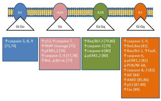
7,8]. The roles played by Ado are complex since its interaction with different receptors achieves different results; in particular, A1 and A3 receptors are coupled with Gi-proteins determining a decrease in cyclic AMP (cAMP), while A2A and A2B receptors are coupled with Gs-proteins causing an increase in intracellular cAMP [9] (Figure 3). Therefore, the role played by CD73 depends on the nature and distribution of the Ado receptors in a particular kind of cell. In addition, CD73 plays also many different roles in cell physiology not related to its catalytic activity [10]. Analysing enzymatic and non-enzymatic functions of CD73, it was concluded that both faces were involved in the aggressive behaviour of cancer cells. The enzymatic function seems to be primarily involved in invasion, whereas the non-enzymatic action of the protein contributes to cell adhesion and migration through activation of focal adhesion kinase [10]. Among the tumours in which CD73 is upregulated, breast cancer is the most studied [11]. In some cases of this type of cancer, an involvement of CD73 enzymatic activity was demonstrated, since supply of Ado could be a substitute for enzyme upregulation in promoting proliferation and motility [12]. In other cases, the involvement of different not yet described mechanisms independent of enzyme activity was demonstrated [13]. In all cases, an activation of the Akt/GSK-3β pathway was found to be involved in the tumour growth and motility promoted by CD73 [13]. In human cervical cancer cells, an increase in cell proliferation and motility was associated to CD73 overexpression, but the mechanism was independent of enzyme activity. In fact, CD73 inhibitors were unable to prevent the increase in proliferation in cells that overexpressed the enzyme. In addition, an increase in Ado, that could be expected when the enzyme is overexpressed, induced a decreased cell proliferation [14]. In this type of tumour, an activation of the Akt pathway was demonstrated as well [14]. In colorectal cancer, a downregulation of miR-30a was shown to determine an increase of CD73 expression in tumour cells, which promoted proliferation and inhibited apoptosis. MiR-30a is one of the most important tumour-suppressor factors in various human cancers and its level is significantly decreased in several tumours [15]. Since, as stated before, many effects exerted by the expression of CD73 on tumours are mediated by the participation of the enzyme activity in the conversion of extracellular ATP into Ado, such effects are dependent on the amount and nature of the Ado receptors expressed by tumour cells and other cells present in the tumour microenvironment. In this regard, in contrast to reports for other cancer types (see above), in an in vivo study on medulloblastoma, overexpression of CD73 reduced tumour growth and vascularization, and also promoted differentiation and initiated apoptosis, supposedly by the accumulation of Ado which interacted with A1 receptor [16]. The level of CD73 expression is very important not only for tumour growth and motility, but also for the success of the therapeutic approach. In fact, during chemotherapy, various immunogenic mediators accumulate in the tumour microenvironment, included ATP. Extracellular ATP, in this case, is released by the cells undergoing intrinsically or extrinsically activated apoptosis, through pannexin-1 channels and functions as a “find me” signal for P2Y family receptors expressed by macrophages and dendritic cells [17]. In this mechanism, the rapid ATP degradation catalysed by several extracellular enzymes is a determinant for the immunogenic activation, since the accumulated AMP, which cannot interact with P2Y receptors, can generate Ado through CD73, which mainly mediates the immune-escape of tumour cells interacting with A2A receptors [18]. Moreover, also in the case of regulatory T-cells (Tregs) in a tumour environment, the presence of several signals triggers apoptosis, and apoptotic Treg cells achieved superior immuno-suppression via an oxidative stress-associated mechanism [19]. Therefore, the induction of apoptosis in a tumour environment in which cells highly expressing CD73 and Ado receptors A2A are present is not always a successful therapeutic approach. Recently, CD73 has been targeted for the synthesis of new inhibitory compounds which prevent extracellular Ado formation from AMP and the consequent immune-escape [20].
Figure 3. Effectors of adenosine receptor-mediated apoptosis. The figure shows the different types of G-proteins associated with the four adenosine receptors and illustrates the apoptosis effectors found in several models by using agonists and antagonists of the receptors, or receptor silencing. The numbers in brackets refer to the respective reference. Note that adenosine receptors can be involved in survival in other cell types [71].

3. Cytosolic 5′-Nucleotidase II

Cytosolic 5′-nucleotidase II (cN-II) (EC 3.1.3.5) is an ubiquitously expressed and highly conserved enzyme that hydrolyses purine nucleoside monophosphates (preferentially IMP and GMP) into their corresponding nucleosides and inorganic phosphate. Its enzymatic activity and biochemical features have been well characterised, and the reader is referred to a recent review for further investigation of these issues [21]. The first observation on its prognostic value in nucleoside analogue-treated patients with acute myeloid leukaemia indicated that patients with high cN-II expression in leukaemic blasts showed a poorer outcome with respect to those with a lower expression [22]. Since then, the implication of cN-II in cancer cells and in the response to anticancer treatment has been extensively demonstrated [23,24]. Indeed, shRNA-based cell models with downregulated cN-II are more sensitive to purine nucleoside and nucleobase analogues, as compared to control cells [25]. The enzyme is highly expressed in tumour cells, and cN-II expression in human neuroblastoma cells and in lung cancer cells correlated with cell proliferation [26,27], whereas its inhibition in human breast cancer cells was associated with a better defence towards reactive oxygen species (ROS) and a better adaptability to glucose deprivation in culture media [28]. In a cell model of lung cancer (A549), an activation of p53 and inactivation of Akt following cN-II partial silencing was demonstrated [27]. Indeed, in an astrocytoma cell line (ADF), transitory cN-II silencing was followed by caspase-3 activation and apoptosis [29]. Furthermore, the siRNA-mediated inhibition of cN-II expression in murine skeletal muscles induced an increase in the AMP/ATP ratio and a subsequent activation of AMP-activated protein kinase (AMPK) [30], even though this was not confirmed in cN-II deficient mice [31]. In parallel to these biological studies, several genetic as well as genome wide association studies have identified the cN-II encoding gene NT5C2 or some genetic variants as being associated to various pathological conditions such as hereditary spastic paraplegia 45, psychiatric disorders, a disturbance of blood pressure, and a decrease in body mass index [32]. Fluctuation of cN-II expression has been demonstrated to influence the concentration of intracellular nucleotides, depending on the type of cell and the technique utilised for enzyme silencing. In some cases, the alteration of nucleotide concentration is not statistically significant, but the effect on metabolic and proliferative features is still present [27,31,33,34]. These observations indicate that cN-II regulates several cellular pathways through a mechanism at least partially independent of its impact on intracellular nucleotide concentration.

4. Adenosine Deaminase

Adenosine deaminase (ADA) (E.C.3.5.4.4) catalyses the deamination of Ado and deoxyadenosine (dAdo) to inosine and deoxyinosine, respectively. There are two isoenzymes of ADA in human tissues, ADA1 and ADA2 [35]. ADA1 is ubiquitous, has a similar affinity for both substrates, and can interact with membrane proteins such as the dipeptidyl dipeptidase-4 (CD26), A1 [36] and A2A receptors [37]. ADA1 also acts as an ectoenzyme which catalyses the deamination of Ado and dAdo in the extracellular space. ADA2 is the main ADA isoenzyme found in human serum [35]. Low ADA activity was found in prostate [38] and gastric tumour tissues [39] and in lymphocytes of patients suffering from different pathologies, such as gynaecological [40], renal [41], head and neck [42], and gastric tumours [43,44], as well as Hodgkin’s lymphoma [45]. Low ADA activity in lymphocytes, as outlined later on in this section, could account for the decreased cellular immune function in cancer patients. Conversely, ADA activity was increased in cancerous tissue from breast [46,47,48], kidney [49], and colorectal tumours [50], in serum of patients with bladder [51] ovarian [52], laryngeal [53], and head and neck squamous cell carcinomas [54,55] and in lymphocytes of patients with chronic lymphocytic leukaemia (CLL) [56]. High ADA activity might be advantageous to the cancer cells by causing, in association with purine nucleoside phosphorylase (PNP), an increase in hypoxanthine, a substrate for the salvage pathway (Figure 2). In addition, increased ADA activity might be a compensatory mechanism against toxic accumulation of its substrates. Indeed, Ado and dAdo are known to induce apoptosis, and ADA inhibition is an antitumoural strategy. Insights from the apoptosis-induced effect of ADA deficiency were obtained from studies regarding severe combined immunodeficiency (SCID) caused by mutations in the ADA1 gene. The primary cause of lymphotoxicity in ADA-SCID is considered to be the accumulation of dAdo and dATP. In the absence of ADA activity, Ado and dAdo accumulate both in the extracellular compartments and inside the cells. dAdo is then phosphorylated by deoxycytidine kinase (dCK) and/or ADK to dAdo monophosphate, which in turn is converted to dAdo triphosphate (dATP). Intracellular dATP might generate DNA strand breaks and inhibit ribonucleotide reductase, leading to DNA synthesis impairment and apoptosis [57]. Deoxycoformycin (dCF), a powerful inhibitor of ADA [58], has been used alone or in combination with other drugs for the treatment of several types of lymphatic leukaemia [59,60,61,62]. dATP accumulation induced by dCF in hairy cell leukaemia led to activation of p53, release of cytochrome c from mitochondria and activation of apoptotic protease-activating factor 1 (Apaf-1), and therefore caspase-9 and caspase-3 activation [63].
The combination of dAdo and dCF was found to be toxic for several tumoural cell lines such as rat hepatoma cells [64], and human colon carcinoma cell lines LoVo and HT29 [65,66,67]. The treatment with dAdo and dCF in combination resulted in the activation of the apoptotic mitochondrial pathway in LoVo, human astrocytoma, and neuroblastoma cell lines [67,68,69,70] with cytochrome c release and caspase-3 activation. Activation of caspase-8, and of both caspase-9 and -8 has also been found in astrocytoma and in neuroblastoma cells, respectively [69,70]. In astrocytoma cells, but not in neuroblastoma cells, a reduction in the production of lactate preceded the effect of dAdo and dCF on cell viability, suggesting a decreased glycolytic capacity. In both cell lines, dAdo must be phosphorylated in order to exert its cytotoxic effect; however, a decrease in the energy charge was observed in astrocytoma, but not in neuroblastoma cells [69,70].
The involvement of the four Ado receptors in apoptosis of cancer cells has been reported and reviewed recently [71,72]. Apoptosis can occur through A1 [73,74,75], A2A [76,77,78], A2B [79,80], and A3 [81,82,83,84,85,86,87,88,89] receptors (Figure 3). Extracellular Ado can enter the cells through Ado transporters, and as intracellular Ado is converted by ADK to AMP which can activate AMPK, an energy sensor of the cells involved both in survival and cancer suppression (for a recent review see [90]). AMPK is responsible for apoptosis in some human gastric [91,92] cancer cells and astrocytoma cells among others [74]. In human mesothelioma cells this pathway led to upregulation of p53 [93]. p53 exerts its pro-apoptotic effect by transcription-dependent and transcription-independent actions. The targets of p53 transactivation are pro-apoptotic members of the Bcl-2 family (Bax, Bid, Puma, Noxa), as well as other apoptotic effector proteins (Apaf-1, caspase-8, caspase-6), cell death receptors, and cell death ligands. The p53 protein also acts directly in the mitochondria facilitating the oligomerization of Bax and Bak and interacting with anti-apoptotic Bcl-2, Bcl-xL, and Mcl-1 proteins [94]. The ADA inhibitor erythro-9-(2-hydroxy-3-nonyl)adenine (EHNA) but not dCF, induced apoptosis in malignant pleural mesothelioma [95] by increasing intracellular Ado that needed to be converted to AMP, since ADK inhibition neutralised the effect. EHNA also inhibits cyclic nucleotide phosphodiesterase [96], therefore, it was postulated that this enzyme might have a role in the cytotoxicity observed in these cells. In addition, Ado-induced apoptosis involves the intrinsic and extrinsic pathways resulting in caspase-3 activation, and caspase-independent pathways leading to accumulation of Apoptosis Inducing Factor (AIF) (or its homologous AIF-homologous mitochondrion-associated inducer of death, AMID) in the nucleus [71,97]. AIF can be released from mitochondria, migrates to the nucleus where it can recruit nucleases, or organize a DNA-degrading complex [98]. In human hepatoma cells, Ado upregulated AMID, and promoted its translocation to the nucleus where it may induce DNA degradation [99]. Ado accumulation also reverses the action of S-adenosylhomocysteine hydrolase, increasing S-adenosylhomocysteine, which inhibits the transmethylation reactions. S-adenosylhomocysteine has been suggested to be involved in Ado-induced apoptosis in hepatoma HepG2 by altering gene expression [100]. It is interesting to note that dCF, through Ado receptor-dependent mechanisms, was also able to decrease the aggressiveness of cancer cells by modulating migration and invasion and by regulating endothelial cell permeability [101].

This entry is adapted from the peer-reviewed paper 10.3390/cancers11091354

This entry is offline, you can click here to edit this entry!