Photodynamic Therapy and Hyperthermia: History
Please note this is an old version of this entry, which may differ significantly from the current revision.

Cancer is one of the leading causes of death in humans. Despite the progress in cancer treatment, and an increase in the effectiveness of diagnostic methods, cancer is still highly lethal and very difficult to treat in many cases. Combination therapy, in the context of cancer treatment, seems to be a promising option that may allow minimizing treatment side effects and may have a significant impact on the cure. It may also increase the effectiveness of anti-cancer therapies. Moreover, combination treatment can significantly increase delivery of drugs to cancerous tissues. Photodynamic therapy and hyperthermia seem to be ideal examples that prove the effectiveness of combination therapy. These two kinds of therapy can kill cancer cells through different mechanisms and activate various signaling pathways. Both PDT and hyperthermia play significant roles in the perfusion of a tumor and the network of blood vessels wrapped around it. 

  • photodynamic therapy (PDT)
  • hyperthermia
  • chemotherapy
  • radiotherapy
  • surgical intervention
  • combination therapy
  • hypoxia
  • drug delivery

1. Introduction: Aims of Anti-Cancer Combination Therapy

Cancer is a disease that is increasingly affecting our society. It is one of the reasons for high mortality across the world. Cancer is a leading cause of death worldwide, and is ranked as first or second in noncommunicable diseases (NCD) in developed countries [1]. In the majority of cases, current cancer treatments are not effective. In 2020, the estimated numbers of new cancer cases amounted to more than 1.8 million, and an estimated 0.6 million cancer-related deaths were predicted [2]. These numbers indicate that 1 of 3 cancer patients die from the disease [2]. Common treatment options include surgical intervention, chemotherapy, and radiotherapy. For some types of cancer, there are new, more popular treatment methods (e.g., immunotherapy for melanoma) [3]. Anti-tumor therapeutic problems include systemic toxicity, pain, reduced immunity, and anemia (it depends on the treatment used). Hence, treatment dosages should be high enough to fight cancer cells, and, at the same time, low enough to prevent serious side effects. The question is: where do cancer cells develop resistance to treatment? The resistance of cancer cells to the administered drugs/radiation and the high toxicity of systemic therapy have contributed to the development of new approaches in cancer treatment. Therefore, attempts have been made to combine various available methods that are used to treat tumors. The main goal of this strategy is to combine different mechanisms of action that lead to sensitizing cells to the next therapeutic factor. Thus, combination therapy is promising for patients, due to the fact that it contributes toward increasing the effectiveness of cancer treatment, and reducing the toxicity for normal cells (as a result of lowering chemotherapeutic doses).

1.1. Combination Treatment–Definition, Pros, and Cons

Combination therapy is defined as a form of therapy where a minimum of two standard forms of therapy are applied. The most common combination therapy is, likely, the application of two chemotherapeutics drugs. However, it is the same type of therapy. The most common combination of different types of therapies is the application of chemotherapy and radiation therapy. Combination therapy offers the most effective treatment results. By using this method, greater treatment effectiveness is achieved, and the toxic effects of chemotherapeutic agents is reduced (Figure 1). The results of the applied combination therapy can be additive or synergistic. Their advantages stem from the fact that anti-cancer treatment is targeted at many biological pathways. In addition to cancer treatment, slowing down tumor development, as well as reducing the resistance of tumors to, e.g., chemotherapy or radiation therapy, is also possible. This approach can have much greater results than using monotherapy. It is postulated that cancer cells are often unable to adapt to the simultaneous harmful effects of two therapeutic methods [4]. This requires answering more questions and having deep knowledge about standard therapy and combinatory results.
Figure 1. Pros and cons of combination treatment.
In addition to killing cancer cells, targeting different pathways through a combination of treatment techniques can reduce the risk of cancer cells becoming more aggressive and resistant [4]. The undoubted advantage of this strategy is the possibility of achieving such an optimization of treatment elements where, in general, the therapy can be targeted against the particular mechanism. Resistance to the drugs used can be reduced, which was already mentioned before, but it also reduces aggressiveness and metastasis, which contributes to the increase in the effectiveness of fighting cancer. The multi-module approach allows consideration of the heterogeneity of cancerous tumors, which increases the chance of killing all cancer cells. Most importantly, it proves a chance to kill cancer stem cell populations that are known to contribute to drug resistance and cancer recurrence after remission in later years [5,6]. Combination therapies can also reduce side effects by lowering doses of individual monotherapies. Unfortunately, combining different treatments can increase harm to the patient [7]. It is worth emphasizing the further side effects of using photodynamic therapy and hyperthermia. Another important issue is the promotion of metastasis due to increased permeability [8]. However, it is not a simple matter. This is a fairly complex problem. It is important to note that a cancer cell must survive after treatment and be able to pass through the blood vessels within the tumor, and then must survive in a completely different niche, which is not a straightforward matter.
This can make it difficult to identify the agent responsible, and may result in the difficulty to assess which agent’s dose should be reduced. If therapeutic agents work similarly and their side effect profiles are similar, the accumulation of side effects can cause more severe clinical symptoms [4]. The problem (and, at the same time, disadvantage) of using combination therapy is the cost of such therapy. It is widely known that the use of monotherapy is much cheaper than the use of two or even more treatment methods. In addition, there are some questions about the method of maximizing the use of more than one therapy. These questions are related to identification of a “therapeutic window”: what time is best to apply the second stage of treatment in order to maximize benefits? Next, which method should be the leading one, and when should we start each therapy in order to achieve the best results? The issue of using combination therapy is an individual matter and it all depends on the tumor vasculature and the type of cancer, but thanks to the individual approach, and combination of various methods suitable for a given type and stage of cancer, we can achieve surprising results. It is especially important to understand the interaction between two or more anti-cancer agents in a combination regimen to obtain maximum efficacy with the least toxicity. To conclude, the new quality of combination treatment emerges from the selected anti-cancer agents for particular tumors and evolves in the time scale. It requires deep knowledge about treatment mechanisms. On the other hand, there are many combinatory protocols already used in clinics, and they are applied successfully without good understanding of the mechanisms of action.

1.2. Action Strategies and the Greatest Successes in the Fight against Cancer

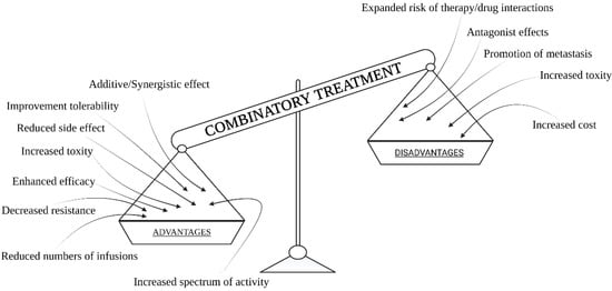
In the context of the use of combination therapies in cancer treatment, there are some very interesting results obtained by the use of chemotherapy, radiotherapy, hyperthermia, photodynamic therapy or surgery, and immunotherapy in various combinations. Currently, a combination treatment of chemotherapy, radiotherapy, and often surgery is used commonly in cancer treatment. With this approach, we can effectively stop cancer development and obtain the best possible results based on current knowledge (but one of three cancer cases still lead to patient death). There is no doubt that combination treatment provides very good results, as opposed to using only one method due to the comprehensiveness of the multi-module treatment. Based on many simulations with the use of computer models, cancer treatment strategies were optimized using a combination of possible treatments [9,10,11]. The crucial elements for optimization are drugs and their doses, as well as the dose of light and radiation, in the context of cancer type and the degree of its aggressiveness. Fractionation of the radiation dose also proved to be more crucial, giving better results [12,13].
In addition, cutting out cancerous tissue, with a margin of healthy tissue, combined with chemotherapy cycles, provides the best results and reduces tumor metastasis. It also allows for targeted treatment and effectiveness increase due to a long-term and selective action. Taking into account the occurrence of cell cycles and cell resistance at various stages of the cell cycle for selected methods, it offers more significant results. Scientific research has also shown that the cells present in the phase that is the most resistant to radiation, i.e., those that are in the S phase (replication phase), are also very sensitive to hyperthermia [14]. Therefore, it was concluded that hyperthermia allows sensitization of cells to X-rays in the further treatment phase [15]. When considering combination therapy, one may come across concepts, such as thermochemotherapy, thermoradiotherapy, and thermochemoradiotherapy. Considering thermoradiotherapy, the general mechanism of action is primarily the possibility of destroying cancer cells that are insensitive to radiation. It also inhibits the process of repairing damage to DNA under the influence of a dose of ionizing radiation. Due to this approach, combination therapy in cancer treatment offers more effective therapeutic results. In turn, analyzing the introduced concept of thermochemotherapy in the treatment of cancer, one may observe an increase in sensitivity and reduction in cancer cell resistance to chemotherapy. With this approach, it is possible to increase the accumulation of chemotherapeutics in the cancer location [15,16]. All of this contributes toward increasing the effectiveness of this complementary method, which, in this context, has been linked to phenomena, such as hyperthermia. Undoubtedly, new classes of chemotherapeutics, as well as various mechanisms of action of drugs, such as the use of drugs that are not dependent on the rate of proliferation, or those that depend on the phase of the cell cycle, and attempts to combine drugs with non-overlapping toxicity, or a use of a combination of drugs that have different mechanisms of action [17], as well as the way of adding drugs, for example using targeted liposomes, significantly improve the effects of treatment and show us the importance of using combination therapy. On the other hand, the use of thermochemoradiotherapy shows that multi-module treatment brings the most beneficial and the best results [18]. Researchers have studied the interaction between hyperthermia and radiation therapy, as well as chemotherapy using chemotherapeutic agents, such as cisplatin. Scientific research has shown that a synergistic effect can be obtained, but only when this three-modular therapy is used on cell lines that are not resistant to cisplatin [19,20]. Considering photodynamic therapy, this combination of therapy with chemotherapy gives quite good results due to direct destruction of cancer cells and strong influence on blood vessels in the tumor surrounding. It was shown that different PDT protocols can significantly damage the vasculature or temporally increase blood flow and tissue oxygenation in the treated area [21]. However, as a result of photodynamic therapy, when not all of the cells are destroyed, the use of chemotherapy in the second wave allows killing the remaining cancer cells that have survived PDT. Additionally, it was shown that PDT can have strong influence on the immune system [22]. With this approach, combining methods can stop the cancer or lead to a complete cure.

2. General Information about Photodynamic Therapy and Hyperthermia

2.1. PDT

In this article we focus on PDT and hyperthermia treatment as a very interesting example of a combination strategy. That is why this section includes basic information about the selected therapies and their modes of action. Photodynamic therapy (PDT) requires three independent factors: photosensitizer (PS), light at proper wavelength to excite PS, and oxygen. We should note that although a photosensibilized reaction can be possible without any oxygen, a photodynamic reaction involves oxygen by definition. The main advantage of PDT as a cancer treatment is based on its double selectivity: PS is a drug that accumulates better in neoplasm tissues, and light can activate only the PS that is localized in an illuminated area. Ideally, PS and light separately are harmless, and in the presence of oxygen, both of them together can create a deadly weapon—reactive oxygen species (ROS). Photosensitizer can be understand as a pro-drug that requires light to become a drug. When we talk about PDT in the context of cancer treatment, the ideal photosensitizer should accumulate selectively only in cancerous tissue, it should have a minimal toxicity before the light exposure and a high toxicity after using light in a given range of wavelengths. At the same time, it must not cause any phototoxic effects on healthy cells. The photosensitizer should absorb light in the range of about 600 to 1200 nm to perform effectively, in the context of light penetration into the tissues (lower light wavelengths are absorbed by endogenous dyes, longer wavelengths are selectively absorbed by water). There are several subgenerations of photosensitizers [158,159,160,161] categorized by their chemical structures. One representative of the first generation photosensitizer is hematoporphyrin. Porphyrins and porphyrin derivatives are examples of second-generation photosensitizers. Moreover, 5-aminolevulinic acid (ALA), which is used to treat skin cancers, is used quite often in PDT. These compounds consist of four pyrrole molecules that are linked together via methylene bridges. Another example of porphyrin derivatives are chlorines. They are chemical compounds that have modified the double bond in their structure and could absorb light in the infrared spectrum more strongly. Due to the fact that chlorines can be excited by infrared light, they are able to penetrate tissue with light more deeply. These chemical compounds are removed quite quickly from a human body–they need about 24–48 h to be cleared, which indicates less toxicity. Bacteriochlorin ale phthalocyanines are also extremely promising photosensitizers in anti-cancer therapies [162]. The can by excited with red/infrared light; pre-clinical and clinical trials show very promising results. Good examples include TOOKAD against prostate cancer [163,164,165,166] and LUZ11 against head and neck cancers [167], photosensitizers with advanced clinical trials. Light is an inseparable element of a photodynamic therapy. The theory that solar radiation can be used to treat many diseases is very old [168,169]. It was argued that solar radiation could cure diseases, such as albinism and psoriasis. In 1903, Niels Finsen received the Nobel Prize for his work on phototherapy. He used UV radiation to treat skin tuberculosis. Later, photodynamic therapy was used to treat skin cancer. The wavelength should be adjusted to the photosensitizer’s absorption spectrum. Different light sources can be used in PDT, e.g., diode, xenon or halogen lasers [170]. It is worth noting that the longer the wavelength, the deeper its beam penetrates into the tissue. Therefore, for about 700 nm, the depth of light penetration is about 1.5 cm (the light power density decreases in relation to the square of the distance).
The last (but not least) necessary element of PDT is oxygen, specifically molecular oxygen, which is dissolved in the tissues. The presence of oxygen allows the formation of reactive oxygen species, such as singlet oxygen, hydroxyl radical, or superoxide anion radical. The effectiveness of therapy is considered a cytotoxic effect under the influence of oxidation. The combination of these three elements is fundamental when it comes to the principle of PDT. Separately, these three factors are believed to be safe, but their combination has an effect on the cells, it contributes to their destruction, making photodynamic therapy effective, and it is increasingly used to treat various diseases. There are two main mechanisms for the formation of free radicals and reactive oxygen species in PDT.
The type I mechanism occurs when the oxygen concentration is low and free radical forms are present. This mechanism involves the transfer of an electron or hydrogen atom between the excited photosensitizer and the irradiated tumor tissue. As a result of the photochemical reaction, radicals or anion radicals are being formed. The type II mechanism occurs when the oxygen concentration is close to the physiological concentration. There is a transfer of energy between the photosensitizer, which is in the triplet state, and between the excited form–singlet oxygen.
To sum up the basic information about photodynamic therapy, the method is an alternative way of treating cancer. Among the advantages there are: lower toxicity, high selectivity of the method, and reduction of side effects as compared to chemotherapy or surgery. However, this method also has some disadvantages. Unfortunately, the dependence on an external light source makes PDT mainly suitable for the treatment of tumors on or just under the skin, and on the lining of internal organs [171].
In addition, intravenous administration of PDT may cause systemic toxicity, and due to abnormal angiogenesis, non-specific interaction with blood components and the presence of fibroblasts in cancer foci, only a part of PDT sensitizing substances can be used successfully in this therapy [171,172,173]. Unfortunately, oxygen consumption within the tumor may exacerbate tumor hypoxia, leading to PDT treatment failure [174,175,176]. This method is also quite expensive. The cost of specialized equipment is particularly high. In addition, the selection of an appropriate photosensitizer is very hard. It must not be toxic to the patient and it should give the least possible side effects. It is also worth mentioning that PDT may cause temporary hypersensitivity to sunlight.

2.2. Hyperthermia

Hyperthermia is a state in which temperature is elevated above the physiological norm. The hyperthermia therapy involves a controlled increase in body tissue locally or selectively to the needed area. Many techniques can be used to achieve it and each has its own pros and cons. For example, contact heating can be the easiest way to heat superficial tissue; however, it will generate significant temperature gradient, which can be responsible for uneven biological effects. On the other hand, different types of the waves can be used, e.g., ultrasound, radio frequency, or infrared–each is effective based on different tissue parameters (considered pros or cons based on the effects needed). Requirements of a high precision device and advanced dosimeters and treatment planning are for this group of heating techniques. Another hyperthermia induction in the tissue involves a combination of nanomaterials, which can accumulate inside the tissue of choice and be the source of heat, after the activation, e.g., gold nanorods could be activated by the near-infrared light. However, this latter way of heating could be the most precise, but also the hardest to perform because of a wide range of influential factors. It is widely known that elevated temperature informs us about inflammation and diseases, and it is one of the immune system defense mechanisms. The normal human body temperature ranges from 36.2 °C to 37.5 °C [177]. The Table 2 shows the temperature range for specific parts of the human body. Human normothermia is reported to be 36.8 °C [178].
Table 2. Temperature ranges for specific parts of the human body.
Temperature Range (°C) Selected Area in the Human Body
36.32–37.76 rectal
35.76–37.52 tympanic
35.61–37.61 urine
35.73–37.41 oral
35.01–36.93 axillary
Generally, we can distinguish between general, systemic hyperthermia, and local site-specific hyperthermia. Furthermore, in both types, hyperthermia can be generated by the organism itself (e.g., fewer, inflammation) or can be induced artificially (e.g., irradiation). From the biological perspective, one more classification is essential that is based on the established tissue temperature. Hyperthermia is defined as a temperature ranging from 39 °C and above, where the selective effect of heat on tumors, cancer tissues, promotes killing cancer cells by the influence of high temperature [179].
The temperature range from 40 °C to 43 °C is most commonly used in the context of hyperthermia in the treatment of cancer [180,181]. Mild hyperthermia is an increase in temperature in the range from 41 °C to 43 °C [182]. Hyperthermia has been used to treat cancer from ancient times [183]. A lot of research has shown that artificial temperature raising can damage cancer cells. Hyperthermia affects the angiogenesis process and can also damage the structure and reduce the tumor size [16,184]. In this aspect, it is a method of a targeted therapy. However, hyperthermia is used in combination with other methods in order to increase their effectiveness, e.g., by improving drug accumulation [185,186,187]. Very often, hyperthermia is combined with chemotherapy or radiation therapy [188].
The main goal of hyperthermia is to sensitize cancer cells to the subsequent treatment method, so it can be understand as a physical adjuvant treatment. It is believed that more sensitive cells should be less resistant to X-rays and chemotherapeutics. For example, cells treated with high temperature may inhibit the repair of drug-induced DNA damage by denaturing the proteins involved in this repair [189]. As mentioned above, hyperthermia can be divided by its location. Local hyperthermia can be carried out using external or internal energy sources, regional hyperthermia through organ perfusion; there is also a whole body hyperthermia [190]. If we consider the principle of local hyperthermia, we locate the tumor area and only heat the area where the tumor is located. To provide the right amount of heat, one may use infrared, ultrasound, or microwave wavelength laser. It is possible to both heat the outside of the skin and introduce appropriate probes with a light source that will generate heat inside the body. In the context of hyperthermia, controlling tissue heating is crucial. To have control over all elements of this therapy, we need to correctly locate a selected heating area, put the heating source in the right place, and constantly monitor the temperature rise. An additional important factor for efficient hyperthermia treatment is the time of the established tissue temperature (plateau) and the time needed to achieve it (ramp). Based on that, the total energy deposition can be calculated. The dose of heat should be safe and should bring the expected results without, e.g., painful burns. It should be highlighted that this method can have various side effects. The most common side effects are burns, swelling, and bleeding. Therefore, hyperthermia needs to be applied carefully and should act very selectively, and the heating area should be estimated very well. It should be mentioned that different kinds of tissue will heat in a different way. Fat will heat and accumulate the temperature differently than a well perfused muscle. In this context, tumors are very special objects to heat because lack of tissue homeostasis results in pathological mechanisms of tissue cooling, e.g., uncontrolled perfusion that slows down heat dispersion, specific vessel structures that affect the possibilities of vein and artery heat enlargement. Hyperthermia may prove to be a successful adjunctive therapy for drug transport in which nanoparticles are used. An example of nanoparticles that can aid drug transport is fullerene, which can target the delivery of drugs to the tumor area [191].
Although hyperthermia is usually used as a supportive method, it can contribute to cell death directly by causing damage to the protein–lipid cell membrane as well as cause denaturation of intracellular proteins [180]. Hyperthermia also leads to dismantling, denaturation, and reorganization of cytoskeleton proteins [192]. As mentioned, hyperthermia can also lead to degradation of proteins responsible for repairing DNA damage caused by chemotherapeutics and, thus, inhibit the repair of DNA double-strand breaks in the process of homologous recombination [193,194]. High-temperature cell death can occur through both apoptosis and necrosis. The putative biological–molecular mechanism of hyperthermia is based, among others, on the expression of heat shock proteins (HSPs), induction and regulation of apoptosis, and signal transduction and modulation of drug resistance in cells. Hyperthermia can lead to cell death directly. Induction of cell death may be dependent on the p53 suppressor protein. It is also believed that apoptosis is mainly caused by activation of procaspase 2, which entails the entire cascade of Bax- or Bak-type proteins [181].
Hyperthermia also affects the immune system. In the case of local tumor hyperthermia, an influx of NK cells and macrophages to the tumor location can be observed where local heating is applied. Moreover, it is said that hyperthermia activates the immune system and generates an immune response. HT helps to increase the level of TNF-alpha. A fairly quick response from the immune system with the secretion of cytokines, such as TNF-alpha and IL-1-beta, contributes to the activation of the immune system defense mechanisms [195]. Hyperthermia is currently being modified to achieve the best results with better safety. It is proposed to use nanogold to monitor the course of hyperthermia. In addition, this technique is being optimized to heat the tumor location more efficiently, and to obtain a correspondingly long therapeutic window to introduce the drug into the tumor area more effectively, or irradiate the tumor area with X-rays. In general, nanogold particles are supposed to absorb infrared light to heat the area in which the nanogold has been located [196]. To achieve better therapeutic results, hyperthermia is being modified and new possibilities of treatment are being created.

3. Proposed Combinations That Are Currently Used in Multimodal Cancer Treatment

Radiotherapy, chemotherapy, and surgical intervention are reliable options for treating cancer [366]. Currently, multi-module treatment is used quite commonly to treat cancer: chemotherapy and radiotherapy are used together very often. In addition, attempts are being made to enhance therapeutic effects by improving the mechanisms on which standard treatments are based. These attempts focus on aspects related to delivery of drugs, photosensitizers, or chemotherapeutic agents in order to minimize side effects and to act selectively. Therefore, drugs can be delivered using liposomes, and chemotherapeutics with a different spectrum of activity can be used to enhance therapeutic effects. In addition, research using gene therapy is being carried out, and new drug are being designed. New mechanisms have been developed to achieve better results without damaging healthy structures, which is why, for example, a radiation-based gamma knife or cyber knife are used.

3.1. Radiotherapy

Radiotherapy is an important part of cancer treatment, and its main goal is to deprive cancer cells of their proliferation potential. Radiation is a physical factor that stores energy in the cells of the tissues it passes through, and then this deposited energy can kill cancer cells or cause genetic changes that lead to cancer cell death [367]. The main mechanism of killing cells by high-energy radiation involves damaging their genetic material of DNA, thereby blocking their ability to further divide and proliferate [367,368]. The goal of improving radiation therapy is to maximize the radiation dose to cancer cells while, at the same time, minimizing the exposure of healthy cells that are adjacent to cancer cells or are exposed to radiation [367]. Radiotherapy is also used in conjunction with other treatments, such as surgery, chemotherapy, or immunotherapy.
Currently, brachytherapy and teleradiotherapy are commonly used. Brachytherapy involves treatment with the use of a radiation source that must be as close to the tumor as possible. The mechanism of brachytherapy is based on the use of radiation in direct contact with the tumor. However, when it comes to teleradiotherapy, the source of radiation is located at a certain distance from the tissue within the area of the cancer. In the context of radiotherapy, the so-called 5R principle that occurs during radiotherapy: redistribution, repopulation, reoxygenation, repair, and radiosensitivity is worth highlighting [369]. Radiation can be classified as ionizing radiation (e.g., X and gamma radiation) or particle radiation with electrons, protons, alpha particles, and neutrons. The biological mechanism of action depends on the type of radiation (e.g., relevance of linear transfer energy and cell damage). On the other hand, cancerous tissue parameters can provoke effects, such as the increase in sensitivity of cancer cells to ionizing radiation if the activity during the division of cancer cells is also greater. However, a higher level of differentiation makes cells less sensitive to radiation. The level of tumor oxygenation is also an important factor. When tumor is more hypoxic, the cancer cells are less sensitive to radiation (oxygen enhancement ratio is 2.5–3 times bigger for a well oxygenized tissue). In context of radiotherapy, there is a very interesting strategy to treat malignant eye tumors—protonotherapy. This technique uses Bragg’s pick to deposit energy into the tumor, and the healthy tissue around the tumor is kept safe at the same time [370]. It was shown that proton radiation can inhibit the metastatic potential of primary cancer cells.
In the outcome of radiotherapy methods and mechanisms, temporary inhibition of tumor growth or growth retardation and tumor regression may take place. There are many factors that have to be considered, although these mechanisms can be influenced by the duration of the cell cycle, the size of the cell growth fraction or the rate of cell loss.
From the perspective of potential needs for combination treatment involving the use of radiotherapy, a few factors should be taken into consideration. Regarding the above-mentioned cell sensitivity to radiation (e.g., cell cycle, oxygen enhancement), the main need is to overcome radioresistance. For example, melanomas are quite resistant to radiation therapy due to their melanin accumulation (accumulation of pigment that acts as a radioprotector). Additionally, cancer cells that contain melanin are hypoxic and, therefore, more resistant to low LET radiation. A high level of cell differentiation also contributes to this resistance [371]. In the treatment of melanoma where melanin is present, its role is to scavenge free radicals. This mechanism allows putting this pigment in the radioprotector category. It protects melanin against the impact of ionizing radiation, for example X-radiation. What is worth emphasizing is that this pigment increases radiation resistance by inactivating free radicals that are formed during the course of radiation. A well-applied complementary treatment can increase melanoma sensitiveness to ROS.
The limitations of radiotherapy, such as insufficient tissue oxygenation can be overcome by using complementary therapies. One such technique is hyperthermia, which is usually a complementary method that involves heating the tumor in order to inhibit the proliferation of cancer cells, destroy them, or make them sensitive to various treatments, including radiation therapy. The combination of hyperthermia and radiotherapy shows a synergistic effect and enhances the killing effect on cancer cells, especially those in the S phase of the cell cycle, which are usually resistant to radiation when applied alone [19]. The synergistic effect of heat and radiation is defined as the thermal enhancement ratio (TER), which defines the magnitude of thermal hypersensitivity to radiation as the survival fraction quotient after irradiation alone and in combination with hyperthermia [372]. The effects of hyperthermia include, but are not limited to, inhibition of the repair of radiation-induced DNA damage, thereby increasing the cytotoxic effect of radiotherapy [372]. Moreover, by reducing the metabolic activity of target cells, heat reduces the oxygen demand of the tumor and increases the oxygenation of the tumor tissue as well, making hyperthermia one of the most powerful radiosensitizers available [373].
Another approach to overcome the limitations of radiation therapy is to combine it with photodynamic therapy. Photodynamic therapy uses photosensitizers that are activated by visible or near-infrared light and transfer energy to molecular oxygen, thus generating reactive oxygen species [373]. Under certain conditions, some photosensitizers can act as radiosensitizers. Combining radiation therapy with photodynamic therapy, i.e., using ionizing radiation tissue penetration and photodynamic therapy can reduce penetration depth problems and can allow radiation dose reduction without decreased clinical efficacy [374], while minimizing damage to healthy tissues. Moreover, the combination of an appropriate photosensitizer with radiation therapy can lead to a significant increase in the cytotoxic and apoptotic death of cancer cells [375]. The combination of photodynamic therapy and radiation therapy, the primary goal of which is to kill cells through nuclear DNA damage, offers the possibility of synergism in killing cells, as photodynamic therapy does also induce several types of DNA damage [376]. In addition, photodynamic therapy does also improve the immune system’s response by inducing inflammation and an immune response against cancer cells. Potential mechanisms of immune stimulation by photodynamic therapy include an acute inflammatory response that can enhance tumor antigen presentation to activate dendritic cells and to guide them to regional and peripheral lymph nodes, ultimately stimulating cytotoxic T lymphocytes and NK cells, accompanied by the formation of immune memory and growth suppression tumor in the future [255].

3.2. Chemotherapy

Chemotherapy is a very broad category of anti-cancer treatment. The induction of apoptosis and the inhibition of mitosis as well as the cell cycle disorder are caused by the use of chemotherapeutic agents. Cytostatic drugs can be grouped into alkylating agents, alkaloids, antibiotics, and antimetabolites. Inhibitor of tyrosine kinases are also very important novel drugs. They affect cell proliferation by targeting cellular DNA or RNA and metabolism with antimetabolites acting on purine or pyrimidine metabolic enzymes, while alkaloids act on the cytoskeleton and mitosis [377]. One of the main problem with chemotherapy is the effective, safe, and selective drug delivery. Chemotherapy is associated with the presence of side effects that include immediate signs of toxicity (effects can be seen on skin and hair, bone marrow and blood, gastrointestinal tract and kidneys, etc.) as well as late signs of chronic toxicity (drug resistance, carcinogenicity) [377]. In order to increase the effectiveness of chemotherapy and reduce side effects, a combination of various drugs with different mechanisms of action is used. Moreover, when several chemotherapeutic agents are used, drug resistance could be counteracted. Overall results of chemotherapy are also improved due to the use of combinations of drugs that do not have overlapping toxicities. Then, we can increase the dose of drugs in the tumor without fearing undesirable negative side effects of the therapy. This approach is used very often to reduce side effects since increasing the dose of only one drug can cause toxicity; therefore, it seems that combining drugs from different groups is less toxic and more effective due to the higher therapeutic dose applied. The same happens when we use drugs that have different mechanisms of action. In this case, cells that are insensitive to one drug are already sensitive to the other drug from this combination. Treating drug-resistant cancers is a significant problem not only in chemotherapy. This mechanism of drug resistance may be influenced by the fact that the chemotherapeutic agent alone is not able to reach the inside of the tumor directly. Still, other challenges face this approach as well, such as improving drug perfusion and increasing the accumulation of the therapeutic compound in the tumor. Hyperthermia, which causes changes in cells and their surroundings due to high temperature, seems to be a good complementary technique. In addition to direct ablation of cancer cells, elevated temperatures can also trigger drug release, especially for heat-sensitive carriers [378]. It is also known that raising the temperature from 37 °C to 43 °C such increases the permeability of the cell membrane, accelerating the absorption of nanoparticles, and it may increase the interaction of carriers with the cell membrane as well [379]. In particular, energy-dependent pathways, such as clathrin- and caveolae-mediated endocytosis are involved in increasing the permeability of the cell membrane at high temperature, thus increasing the internalization of drugs and carriers [378,379]. Hyperthermia also affects blood flow and, hence, changes the drug distribution. Certainly, the heat-induced change in blood flow in tumors differs from the one that occurs in regular tissues because the tumor vasculature is less able to dissipate heat and is more susceptible to damage during hyperthermia treatment. However, it is worth noting that mild heat increases blood flow in the tumor, allowing chemotherapy to have a greater effect on cancer cells [373]; thus, in some types of tumors, blood flow increases when heated to relatively low temperatures. On the other hand, higher temperatures (43 °C or 44 °C) result in stronger and longer-lasting vascular closure [380].
In addition to allowing more efficient drug delivery, heat can also modify the cytotoxicity of many chemotherapeutic agents. In many cases, synergism can be seen as a continual change in the rate at which the drug kills cells as temperature increases. The cytotoxicity of most alkylating agents, platinum compounds, and also nitrosoureas increases linearly with the temperature increase, typically by thermal enhancement, including increased alkylation rate constants, increased drug absorption, and inhibition of repair of lethal or sublethal drug-induced damage [381].
Additionally, hyperthermia may increase the killing effect on tumor cells located at the hypoxic centers of tumors that are relatively resistant to chemotherapy due to poor drug delivery. In addition, some chemotherapy drugs also require oxygen to generate free radicals in order to induce tumor cytotoxicity. It is known that elevated temperatures can increase the rate of biochemical reactions, increasing cellular metabolism, which can result in an increased oxidative stress. The level of reactive oxygen species may increase after exposure to elevated temperatures, possibly due to dysfunction of the mitochondrial respiratory chain or by increased activity of the enzymes NADPH oxidase and xanthine oxidase [382].

3.3. Surgical Intervention

Surgery is one of the three most popular treatments for cancer today, and also the oldest one. It plays the most important role in the treatment of cancer. Its application ranges from the diagnosis of lesions, i.e., taking biopsies for diagnosis, through reducing the tumor mass, to radical treatment in order to completely excise the lesion together with a margin of healthy tissues. Complete tumor excision with cutting out a healthy tissue margin is the most effective form of treatment in the case of early stage neoplasms, especially when there are no metastases yet. Such achievement of a sufficiently negative margin during oncological surgery minimizes the risk of adverse treatment outcomes and recurrence of the disease [383]. In some cases, the unfavorable location of the tumor or the presence of disseminated metastases can make it impossible to remove the tumor. Hence, it is not always possible to obtain the necessary margin, in particular in case of surgery in the vascular system area, in case of other critical areas, and in case of tumor involvement in adjacent tissues. This problem applies to such neoplasms as hepatocellular carcinoma, pancreatic ductal adenocarcinoma (PDAC), neuroblastoma, or neuroendocrine tumors of the gastrointestinal tract. Involvement of a blood vessel can sometimes be resolved by surgical resection and reconstruction of the affected vessel, such as a vein or artery, but these procedures are associated with an increased risk for the patient, especially in the case of arteries [384,385].
In conclusion, failure to obtain adequate surgical margins increases both the surgical and oncological risk of poor prognosis, which is usually the case when tumors have invaded large blood vessels [385]. If it is impossible to remove the entire neoplastic lesion, the combination of surgery and radiation therapy is often used. Postoperative radiotherapy reduces the risk of cancer recurrence and also helps destroy any remaining cancer cells, especially when only the tumor was removed and only a small amount of regular tissue around it was removed, or a margin is left that is positive for cancer cells. Indications for postoperative radiotherapy include not only insufficient resection margin, but also uncertain radicality of resection, infiltration of tissue with diffuse cancer foci, and low tumor differentiation. Therefore, in most cases, only combinations of treatments such as surgery combined with radiation therapy are usually the only way to destroy cancer cells [373].
Returning to the issue of tumor areas that cannot be surgically excised due to the fact that their location is too close to inoperable vessel, there is one interesting complementary method: the use of local, mild hyperthermia. By applying uniform and gentle heating, we can destroy tumors that surround the vessels and, at the same time, protect these sensitive structures from damage [385]. It has been shown (on the example of pancreatic ductal adenocarcinoma) that an increase in temperature in the range of 41–46 °C leads to killing cancer cells, including the elimination of cancer stem cells, as well as changes in the proteomic profiles of cancer cells, with simultaneous protection of regular cells [385].
Another method that is complementary to surgery is photodynamic therapy, which is based on the complex cell killing phenomenon resulting from the interaction of a chemical compound (photosensitizer), light, and oxygen. Photodynamic therapy is recognized as a safe and effective method, and this is why it plays a unique role in the treatment of cancer with its targeting precision. It does not damage healthy structures surrounding the treated lesions, and it is applied in the treatment of cancerous tumors with limited access [386]. In systemic photodynamic therapy there is a wide distribution of photosensitizer, but there is also a higher potential for accumulation in cancerous tissues than in healthy tissues, and multi-site deep light, which can be performed intraoperatively in combination with standard or minimal surgical access [386]. Consequently, by using the preferential accumulation of photosensitizers in cancer cells with the appropriate selection of irradiation, it is possible to eliminate the remaining tumor fragments that cannot be surgically removed. Photodynamic therapy may be used in some cases before surgery as a neoadjuvant therapy to alleviate cancer.
In addition to destroying cancer cells, photodynamic therapy is closely related to the fluorescence phenomenon used in photodiagnostics to detect lesions. As noted, the optical properties of healthy and abnormal tissues in the ultraviolet and visible spectrum differ from each other. Due to endogenous chromophores, some tissues exhibit a characteristic fluorescence emission band that changes when the disease process occurs, changing these chromophore components [386]. Thus, using the appropriate wavelength, an image can be obtained that is used as the basis of autofluorescence photodiagnosis [386,387]. This method may be helpful in determining the optimal biopsy site for histological diagnosis. In addition to using endogenous chromophores, photosensitizers can be used as exogenous fluorophores to enhance fluorescence [386]. The use of such enhanced photodiagnostics is especially valuable in surgical operations where it can indicate residual neoplastic infiltration, which may be present at the margin of resection. In addition, it can be used in brain surgeries, in the removal of tumor remains that are invisible to the naked eye or with the use of microscopic surgical instruments [386,388].

This entry is adapted from the peer-reviewed paper 10.3390/pharmaceutics13081147

This entry is offline, you can click here to edit this entry!
Video Production Service