Cytotoxicity (cell-mediated killing) is the process in which certain cells secrete “toxins” that lysate and neutralize unwanted target cells (i.e., pathogens, viral-infected cells, tumoral cells, foreign transplanted cells). Cooper has defined that through evolution, immune-mediated cytotoxicity could be classified into level I and level II cytotoxicity. Level I in mammals is mediated by macrophages and neutrophils which release toxic granules in the inflammatory area. They are considered the most basic manifestations of cytotoxicity and are present in invertebrates (sponges, coelenterates, sipunculids, annelids, mollusks, arthropods, echinoderms and protochordates). In contrast, level II or acquired cytotoxicity is mediated by cells that require a specific induction to be activated, through specific recognition, such as allogeneic responses, or induced by adaptive molecules. These include cytotoxic T lymphocytes (CTL), natural killer (NK), and antibody-dependent cell-mediated cytotoxicity (ADCC). It seems that these cells appear later in evolution [
2,
48], but the characterization of specific allogeneic responses suggests that they might be in more animal groups than what is currently known. Since the work of Cooper in 1980, NK cells were shown to have specificity and recognition of targets for cytotoxicity trough immunological synapse without the killing of adjacent cells [
49,
50], for that reason we moved NK-like recognition based cytotoxicity mechanism to level II cytotoxicity, compared to the original definition by Cooper [
48].
2.1. Effector Cells
All animals have several immune cells with cytotoxic activity to lyse host and foreign cells. Regarding level I cytotoxicity, those cells have been found in different invertebrate organisms as probably the most ancient type of cytotoxic immune response. For example, killer cells from sipunculid worms exert cytotoxic effects on allogeneic erythrocytes, but not on erythrocytes from worms that reside closely [
52]. In the freshwater pulmonated mollusk
Planorbarius corneus, a class of hemocyte that has been morphologically characterized as round hemocytes (RH) exerts cytotoxic activity on the human erythroleukemia K562 cells thus participating in the graft rejection of allo- and xeno-grafts [
51]. In arthropods, the amoeboid hemocytes of
Limolus polyphemus are stimulated by bacterial lipopolysaccharide (LPS) endotoxins to secrete great amounts of clottable protein and to produce nitric oxide (NO) synthesis as a cytotoxic mechanism to protect the host from invading pathogens [
53]. Additionally, in in vitro experiments, crayfish (
Astacus astacus) granular and semi granular hemocytes with phenoloxidase and laccase activity display cytotoxic effects towards different mammalian tumor and non-tumor cells [
54,
55]. Coelomocytes from the sea urchin
Arbacia punctulata exert cytotoxic activity against human and murine target cells. A particular population of these phagocytic cells is the one with the highest cytotoxic activity which are positive to the human NK markers CD14, CD56 and CD158b [
56]. These cytotoxic cells have also found in
Strongylocentrotus droebachiensis, S. pallidus and
Echinus esculentus in which targeted-cell death is characterized by cell detachment, disintegration and the formation of a multinuclear non-cellular protoplasm [
57].
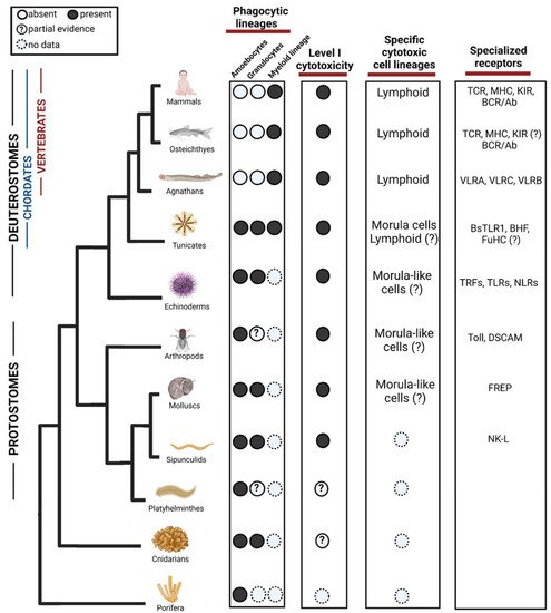
At this point we do not think that phagocytosis of damaged self-cells should be considered as level 1 cytotoxicity. Although, we do think that where phagocytosis of damaged self-cells is found, it can be suggestive of potential level 1 cytotoxicity (as the mechanisms of altered-self recognition exist) and should be further investigated (
Figure 1). For instance, the recent discovery of phagocytosis of heat-stressed cells in ex-vivo experiments in Hexacorallians [
10].
Figure 1. Evolution of phagocytic and cytotoxic cell lineages in the immune system. Among phagocytic lineages, amoebocytes and granulocytes are present in invertebrates, while myeloid lineage seems to evolve in the ancestor of tunicates and vertebrates. Cells representing the Level I cytotoxicity exist in Protostomes but might appear in cnidarian. Among specific cytotoxic cell lineages, morula cells have been found in tunicates although partial evidence suggests that they may evolve in earlier ancestors. Lymphoid cells are found in agnathans and have diversified in vertebrates, there are suggestions of their form in Tunicates but not validated yet. In the third column, we show examples of diversification of specialized receptors acting in both phagocytosis and cytotoxicity. TLR = Toll-like receptors, TCR = T-cell receptor, MHC = Major histocompatibility complex, KIR = Killer Inhibitory Receptors, BCR = B cell receptor, VLR = Variable lymphocyte receptor, BHF = Botryllus histocompatibility factor, TRF = Transformer proteins, NLR = NOD like receptors, DSCAM = Down syndrome cell adhesion molecule, FREP = Fibrinogen-related proteins, NK-L = Natural killer-like receptor. ?—Represents missing functional or molecular validation. Created with BioRender.com. Data sources are summarized in Table 1.
Table 1. Data sources for Figure 2.
| Animal Group |
Effector Cells |
Receptors and Effector Molecules |
| Mammals |
(Wolff and Humeniuk 2013) [105] |
(Vandendriessche, Cambier et al., 2021) [19] |
| (Rosental, Appel et al., 2012) [50] |
| (Hirayama, Iida et al., 2017) [20] |
| (Geffner 2005) [71] |
| Osteichthyes |
(Potts and Bowman 2017) [106] |
(Wei, Zhou et al., 2007) [107] |
| (Moss, Monette et al., 2009) [108] |
(Kasahara and Flajnik 2019) [90] |
| (Tang, Iyer et al., 2017) [109] |
(Flajnik and Du Pasquier 2004) [75] |
| (Flajnik 2018) [65] |
|
| Agnathans |
(Han, Das et al., 2015) [110] |
(Pancer, Amemiya et al., 2004) [83] |
| (Hirano, Guo et al., 2013) [111] |
| (Das, Li et al., 2015) [61] |
| (Mayer, Uinuk-ool et al., 2002) [62] |
| Tunicates |
(Rosental, Kowarsky et al., 2018) [16] |
(Voskoboynik, Newman et al., 2013) [59] |
| (Rosental, Raveh et al., 2020) [17] |
(Nyholm, Passegue et al., 2006) [112] |
| |
(Peronato, Franchi et al., 2020) [25] |
| Echinoderms |
(Arizza, Giaramita et al., 2007) [113] |
(Yakovenko, Donnyo et al., 2021) [32] |
| (Cooper 1980) [48] |
(Chou, Lun et al., 2018) [33] |
| (Lin, Zhang et al., 2001) [56] |
|
| Arthropods |
(Muñoz-Chápuli, Carmona et al., 2005) [114] |
(Melcarne, Lemaitre et al., 2019) [115] |
| (Lanot, Zachary et al., 2001) [116] |
| (Meister and Lagueux 2003) [117] |
| (Cattenoz, Sakr et al., 2020) [15] |
| (Csordás, Gábor et al., 2021) [14] |
| (Cárdenas, Dankert et al., 2004) [78] |
| Molluscs |
(Franceschi, Cossarizza et al., 1991) [51] |
(Schultz, Bu et al., 2018) [76] |
| (Nakayama, Nomoto et al., 1997) [118] |
(Loker, Adema et al., 2004) [77] |
| Sipunculids |
(Boiledieu and Valembois 1977) [52] |
(Flajnik and Du Pasquier 2004) [75] |
| Platyelminthes |
(de Oliveira, Lopes et al., 2018) [119] |
(Peiris, Hoyer et al., 2014) [28] |
| (Morita 1991) [11] |
| Cnidarians |
(Snyder G 2021) [10] |
|
| Porifera |
(Mukherjee, Ray et al., 2015) [9] |
(Pita, Hoeppner et al., 2018) [120] |
| (Yuen, Bayes et al., 2014) [21] |
| (Wiens, Korzhev et al., 2005) [22] |
For level II of specific cytotoxicity, in the tunicate
B. schlosseri, morula cells (MC) have been characterized as cytotoxic cells. They contain phenoloxidase, accumulate in rejection points and morphologically resembles NK cells. While 15% of their genes are shared with cytotoxic lymphocytes of vertebrates, MC express 85% tunicate-specific gene repertoire [
16,
17], suggestive of convergent lineage more resembling other invertebrates phenoloxidase based immune cells (
Figure 1). The cytotoxic activity of MCs is involved in a natural process of allorecognition that occurs when two
B. schlosseri colonies with different genotypes touch [
58]. This self–nonself recognition is regulated by a single polymorphic histocompatibility gene: the Botryllus Histocompatibility Factor (BHF) [
59]. The sharing of at least one BHF allele is a determinant for the fusion or rejection of the colonies [
16,
17]. Interestingly, MC express high levels of the polymorphic gene FuHC which could be a base for BHF recognition mechanism [
17,
60] (
Figure 1).
Transcriptome analysis and cellular characterization studies in the jawless vertebrates, lampreys, have proposed that two T-like cell populations expressing the variable lymphocyte receptors (VLRs) VLRA and VLRC, respectively, carries cytotoxic activity [
61,
62]. These cells express several genes including cell surface receptors (i.e., CD45, TCR, VpreB, paired-Ig-like receptors), cytokines (IL17) and transcription factors (i.e., GATA 2/3, c-Rel, BCL11b) resembling those that T lymphocytes use to migrate, proliferate and differentiate in jawed vertebrates [
63]. Lymphocyte lineages have also been found in the hagfish, however these have not been fully characterized [
61,
64]. This shows a whole convergent parallel adaptive immune system of lymphocytes, which is based on LLRs instead of Ig superfamily like the jawed vertebrates (
Figure 1).
In jawed vertebrates, the cytotoxic cells NK and CTLs belong to the lymphoid lineage. NK cells do not express antigen-specific receptors but are highly capable to recognize and kill tumor and viral-infected cells [
40]. NK cell function has been recognized in representatives of all vertebrate classes [
65]. Comparative studies using high-quality genome assemblies have shown that NK cells diversity in mammals has a big influence on species-specific immune responses and outcomes of pathogen infection [
66]. NKT-like cells have been identified in fishes [
65,
67] the frog
Xenopus laevis [
68] and through the identification of CD1 molecules, it is suggested that this kind of cells are also present in reptiles and birds [
65].
CTLs have three important elements to recognize antigens expressed by viral-infected cells or cancer tissue: (1) a diverse repertoire of Ig domain-based receptors, (2) T cell receptor (TCR) and (3) major histocompatibility complex (MHC). Genes of these elements and the presence of CTLs is a common feature found in all jawed vertebrates which include cartilaginous and bony fishes, amphibians, reptiles, birds, and mammals. Moreover, these cells were also found in the extinct placoderms [
65,
69]. In mammals, also activated macrophages could selectively lyse malignant cells in a contact-dependent and non-phagocytic process, having important roles in the restoration of tissue homeostasis [
70]. Finally, ADCC is well characterized in mammals by eosinophilic and neutrophilic granulocytes, NK cells, and CD16
+ blood monocytes [
71]. Their main effects are associated with graft rejection, autoimmune diseases, tumor surveillance, antiviral and antiparasitic defense [
71,
72,
73,
74].
We think that in cases where allogeneic specificity is found in cytotoxicity, this should be considered and further investigated as potential for level II cytotoxicity. For instance, the example mentioned earlier of sipunculid worms showing different cytotoxic effects between levels of allogeneic proximity of the target erythrocytes is suggestive of specificity recognition by the cytotoxic effector cells [
52]. Characterization of the cells and those recognition mechanisms would shed light if there were a presence of level II specific cytotoxicity (
Figure 2). Moreover, receptor recombination as an example of Trfs in urchins could also suggest specific cytotoxicity, at this stage it was only tested against bacterial opsonization [
32,
33], but if it is effective against foreign cells or abnormal self, then could be considered as level II specific lysis.
2.2. Receptors
Killer cells possess diverse receptors for triggering their cytotoxic functions, these receptors seem to appear early in evolution although without high specificity. In mollusks, the Fibrinogen-related proteins (FREPs) bind to parasites and has been suggested as potential adaptive defense molecules [
75,
76,
77]. Hemocyte membranes of the arthropod
P. bicarinatus have trypsin-labile receptors and nonspecific cytotoxic cell receptors protein 1 (NCCRP-1) which appear to be mediators of allorecognition [
78]. Additionally, haemocytes from
Prodenia eridania express non-specific receptors for foreign particles [
48]. NK cells from mammals have Killer Inhibitory Receptors (KIRs) which inhibit the activation of cytotoxic programs when recognize self-MHC, according to the “missing self” hypothesis NK cells kill in this manner allogeneic foreign cells that do not express self-MHC or that have altered expression of MHC molecules [
79,
80,
81]. In humans, CD94, NKG2 and NKR-P1 are examples of these receptors, while rodents express a group of Ly49 receptors. Blood cells of the tunicate
B. schlosseri also express a gene coding for a type II transmembrane protein with high similarity to vertebrates CD94 and NKR-P1, which is upregulated during allorecognition [
82]. VLRA, VLRB and VLRC genes have been identified in lampreys and hagfish [
61,
62]. These receptors are highly diverse due to a somatic diversification strategy based on the insertion of LRR sequences in a single germline gene [
83]. The locus VLRA has >500 LRR cassettes, while there are >800 LRR cassettes in the VLRB locus and >200 LRR cassettes in the VLRC locus [
84,
85]. This evolutionary mechanism to diversify lymphocyte receptors at a single cell level is similar, but not identical, to the multigene recombinatorial strategy of immunoglobulin gene segments and TCR molecules in jawed vertebrates [
83,
86].
In all jawed vertebrates, CTLs recognize peptides bound to MHC molecules by expressing the T cell receptor complex (TCR). Peptides are derived from viral or bacterial proteins during infection [
87], and MHC I nonself recognition also occurs after allotransplantation [
88]. In this sense, the expression of TCRs is clonotypic since one T cell expresses a single TCR specificity that is determined by both the antigen peptide and the MHC determinants. TCR diversity, which is generated by mechanisms of genomic rearrangement, and the fact that MHC molecules are highly polymorphic allow organisms to counteract pathogen evasion of T cell responses [
89]. While TCR is the major CTL marker, the glycoproteins CD3, CD8 and CD57 are other important markers which bind MHC molecules [
89]. The structure and function of immunoglobulin M (IgM), the classic TCRs (αβ chains) and MHC class II are conserved in jawed vertebrates from cartilaginous and bony fishes, to amphibians, reptiles, birds, and mammals. However, other immune receptors as γδ TCR, NKRs and nonclassical MHCs are present but with diverse function, which in most cases is still unexplored [
65,
90,
91,
92].