Basic Amino Acids and SARS-CoV-2: History
Please note this is an old version of this entry, which may differ significantly from the current revision.
Subjects: Virology
Contributor:

Amino acids have been implicated with virus infection and replication. Here, we demonstrate the effects of two basic amino acids, arginine and lysine, and their ester derivatives on infection of two enveloped viruses, SARS-CoV-2, and influenza A virus. We found that lysine and its ester derivative can efficiently block infection of both viruses in vitro. Furthermore, the arginine ester derivative caused a significant boost in virus infection. Studies on their mechanism of action revealed that the compounds potentially disturb virus uncoating rather than virus attachment and endosomal acidification. Our findings suggest that lysine supplementation and the reduction of arginine-rich food intake can be considered as prophylactic and therapeutic regimens against these viruses while also providing a paradigm for the development of broad-spectrum antivirals.

  • SARS-CoV-2
  • COVID-19
  • influenza A virus
  • lysine
  • arginine
  • disease prevention
  • antiviral therapy

1. Introduction

Coronavirus disease 2019 (COVID-19) is a global pandemic and has burdened the world since late 2019. With a reproductive number of around 3.28 [1], severe acute respiratory syndrome coronavirus 2 (SARS-CoV-2), the etiologic agent of COVID-19, has infected approximately 158 million people worldwide; while 3.29 million individuals have succumbed to the virus by April 2021 (Source: World Health Organization). Thus far, there are seven coronaviruses (CoVs) that can infect humans, and only three CoVs, including SARS-CoV, Middle East respiratory syndrome coronavirus (MERS-CoV), and SARS-CoV-2, cause severe symptoms and even death [2]. Recovered patients from SARS-CoV, which shares 82.45% homology with SARS-CoV-2 [3], were incapable of fully gaining back their lung function even after two years post-infection [4]. COVID-19, however, has a lower mortality rate yet shows similar clinical manifestations as SARS-CoV infection. Moreover, it has reports show that even after months of being cleared of SARS-CoV-2 viral antigen, people (called “long haulers”) were still experiencing the severe effects of COVID-19, such as extreme fatigue and brain fog [5]. Currently, no effective treatment against SARS-CoV-2 is available.
The influenza A virus (IAV), a causative agent of the common flu, is another respiratory virus that causes annual epidemics and recurring pandemics that threaten public health and the global economy. Though vaccines and antivirals against IAV are in development and production, its rapid evolution still poses a cause for concern [6]. Furthermore, it has a high rate of co-infection with SARS-CoV-2 [7]. Pre-infection of IAV augments SARS-CoV-2 infectivity, increases SARS-CoV-2 viral load, and induces more severe lung pathogenic changes [8][9].
Both of these enveloped viruses enter the cells via receptor-mediated endocytosis. Their virus surface proteins, SARS-CoV-2 Spike protein and IAV Hemagglutinin (HA), recognize and bind to the cell surface receptors ACE2 and sialic acid-containing receptors, respectively. The host cell then engulfs the virus by endocytosis. During endocytosis, the endosomes have low pH (pH 6.5-4.8) that triggers the viral attachment proteins to induce conformational changes of fusing with the endosomal membrane and proceed to uncoat and release its viral genome into the cytoplasm [10][11][12][13][14][15]. Another cell entry mechanism of SARS-CoV-2 is by plasma membrane fusion, wherein TMPRSS2, a cell surface protease, cleaves SARS-CoV-2 Spike to promote viral and cell membrane fusion; now permitting SARS-CoV-2 to release its viral genome into the cytoplasm [16][17][18].
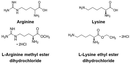
A high mutation rate is a significant challenge in developing antivirals against RNA viruses. These mutations may benefit virus life cycles, such as evading immune response, increasing virus transmissibility, or enhancing pathogenesis. Hence, the need to discover broad-spectrum antiviral agents for current and future virus diseases is undisputed [19]. Previous research has shown that the amino acids arginine and lysine play roles in virus infection and replication [20][21][22][23][24][25][26]. Lysine, an essential amino acid, has long been prescribed against herpes simplex virus (HSV) infection [25][26][27]. A high lysine/arginine ratio has decreased HSV plaque formation in vitro [22][23][24]. Clinical trials have exhibited a reduction of recurrent HSV attacks, fewer healing days, and milder symptoms during a six-month period of L-lysine monochloride therapy [27][28]. Therefore, we investigated whether these basic amino acids could also affect SARS-CoV-2 and IAV infection. In this study, we used an in vitro system to evaluate the effects of lysine, arginine, and their derivatives, L-Arginine methyl ester dihydrochloride (Arg-ester), and L-Lysine ethyl ester dihydrochloride (Lys-ester) (Figure 1), on SARS-CoV-2 and IAV infection.
Viruses 13 01301 g001 550
Figure 1. Chemical structure of the compounds.

2. Effects of Basic Amino Acids and Their Derivatives on SARS-CoV-2 and Influenza-A Virus Infection

2.1. Lysine and Its Derivative Attenuate SARS-CoV-2 Vpp Infection
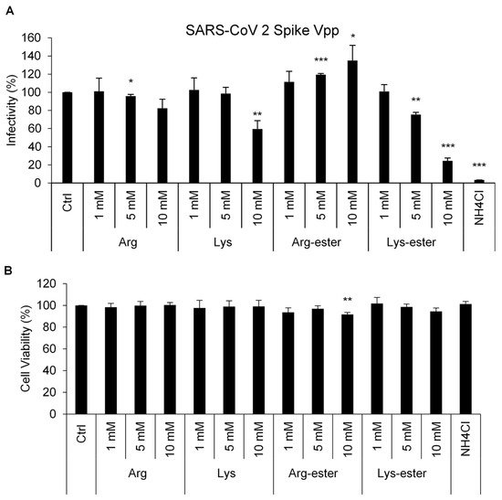
Weak bases, like chloroquine analogs, have been investigated to impede viral entry by interfering with endosome acidification [29]. Given that arginine and lysine are basic amino acids, we hypothesized that these compounds may affect SARS-CoV-2 entering into cells. To prove this hypothesis, we utilized an established luciferase-based pseudotyped viral particle (Vpp) system for rapid quantitation of infected cells. These Vpps carry SARS-CoV-2 spike (S) protein and can mimic natural SARS-CoV-2 virus entry. Upon entering target cells, the luciferase reporter is expressed, and its intensity corresponds to the number of infected cells [12]. Human embryonic kidney 293 (HEK293T) cells were infected with SARS-CoV-2 Vpp in the presence of the compounds, and the cell infection rate was measured by the detection of luciferase activity (Figure 2A). As expected, treatment with NH4Cl, a lysosomotropic weak base that blocks virus entry [16][30][31][32], diminished SARS-CoV-2 Vpp infection. Among the four compounds tested, lysine and Lys-ester remarkably reduced Vpp entry in a dose-dependent manner. 10 mM of lysine and Lys-ester could inhibit 40% and 75% of viral entry, respectively. In contrast, 10 mM Arg-ester significantly increased Vpp infectivity (1.35-fold). Treatment and infection in Huh7 cells also resulted in a significant decrease in Vpp infection in lysine and Lys-ester-treated cells (Supplementary Figure S1).
Viruses 13 01301 g002 550
Figure 2. Compound effects on SARS-CoV-2 Spike Vpp infection in HEK293T cells. HEK293T cells were pre-treated with different concentrations of the compounds for 1 h and transduced with SARS-CoV-2 Spike Vpp for 1 h in the presence of the compounds in the media. (A) Infectivity and (B) cell viability were measured at 3 days post-infection using luciferase assay and MTS assay, respectively. The values represent the means ± standard deviation (SD) of data from three independent experiments. Ctrl: medium only. *, p < 0.05; **, p < 0.01; and ***, p < 0.001 compared with controls (n = 3).
Furthermore, to understand the cytotoxicity of these compounds, a cell viability assay was conducted. As shown in Figure 2B, only treatment with 10 mM Arg-ester showed significant cytotoxicity in HEK293T cells. These altogether suggest that lysine and Lys-ester control SARS-CoV-2-S-mediated infection at non-cytotoxic doses.

2.2. The Compounds Do Not Interfere the Interaction between Spike and ACE2
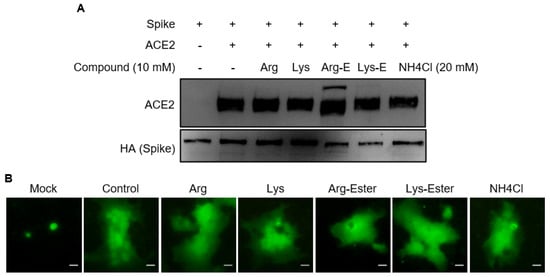
SARS-CoV-2 enters cells by binding its S protein to the ACE2 receptor protein of the host cell [16]. To evaluate whether the compounds affect the interaction between the S and ACE2, HA-tagged-S and ACE2 protein were separately expressed in HEK293T cells and their lysates were then used for immunoprecipitation assay in the presence of the compounds. The results indicated that no compound interfered with the interaction of Spike and ACE2 (Figure 3A).
Viruses 13 01301 g003 550
Figure 3. Effects of compounds on the interaction between SARS-CoV-2 Spike and ACE2. (A) HEK293T cells were transfected with either HA-tagged SARS-CoV-2 Spike or ACE2. Equal amounts of Spike-expressing and ACE2-expressing cell lysates were mixed, incubated with or without the compounds, and were used for immunoprecipitation assay using anti-HA agarose. The immunoprecipitates were resolved on SDS-PAGE and immunoblotted with anti-ACE2 or anti-HA antibodies. (B) Effects of compounds on SARS-CoV-2 Spike-mediated cell-cell fusion. Mock- or ACE2-transfected H1650 cells were co-cultured with spike and GFP-coexpressing H1650 cells for 4 h in the presence of the indicated compounds (10 mM) or NH4Cl (20 nM). Scale bar = 25 μM.
After binding to ACE2, SARS-CoV-2 enters cells via fusion with cell membrane or endocytosis. We further investigated whether the compounds inhibited entry by interfering with the fusion of the viral membrane and the host membrane using a cell–cell fusion assay. In brief, we overlaid S protein and green fluorescent protein (GFP)-expressing human lung cancer H1650 cells on ACE2-transfected cells in the presence of the compounds. As shown in Figure 3B, large syncytia were formed despite compound treatment, implying that all tested compounds do not influence cell–cell fusion. These data collectively suggest that the compounds affect SARS-CoV-2-S Vpp infection by targeting a post-binding step.

2.3. Lysine and Lys-Ester Inhibit IAV Replication
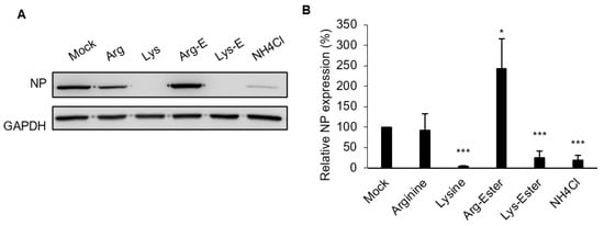
Next, we evaluated the effect of compounds on IAV infection due to it being the cause of outbreaks for decades worldwide and having a high prevalence of co-infection with SARS-CoV-2 [7]. As shown in Figure 4A, treatment of A549 cells with lysine and Lys-ester inhibited virus replication at 6 h post-infection (hpi), as evidenced by the reduction of nucleoprotein (NP) expression. This result was further verified in Figure 4B, wherein viral RNA was reduced to 4% and 25% by lysine and Lys-ester, respectively. Similar to the effect on SARS-CoV-2 Vpp infection, IAV viral RNA also increased in the presence of Arg-ester. These indicate that lysine and Lys-ester also have inhibitory effects on IAV infection, while Arg-ester has the opposite effect.
Viruses 13 01301 g004 550
Figure 4. Compound effects on IAV infection in A549 cells. A549 cells were pre-treated with different concentrations of the compounds for 1 h and infected with IAV for 6 h in the presence of the compounds in the media. Cells treated with the medium were used as Mock control. (A) Cell lysates were harvested for Western blotting using anti-IAV NP antibody. GAPDH was used as a loading control. (B) Total RNA was extracted and used for RT-qPCR analysis. The levels of viral RNA were normalized with GAPDH RNA. The concentrations of the compounds were 10 nM, except for NH4Cl, 20 mM. The values represent the means ± standard deviation (SD) of data from three independent experiments. *, p < 0.05; and ***, p < 0.001 compared with controls (n = 3).

2.4. The Compounds Do Not Affect IAV Binding and Internalization

The inhibition of SARS-CoV-2 Vpp infection by lysine and Lys-ester suggests that these compounds may affect virus entry (Figure 2A). For most enveloped viruses, virus entry steps include binding, internalization, endocytosis, and uncoating. To reveal the stage of the IAV replication cycle interfered with via the compounds, we first examined the effects of compounds on the binding and internalization of a virus. At 4 °C, the virus binds to the cell surface without internalization. At 37 °C, the virus can bind and be internalized into the cells [33]. Here, we detected virus particles by immunoblotting viral M1 protein, the most abundant structural protein of IAV particles. Compared to mock treatment, no significant change in the amount of bound (Figure 5A) or internalized (Figure 5B) M1 could be observed in the compound-treated cells, suggesting that the compounds most likely function at a step after internalization of virus particles.
Viruses 13 01301 g005 550
Figure 5. Compound effects on IAV binding and internalization. A549 cells were treated with the compounds for 1 h, then infected with IAV (MOI 5) at 4 °C for 1 h in the presence of the compounds. (A) After virus infection, the cells were washed with cold PBS to remove the unbound virus and then harvested. (B) Following virus infection, the cells were refreshed with a warm medium and incubated at 37 °C for an additional 1 h with the compounds. Cell lysates were harvested and analyzed by Western blotting with anti–M1 antibody. The concentrations of the compounds were 10 nM, except for NH4Cl, 20 mM. Mock: medium only.

2.5. The Compounds Do Not Reduce Endosomal Acidification
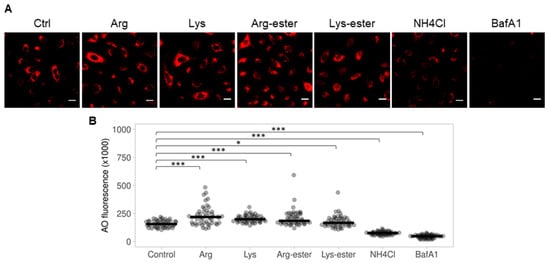
Soon after receptor recognition and attachment, the virus is endocytosed into the cell to proceed infection. SARS-CoV-2 and IAV require the acidic pH of the endosomes for a fusion of the viral membrane with the endosomal membrane to release its genome into the cytoplasm [11][13][14]. To examine whether the compounds reduce endosomal acidification, we performed acridine orange staining, which accumulates in acidic compartments where it fluoresces bright red [34]Figure 6 illustrates that pre-treatment of cells with the compounds did not lessen the formation of acidic vacuoles. A marked increase in red fluorescence was observed after compound treatment (Figure 6B). However, cells treated with NH4Cl and bafilomycin A1, which are known to block endosomal acidification and are commonly used to demonstrate the requirement of acidic vacuoles in viral infectivity [35][36], exhibited lower fluorescence. In addition, we also performed LysoTracker Red staining to confirm our findings. LysoTracker Red staining demonstrated similar results with acridine orange staining (Supplementary Figure S2). These data altogether imply that the compounds did not inhibit virus infection by reducing endosomal acidification.
Viruses 13 01301 g006 550
Figure 6. Acridine orange staining of A549 cells following compound treatment for 1 h. Cells were treated with the compounds (10 mM), NH4Cl (20 mM), or bafilomycin A1 (40 nM) for 1 h and stained with 1 μg/mL AO for 15 min. (A) Acidic vacuoles (orange-red) were observed using fluorescence microscopy. Scale bar = 100 μM. (B) Red fluorescence from 50 cells were calculated and plotted using ImageJ and PlotsOfData, respectively. Black horizontal lines represent the medians ±95% confidence intervals. *, p < 0.05; and ***, p < 0.001 compared with controls.

2.6. Lysine and Lys-Ester Reduce Nuclear Distribution of IAV

Having demonstrated that the compounds might affect a step following endocytosis, we next examined if the inhibitory effects of lysine and Lys-ester were associated with virus release from the endosomes. Regarding the influenza virus, once the viral RNA is released into the cytoplasm, it is subsequently translocated to the nucleus. Thus, we evaluated the trafficking of the internalized virus by observing the nuclear localization of viral ribonucleoprotein (vRNP), which is composed of viral RNA, NP, and viral RNA-dependent RNA polymerase (RdRp). We found that lysine and Lys-ester treatment, similar to NH4Cl treatment, showed almost no NP signal in the nucleus (Figure 7). Combining all of the results, lysine and Lys-ester may most likely affect the stage after endocytosis, such as viral uncoating and nuclear import.
Viruses 13 01301 g007 550
Figure 7. Compound effects on nuclear transport of IAV NP protein. A549 cells were treated with the compounds for 1 h and then infected with IAV (MOI 5) for 1 h at 37 °C in the presence of the compounds. After 3 hpi, cells were fixed and subjected to immunofluorescence staining with anti-NP antibody and Alexa Fluor 488 goat anti-mouse antibodies. Nuclei were counterstained with DAPI and cells were visualized with a fluorescence microscope. Scale bar = 20 μM.

3. Conclusions

Lysine and Lys-ester can prevent SARS-CoV-2 and IAV infection, particularly in the entry stage. In contrast to that, Arg-ester can potently boost infection of both viruses. It would therefore be beneficial to consider the nutrient intake of COVID-19 and flu patients. We recommend the inclusion of lysine supplementation in addition to a reduced arginine intake for the prevention and treatment of SARS-CoV-2 and IAV infections.

This entry is adapted from the peer-reviewed paper 10.3390/v13071301

References

  1. Liu, Y.; Gayle, A.A.; Wilder-Smith, A.; Rocklöv, J. The reproductive number of COVID-19 is higher compared to SARS coronavirus. J. Travel Med. 2020, 27, taaa021.
  2. Hu, T.; Liu, Y.; Zhao, M.; Zhuang, Q.; Xu, L.; He, Q. A comparison of COVID-19, SARS and MERS. PeerJ 2020, 8, e9725.
  3. Kaur, N.; Singh, R.; Dar, Z.; Bijarnia, R.K.; Dhingra, N.; Kaur, T. Genetic comparison among various coronavirus strains for the identification of potential vaccine targets of SARS-CoV2. Infect. Genet. Evol. 2021, 89, 104490.
  4. Ngai, J.C.; Ko, F.W.; Ng, S.S.; To, K.-W.; Tong, M.; Hui, D.S. The long-term impact of severe acute respiratory syndrome on pulmonary function, exercise capacity and health status. Respirology 2010, 15, 543–550.
  5. Rubin, R. As their numbers grow, COVID-19 “Long Haulers” stump experts. JAMA 2020, 324, 1381–1383.
  6. Arbeitskreis Blut, U.B.B.K. Influenza virus. Transfus. Meds. Hemother. 2009, 36, 32–39.
  7. Hashemi, S.A.; Safamanesh, S.; Ghasemzadeh-moghaddam, H.; Ghafouri, M.; Azimian, A. High prevalence of SARS-CoV-2 and influenza A virus (H1N1) coinfection in dead patients in Northeastern Iran. J. Med. Virol. 2021, 93, 1008–1012.
  8. Bai, L.; Zhao, Y.; Dong, J.; Liang, S.; Guo, M.; Liu, X.; Wang, X.; Huang, Z.; Sun, X.; Zhang, Z.; et al. Co-infection of influenza A virus enhances SARS-CoV-2 infectivity. bioRxiv 2020.
  9. Zhang, A.J.; Lee, A.C.; Chan, J.F.; Liu, F.; Li, C.; Chen, Y.; Chu, H.; Lau, S.Y.; Wang, P.; Chan, C.C.; et al. Co-infection by severe acute respiratory syndrome coronavirus 2 and influenza A(H1N1)pdm09 virus enhances the severity of pneumonia in golden Syrian hamsters. Clin. Infect. Dis. 2020, 72, e978–e992.
  10. Lakadamyali, M.; Rust, M.J.; Zhuang, X. Endocytosis of influenza viruses. Microbes Infect. 2004, 6, 929–936.
  11. Li, S.; Sieben, C.; Ludwig, K.; Höfer, C.T.; Chiantia, S.; Herrmann, A.; Eghiaian, F.; Schaap, I.A.T. pH-Controlled two-step uncoating of influenza virus. Biophys. J. 2014, 106, 1447–1456.
  12. Ou, X.; Liu, Y.; Lei, X.; Li, P.; Mi, D.; Ren, L.; Guo, L.; Guo, R.; Chen, T.; Hu, J.; et al. Characterization of spike glycoprotein of SARS-CoV-2 on virus entry and its immune cross-reactivity with SARS-CoV. Nat. Commun. 2020, 11, 1620.
  13. Helenius, A. Virus entry: What has pH got to do with it? Nat. Cell Biol 2013, 15, 125.
  14. V’kovski, P.; Kratzel, A.; Steiner, S.; Stalder, H.; Thiel, V. Coronavirus biology and replication: Implications for SARS-CoV-2. Nat. Rev. Microbiol. 2021, 19, 155–170.
  15. Repnik, U.; Česen, M.H.; Turk, B. The endolysosomal system in cell death and survival. Cold Spring Harb. Perspect. Biol. 2013, 5, a008755.
  16. Hoffmann, M.; Kleine-Weber, H.; Schroeder, S.; Krüger, N.; Herrler, T.; Erichsen, S.; Schiergens, T.S.; Herrler, G.; Wu, N.-H.; Nitsche, A.; et al. SARS-CoV-2 cell entry depends on ACE2 and TMPRSS2 and is blocked by a clinically proven protease inhibitor. Cell 2020, 181, 271–280.e278.
  17. Shang, J.; Wan, Y.; Luo, C.; Ye, G.; Geng, Q.; Auerbach, A.; Li, F. Cell entry mechanisms of SARS-CoV-2. Proc. Natl. Acad. Sci. USA 2020, 117, 11727–11734.
  18. Tang, T.; Bidon, M.; Jaimes, J.A.; Whittaker, G.R.; Daniel, S. Coronavirus membrane fusion mechanism offers a potential target for antiviral development. Antivir. Res. 2020, 178, 104792.
  19. Chitalia, V.C.; Munawar, A.H. A painful lesson from the COVID-19 pandemic: The need for broad-spectrum, host-directed antivirals. J. Transl. Med. 2020, 18, 390.
  20. Becht, H. Induction of an arginine-rich component during infection with influenza virus. J. Gen. Virol. 1969, 4, 215–220.
  21. Eaton, M.D.; Scala, A.R.; Low, I.E. Amino acid imbalance and incomplete viral replication. Arch. Für Die Gesamte Virusforsch. 1964, 14, 583–598.
  22. Griffith, R.S.; DeLong, D.C.; Nelson, J.D. Relation of arginine-lysine antagonism to herpes simplex growth in tissue culture. Chemotherapy 1981, 27, 209–213.
  23. Rossi, M.; Jacobs, B. Chapter 20—Herpes simplex virus. In Integrative Medicine (Fourth Edition); Rakel, D., Ed.; Elsevier: Amsterdam, The Netherlands, 2018; pp. 191–197.e192.
  24. Luiking, Y.C.; Deutz, N.E.P. Biomarkers of arginine and lysine excess. J. Nutr. 2007, 137, 1662S–1668S.
  25. Mailoo, V.J.; Rampes, S. Lysine for herpes simplex prophylaxis: A review of the evidence. Integr. Med. (Encinitas) 2017, 16, 42–46.
  26. Griffith, R.S.; Norins, A.L.; Kagan, C. A multicentered study of lysine therapy in Herpes simplex infection. Dermatologica 1978, 156, 257–267.
  27. Griffith, R.S.; Walsh, D.E.; Myrmel, K.H.; Thompson, R.W.; Behforooz, A. Success of L-lysine therapy in frequently recurrent herpes simplex infection. Treatment and prophylaxis. Dermatologica 1987, 175, 183–190.
  28. Thein, D.J.; Hurt, W.C. Lysine as a prophylactic agent in the treatment of recurrent herpes simplex labialis. Oral Surg. Oral Med. Oral Pathol. 1984, 58, 659–666.
  29. Al-Bari, M.A.A. Targeting endosomal acidification by chloroquine analogs as a promising strategy for the treatment of emerging viral diseases. Pharm. Res. Perspect 2017, 5, e00293.
  30. Dabydeen, S.A.; Meneses, P.I. The role of NH4Cl and cysteine proteases in Human Papillomavirus type 16 infection. Virol. J. 2009, 6, 109.
  31. Matlin, K.S. Ammonium chloride slows transport of the influenza virus hemagglutinin but does not cause mis-sorting in a polarized epithelial cell line. J. Biol. Chem. 1986, 261, 15172–15178.
  32. Oomens, A.G.P.; Wertz, G.W. The baculovirus GP64 protein mediates highly stable infectivity of a human respiratory syncytial virus lacking its homologous transmembrane glycoproteins. J. Virol. 2004, 78, 124–135.
  33. Su, W.-C.; Chen, Y.-C.; Tseng, C.-H.; Hsu, P.W.-C.; Tung, K.-F.; Jeng, K.-S.; Lai, M.M.C. Pooled RNAi screen identifies ubiquitin ligase Itch as crucial for influenza A virus release from the endosome during virus entry. Proc. Natl. Acad. Sci. USA 2013, 110, 17516–17521.
  34. Krolenko, S.A.; Adamyan, S.Y.; Belyaeva, T.N.; Mozhenok, T.P. Acridine orange accumulation in acid organelles of normal and vacuolated frog skeletal muscle fibres. Cell Biol. Int. 2006, 30, 933–939.
  35. Bayer, N.; Schober, D.; Prchla, E.; Murphy, R.F.; Blaas, D.; Fuchs, R. Effect of bafilomycin A1 and nocodazole on endocytic transport in HeLa cells: Implications for viral uncoating and infection. J. Virol. 1998, 72, 9645–9655.
  36. Shulla, A.; Heald-Sargent, T.; Subramanya, G.; Zhao, J.; Perlman, S.; Gallagher, T. A transmembrane serine protease is linked to the severe acute respiratory syndrome coronavirus receptor and activates virus entry. J. Virol. 2011, 85, 873–882.
More
This entry is offline, you can click here to edit this entry!