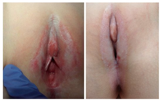
Vulvar lichen sclerosus (VLS) is a chronic inflammatory condition affecting the anogenital region, which may present in a prepubertal or adolescent patient. The most popular theories are its autoimmune and genetic conditioning, although theories concerning hormonal and infectious etiology have also been raised. The most common presenting symptoms of VLS is vulva pruritus, discomfort, dysuria and constipation. The lesions initially are white, flat-topped papules, thin plaques, or commonly atrophic patches. Purpura is a hallmark feature of VLS. The treatment includes topical anti-inflammatory agents and long-term follow-up, as there is a high risk of recurrence and an increased risk of vulvar cancer in adult women with a history of lichen sclerosus.
- vulvar lichen sclerosus
- pediatric
- adolescent
1. Introduction
VLS manifests in lesions in vulvar mucosa, which often spreads to the skin of the anus [1]. The symptoms of this condition may include whitening of the perineal area, but also itching, burning, discomfort, vaginal bleeding, and dysuria, which in sexually active girls may be mistaken for symptoms of urogenital infection [2]. In some cases, due to anorectal lesions, additional symptoms may occur, such as constipation or painful defecation, without any gastrointestinal problems in the patient’s medical history [3]. The prevalence of VLS in underaged girls is understated, due to the misreading of the symptoms by GPs (general practitioners), and the delayed access to specialists in the field of pediatric gynecology or dermatology [4].
2. Epidemiology
VLS can occur at any age or in any sex, although the highest values can be observed in women aged 40–60 years old, and in pre-pubertal girls. There is a clear peak of incidence in girls aged four to six years old, which represents 7–15% of all vulvar lichen sclerosus cases [5].
It is estimated that VLS can be observed in 1:900 of premenarchal girls [4][6]. The first symptoms are usually very non-specific and misdiagnosed by non-gynecologist and non-dermatologist doctors. Some of the symptoms can spontaneously recede after the menarche, and the course of the disease can be latent. This is why the epidemiology of VLS is underestimated [6].
3. Etiopathogenesis
3.1. Immunological Theory
3.2. Genetic Theory
3.3. Hormonal Theory
3.4. Trauma Theory
3.5. Infectious Theory
3.6. Drug-Induced Theory
4. Symptoms
5. Complications in the Course of Vulvar Lichen Sclerosus
In the entire population of VLS patients, 2.6–6.7% will undergo neoplastic transformation, which is the result of chronic inflammation, altered expression of the p53 oncogene, and oxidative stress [34][35]. It should be emphasized that in pediatric patients, the incidence of vSCC is lower than in the rest of patients with VLS, and it amounts to approx. This occurs due to the pathogenesis of the neoplasm and additional risk factors—mainly age. However, the study by Halonen et al. showed that the early age of VLS diagnosis is an additional risk factor for this complication [36].
In patients with vulvar lichen sclerosus, scars and deformities of the affected areas are observed more often. It has not been determined whether the early treatment initiation improves the prognosis for the occurrence of these complications. The described changes may coexist with chronic pain and itching, significantly worsening the quality of life. In sexually active patients, penetration difficulties and dyspareunia may appear [21][36].
The narrowing of the vulval vestibule may occur as a result of the fusion of the labia. If this produces the inability to have sexual intercourse or causes problems with urination, surgery may be necessary. Topical steroids and dilators can help in the postoperative period, by preventing re-deformation. Occasionally, the clitoral adhesions result in the formation of a painful pseudocyst, which is also a condition requiring surgical intervention [37].
As a result of the inflammatory condition in the course of VLS, sensorial disturbances in the vulva may develop and lead to vulvodynia. These symptoms may persist during and after the treatment. Pain sensation is not reduced by treatment with glucocorticosteroids. In such a case, the patient should be offered a treatment aimed at neuropathic pain [37].
6. Diagnosis
The differentiation of symptoms in the course of vulvar lichen sclerosus causes great diagnostic difficulties. In only 16% of cases, girls are diagnosed with vulvar lichen sclerosus in the initial stage of the disease. Changes in the course of VLS may imitate the clinical symptoms of many dermatoses. The differential diagnosis of vulvar lichen sclerosus includes lichen simplex chronicus, lichen planus, eczema, psoriasis, atopic dermatitis, seborrheic skin lesions, vitiligo, sexual harassment, vulvar injuries, and linear IgA disease of the childhood (chronic bullous dermatosis) in cases when VLS presents with bullae [38].
In most cases, the diagnosis is made on the basis of a thorough medical history interview with the patient/guardian, and as the result of a physical examination. This is particularly true in children and men. However, the histological examination is recommended if there are atypical features or diagnostic uncertainty, and it is essential if there is any suspicion of a neoplastic lesion. It is important to remember that vulva biopsies are reserved for doubtful cases, especially when there is no improvement after treatment [21][37][38].
According to the guidelines of the British Dermatological Society, skin biopsy should be performed in the following cases (taking material for histopathological examination, with particular emphasis on hyperkeratotic lesions):The disease fails to respond to an adequate treatment, or an alternative/additional therapy with a potent topical steroid is to be implemented;There is an extragenital lichen sclerosus that has features mimicking the morphoea;There are pigmented areas to exclude abnormal melanocytic proliferation;There is a suspicion of neoplastic lesion. These are usually lesions with a persistent area of hyperkeratosis, erosion or erythema, or new warty or papular lesions. Several mapping biopsies may be required if there is an extensive abnormality. If there are any lesions that are highly suspicious of an SCC, the patient should be referred urgently to a gynecologist for the excision of the whole lesion for an adequate staging [37].
As we mentioned before, in children, a vulval biopsy is usually not performed, because it may be very traumatic for the child. It should be reserved only for cases with an uncertain diagnosis, and for those who fail to respond to treatments [39]. The typical histological features of VLS are orthohyperkeratosis, epidermal atrophy, basal cell degeneration, dermal hyalinization, and a band-like lymphocytic infiltrate [21].
7. Pharmacotherapy
| Treatment | Effects | Side Effects |
|---|---|---|
| High-potency corticosteroids |
|
Prolonged use of topical steroids can be associated with:
|
| Calcineurin inhibitors—tacrolimus, pimecrolimus | Tacrolimus 0.03% ointment:
|
|
| Retinoids |
|
No report. |
| Topical sex hormons | No report in children. | No report. |
| Cyclosporine | In patients with refractory VLS with symptomatic improvement and decrease in erythema and erosions after one month of therapy. |
|
| Phototherapy | No report in children. | No report. |
| Vitamins D, A and E | Additional data are needed to assess the usefulness of vitamin supplementation in the treatment. | No report. |
This entry is adapted from the peer-reviewed paper 10.3390/ijerph18137153
References
- Jensen, L.S.; Bygum, A. Childhood lichen sclerosus is a rare but important diagnosis. Dan. Med. J. 2012, 59, A4424.
- Topal, İ.O.; Sayılgan, A.T.; Kalçın, S. An uncommon cause of vulval pruritus in childhood: Lichen sclerosus. Turk. Pediatri. Ars. 2014, 49, 86–87.
- Maronn, M.L.; Esterly, N.B. Constipation as a feature of anogenital lichen sclerosus in children. Pediatrics 2005, 115, 230–232.
- Powell, J.; Wojnarowska, F. Childhood vulvar lichen sclerosus: An increasingly common problem. J. Am. Acad. Dermatol. 2001, 44, 803–806.
- Nerantzoulis, I.; Grigoriadis, T.; Michala, L. Genital lichen sclerosus in childhood and adolescence-a retrospective case series of 15 patients: Early diagnosis is crucial to avoid long-term sequelae. Eur. J. Pediatr. 2017, 176, 1429–1432.
- Dinh, H.; Purcell, S.M.; Chung, C.; Zaenglein, A.L. Pediatric Lichen Sclerosus: A Review of the Literature and Management Recommendations. J. Clin. Aesthet. Dermatol. 2016, 9, 49–54.
- Klaber, R. Lichen Sclerosus et Atrophicans (Hallopeau). Proc. R. Soc. Med. 1937, 30, 977–979.
- Farrell, A.M.; Dean, D.; Millard, P.; Charnock, F.M.; Wojnarowska, F. Cytokine alterations in lichen sclerosus: An immunohistochemical study. Br. J. Dermatol. 2006, 155, 931–940.
- Simpkin, S.; Oakley, A. Clinical review of 202 patients with vulval lichen sclerosus: A possible association with psoriasis. Australas. J. Dermatol. 2007, 48, 28–31.
- Cooper, S.M.; Ali, I.; Baldo, M.; Wojnarowska, F. The association of lichen sclerosus and erosive lichen planus of the vulva with autoimmune disease: A case-control study. Arch. Dermatol. 2008, 144, 1432–1435.
- Sherman, V.; McPherson, T.; Baldo, M.; Salim, A.; Gao, X.H.; Wojnarowska, F. The high rate of familial lichen sclerosus suggests a genetic contribution: An observational cohort study. J. Eur. Acad. Dermatol. Venereol. 2010, 24, 1031–1034.
- Doulaveri, G.; Armira, K.; Kouris, A.; Karypidis, D.; Potouridou, I. Genital vulvar lichen sclerosus in monozygotic twin women: A case report and review of the literature. Case Rep. Dermatol. 2013, 5, 321–325.
- Singh, N.; Ghatage, P. Etiology, Clinical Features, and Diagnosis of Vulvar Lichen Sclerosus: A Scoping Review. Obstet. Gynecol. Int. 2020, 2020, 7480754.
- Chakhtoura, Z.; Vigoureux, S.; Courtillot, C.; Tejedor, I.; Touraine, P. Vulvar lichen sclerosus is very frequent in women with Turner syndrome. J. Clin. Endocrinol. Metab. 2014, 99, 1103–1104.
- Clifton, M.M.; Garner, I.B.; Kohler, S.; Smoller, B.R. Immunohistochemical evaluation of androgen receptors in genital and extragenital lichen sclerosus: Evidence for loss of androgen receptors in lesional epidermis. J. Am. Acad. Dermatol. 1999, 4, 43–46.
- Ismail, D.; Owen, C.M. Paediatric vulval lichen sclerosus: A retrospective study. Clin. Exp. Dermatol. 2019, 44, 753–758.
- Poindexter, G.; Morrell, D.S. Anogenital pruritus: Lichen sclerosus in children. Pediatr. Ann. 2007, 36, 785–791.
- Edwards, L.R.; Privette, E.D.; Patterson, J.W.; Tchernev, G.; Chokoeva, A.A.; Wollina, U.; Lotti, T.; Wilson, B.B. Radiation-induced lichen sclerosus of the vulva: First report in the medical literature. Wien. Med. Wochenschr. 2017, 167, 74–77.
- Kirtschig, G. Lichen Sclerosus-Presentation, Diagnosis and Management. Dtsch. Arztebl. Int. 2016, 113, 337–343.
- Chattopadhyay, S.; Arnold, J.D.; Malayil, L.; Hittle, L.; Mongodin, E.F.; Marathe, K.S.; Gomez-Lobo, V.; Sapkota, A.R. Potential role of the skin and gut microbiota in premenarchal vulvar lichen sclerosus: A pilot case-control study. PLoS ONE 2021, 16, e0245243.
- Kirtschig, G.; Becker, K.; Günthert, A.; Jasaitiene, D.; Cooper, S.; Chi, C.C.; Kreuter, A.; Rall, K.K.; Aberer, W.; Riechardt, S.; et al. Evidence-based (S3) Guideline on (anogenital) Lichen sclerosus. J. Eur. Acad. Dermatol. Venereol. 2015, 10, e1–e43.
- Pranteda, G.; Muscianese, M.; Grimaldi, M.; Fidanza, L.; Pranteda, G.; Narcisi, A.; Nistico, S.; Bottoni, U. Lichen sclerosus et atrophicus induced by carbamazepine: A case report. Int. J. Immunopathol. Pharmacol. 2013, 26, 791–794.
- Baldo, M.; Ali, I.; Wojnarowska, F. The contribution of drugs to lichen sclerosus. Clin. Exp. Dermatol. 2014, 39, 234.
- Gibbon, K.L.; Bewley, A.P.; Salisbury, J.A. Labial fusion in children: A presenting feature of genital lichen sclerosus? Pediatr. Dermatol. 1999, 16, 388–391.
- Lagerstedt, M.; Karvinen, K.; Joki-Erkkilä, M.; Huotari-Orava, R.; Snellman, E.; Laasanen, S.L. Childhood lichen sclerosus--a challenge for clinicians. Pediatr. Dermatol. 2013, 30, 444–450.
- Dendrinos, M.L.; Quint, E.H. Lichen sclerosus in children and adolescents. Curr. Opin. Obstet. Gynecol. 2013, 25, 370–374.
- Arlen, A.M.; Wang, M.; Vash-Margita, A. Lichen Sclerosus in Prepubertal Girls: An Uncommon but Treatable Cause of Lower Urinary Tract Symptoms. Urology 2020, 137, e1–e2.
- Ellis, E.; Fischer, G. Prepubertal-Onset Vulvar Lichen Sclerosus: The Importance of Maintenance Therapy in Long-Term Outcomes. Pediatr. Dermatol. 2015, 32, 461–467.
- Friedland, R.; Ben-Amitai, D.; Didkovsky, E.; Feinmesser, M.; Zvulunov, A. Vascular lesions in genital lichen sclerosus in pediatric patients. Pediatr. Dermatol. 2020, 37, 849–852.
- Di Altobrando, A.; Patrizi, A.; Bassi, A.; Virdi, A.; Besagni, F.; Sacchelli, L.; Neri, I. Lichen sclerosus with enlarged vessels: A variant of lichen sclerosus in young girls. Pediatr. Dermatol. 2021, 38, 318–319.
- Vash-Margita, A.; Smith, Y.R.; Rabah, R.; Quint, E.H. Adolescent Vulvar Angiokeratoma Associated with Lichen Sclerosus. J. Pediatr. Adolesc. Gynecol. 2019, 32, 440–442.
- Belotto, R.A.; Chavantes, M.C.; Tardivo, J.P.; Euzébio Dos Santos, R.; Fernandes, R.C.M.; Horliana, A.C.R.T.; Pavani, C.; Teixeira da Silva, D.F. Therapeutic comparison between treatments for Vulvar Lichen Sclerosus: Study protocol of a randomized prospective and controlled trial. BMC Womens Health 2017, 17, 61.
- Olejek, A.; Steplewska, K.; Gabriel, A.; Kozak-Darmas, I.; Jarek, A.; Kellas-Sleczka, S.; Bydliński, F.; Sieroń-Stołtny, K.; Horak, S.; Chełmicki, A.; et al. Efficacy of photodynamic therapy in vulvar lichen sclerosus treatment based on immunohistochemical analysis of CD34, CD44, myelin basic protein, and Ki67 antibodies. Int. J. Gynecol. Cancer 2010, 20, 879–887.
- Salomon-Perzyńska, M. Lichen sclerosus—Algorytm postępowania diagnostyczno-terapeutycznego. Forum Ginekol. 2018, 40, 42–48.
- Bigby, S.M.; Eva, L.J.; Fong, K.L.; Jones, R.W. The natural history of vulvar intraepithelial neoplasia, differentiated type: Evidence for progression and diagnostic challenges. Int. J. Gynecol. Pathol. 2016, 35, 574–584.
- Morrel, B.; van Eersel, R.; Burger, C.W.; Bramer, W.M.; Ten Kate-Booij, M.J.; van der Avoort, I.A.M.; Pasmans, S.G.M.A. The long-term clinical consequences of juvenile vulvar lichen sclerosus: A systematic review. J. Am. Acad. Dermatol. 2020, 82, 469–477.
- Lewis, F.M.; Tatnall, F.M.; Velangi, S.S.; Bunker, C.B.; Kumar, A.; Brackenbury, F.; Mohd Mustapa, M.F.; Exton, L.S. British Association of Dermatologists guidelines for the management of lichen sclerosus, 2018. Br. J. Dermatol. 2018, 178, 839–853.
- Bercaw-Pratt, J.L.; Boardman, L.A.; Simms-Cendan, J.S. North American Society for Pediatric and Adolescent Gynecology. Clinical recommendation: Pediatric lichen sclerosus. J. Pediatr. Adolesc. Gynecol. 2014, 27, 111–116.
- Neill, S.M.; Lewis, F.M.; Tatnall, F.M.; Cox, N.H. British Association of Dermatologists. British Association of Dermatologists’ guidelines for the management of lichen sclerosus 2010. Br. J. Dermatol. 2010, 163, 672–682.