Molecular Hydrogen as Radioprotective Agent: History
Please note this is an old version of this entry, which may differ significantly from the current revision.
Subjects: Biophysics
Contributor:

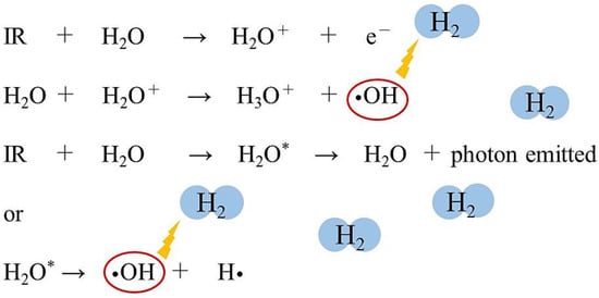
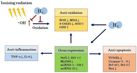
Molecular hydrogen (H2) has the potential to be a radioprotective agent because it can selectively scavenge •OH, a reactive oxygen species with strong oxidizing power. Animal experiments and clinical trials have reported that H2 exhibits a highly safe radioprotective effect.

  • molecular hydrogen
  • radiation-induced damage
  • medical application
  • radioprotective agent
  • non-DNA target
  • intracellular response
  • oxidation
  • inflammation
  • apoptosis
  • gene expression

1. Introduction

Ionizing radiation (radiation) is commonly used for medical diagnosis and cancer treatment. Amongst these uses, radiation therapy is known to be one of the most effective treatments for cancer. It is difficult to control radiation-induced damage with conventional radiation therapy; therefore, intensity-modulated radiation therapy (IMRT) has recently been used [1]. However, various radiation damages can also occur with IMRT. The harmful effects of radiation on the living body can be classified into direct and indirect effects. Direct effects are caused by the direct absorption of radiation energy into nucleic acids (DNA), proteins, and lipids [2][3][4][5]. Indirect effects are caused by free radicals, such as hydroxyl radicals (•OH), and molecular products generated in the process of water radiolysis [2][3][4][5]. In addition to the direct damage on DNA, secondary damages to non-DNA targets cannot be ignored because low-dose radiation damage is mainly caused by these indirect effects. Secondary damages include oxidation, inflammation, apoptosis, and effects on gene expression related to intracellular responses.
Medical applications of H2 were first reported by Dole et al. in 1975 [6]. They reported that the inhalation of hyperbaric H2 caused a marked regression in squamous cell carcinoma in mice induced by UV radiation. With the exception of a few studies, however, H2 has not been extensively studied for medical applications. In 2007, Ohsawa et al. reported that the inhalation of H2 gas ameliorated ischemia-reperfusion injury in a rat model with cerebral infarction [7]. In this paper, they showed that H2 is an antioxidant that selectively reduces highly oxidative reactive oxygen species (ROS) and reactive nitrogen species (RNS), such as •OH and peroxynitrite (ONOO), respectively, but does not react with other ROS such as superoxide anions (O2) and hydrogen peroxide (H2O2). However, we need to reacquaint ourselves with the pioneering paper on the antioxidant effects of H2 by Yanagihara et al. [8], published two years before the study by Ohsawa et al. [7]. They reported that the ingestion of neutral H2-rich water produced by water electrolysis alleviated liver damage in rats induced by chemical oxidants. These papers have led to global research on the medical applications of H2. We recently showed that although H2 is an inactive substance, compared to other antioxidants, it is the only molecule with mitochondrial permeability and an ability to reduce •OH, which is promising for future medical applications [9][10]. Selective •OH scavengers may have potential medical applications as radioprotective agents. The efficacy of H2 against various diseases and disease models have been reported, and there are now more than 1000 papers on the medical applications of H2, including 80 clinical trials.
The use of a safer and more effective radioprotective agent in clinical practice is of great importance. Many drugs have been evaluated in a variety of ways. For instance, the radioprotective effects of many synthetic and natural compounds have been investigated. Cytokines such as granulocyte-macrophage colony-stimulating factor (GM-CSF), interleukin (IL)-1, IL-12, and natural compounds such as vitamin C, vitamin D, vitamin E, melatonin, succinate, alpha lipoic acid, and N-acetyl cysteine (NAC) have been reported to exhibit radioprotective effects in animal studies [11][12][13][14][15][16]. Many drugs are in various stages of evaluation, but many are far from being ideal radioprotective agents. However, amifostine (WR2721) has been developed as a radioprotective agent with free radical scavenging properties, such as against •OH, and is the only radioprotective agent approved by the U.S. FDA for clinical use [17][18][19][20][21][22]. However, this drug has not been widely considered as a useful radioprotective agent of choice because of its dose-dependent side effects such as hypotension, nausea, and vomiting [20]. Therefore, it is not an exaggeration to say that there are no clinically usable radioprotective agents with high efficacy and few side effects.
On the other hand, H2 has been reported to show radioprotective effects in many animal studies, and because H2 has also shown to have no side effects in clinical studies, it may be a clinically reliable radioprotective agent. As for its radioprotective effects in clinical trials, Kang et al. reported that H2-rich water improved the quality of life (QOL) of liver cancer patients receiving radiotherapy [23]. We recently reported that the inhalation of H2 gas reduced bone marrow damage in end-stage cancer patients receiving IMRT without compromising the antitumor effects [24][25].

2. Biological Effects of Radiation

Exposure to radiation induces many detrimental effects, including genetic mutation, cell death, and carcinogenesis. The most radiation-sensitive organs are in the hematopoietic, digestive, reproductive, and skin systems, consisting of those with high cell proliferation [26][27]. Radiation damage occurs at the cellular level, either directly or indirectly. Thus, harmful effects of radiation on living organisms can be divided into direct and indirect effects [2][3][4][5].
Direct damages occur when radiation energy is directly absorbed by the target molecule, DNA. This direct action excites or ionizes the DNA, making it unstable because of the extra energy that is accumulated. In the process of releasing this extra energy, the ionization of DNA directly breaks chemical bonds in the DNA [2][3][4][5]. On the other hand, there are also indirect effects, which occur when molecules other than the target absorb radiation energy and produce active bodies, such as radicals, which eventually react with the target molecule. In aqueous solutions, radiation is first absorbed by water molecules to produce radicals and molecular products such as •OH, hydrogen radicals (H•), hydration electrons (eaq), H2, and H2O2 [4] (Figure 1). These active substances then move through the water and induce chemical reactions with DNA.
Figure 1. Ionizing radiation (IR) acts on water, a component of living organisms, ionizing and exciting the water molecules. Short-lived radical-cations (H2O+) are very unstable and decompose to produce hydroxyl radicals (•OH) and hydronium (H3O+). Electronically excited water molecules (H2O*) cleave to produce •OH and hydrogen radicals (H•). Molecular hydrogen (H2) can selectively eliminate the •OH by the following chemical reaction: •OH + H2 → H• + H2O.
In other words, radiation acts on water, which is a constituent of cells, and causes the ionization and excitation of water molecules. The water molecule ion (H2O+) is highly unstable and produces •OH and hydronium (H3O+). Excited water molecules (H2O*) cleave to produce •OH and H•. The electrons from the water molecules are trapped between other water molecules and produce eaq [4] (Figure 1). Approximately 60–70% of DNA damage is induced by the indirect action of free radicals [3].
The •OH produced during water radiolysis causes the oxidation of DNA, lipids, amino acids, and saccharides, and the oxidation of these biological materials leads to the formation of various secondary free radicals [26][27]. DNA is one of the major targets of free radicals. The compound 8-hydroxydeoxyguanosine (8-OHdG) is produced by •OH from deoxyguanosine in DNA and is considered to be one of the biomarkers of DNA damage and carcinogenesis [28][29]. Structural changes in proteins are induced by •OH and other free radicals, leading to functional changes in proteins [30]. Lipids in cell membranes are one of the major targets of •OH and other free radicals. Lipid peroxides such as malondialdehyde (MDA) and 2-thiobarbituric acid reactive substances (TBARS) are indicators of lipid damage [31]. These lipid peroxides induce changes in cell membrane permeability [32].
On the other hand, as an indirect effect of radiation, the molecular products generated by water radiolysis, such as eaq, H2 and H2O2, also cause chemical reactions in biomolecules [4]. In particular, low doses of radiation induce modifications of intracellular molecules, leading to effects on oxidation, inflammation, apoptosis, and gene expression. It has been reported that there is also a bystander effect, in which information can be transmitted from exposed cells to unexposed cells, transferring radiation damage to these unexposed cells [33], as well as an abscopal effect, in which the local radiation therapy of a tumor can also shrink distant untreated tumors [34]. The involvement of radiation in cellular responses and the immune system has also been considered. Furthermore, the effects of radiation on epigenetic effects, i.e., changes in gene expression or cellular phenotypes that are inherited after cell division without changes in DNA sequence, have also been pointed out [35].

3. Radioprotective Effects of H2 in Animal Models

As for the radioprotective effects of H2 in animal models, protective effects on cognitive function, the immune system, lungs, heart, digestive organs, hematopoietic organs, testis, skin, and cartilage disorders have been reported [36][37][38][39][40][41][42][43][44][45][46][47][48][49][50][51][52][53]. An inhibitory effect on thymic lymphoma caused by radiation has also been reported [54]. The following is a summary of the literature that reports specific examples of the protective effects of H2 against different radiation disorders (Table 1).
Table 1. Radioprotective effects of H2 in cell free system, cells, animal models and clinical trials.

Damages/Damage Models

Species/Cells

Effects of H2

Ref. No.

Cell-free system


•OH is produced by the Fenton reaction and water radiolysis, and it was reduced by H2.

[48]

Cognitive impairment

Rats

Radiation-induced cognitive dysfunction was protected by H2-rich water.

[36]

Immune dysfunction

AHH-1 cells

Pretreatment with H2-rich PBS prior to radiation reduced the levels of MDA and 8-OHdG.

[37]

AHH-1 cells

Pretreatment with H2-rich saline increased the viability of AHH-1 cells and inhibited apoptosis.

[38]

AHH-1 cells

Pretreatment with H2-rich medium reduced •OH induced by radiation.

[39]

Mice

H2-rich saline protected immunocytes from radiation-induced apoptosis.

[39]

Mice

H2-rich saline protected against radiation-induced immune dysfunction.

[40]

Lung damage

A549 cells

H2-rich PBS suppressed ROS production, and improved oxidative stress and apoptosis markers.

[41]

Mice

H2 gas inhibited not only acute lung damage, but also chronic lung damage.

[41]

Myocardial damage

Mice

H2-rich water protected against radiation-induced myocardium damage.

[42]

Rats

H2-rich water protected against radiation-induced myocardium damage.

[43]

Gastrointestinal damage

HIEC

H2-rich PBS inhibited apoptosis and increased the cell viability of HIEC.

[37]

Mice

H2-rich saline protected against radiation-induced gastrointestinal disorders.

[38]

Mice

H2 water ameliorated radiation-induced gastrointestinal toxicity.

[44]

IEC-6 cells

H2-rich medium improved survival and inhibited ROS production.

[45]

Mice

H2-rich saline improved mouse survival and intestinal mucosal damage and function.

[45]

Hematopoietic cell injury

Mice

H2-rich water ameliorated radiation-induced hematopoietic stem cell injury.

[46]

Spermatogenesis and hematopoiesis disorders

Mice

H2-rich saline protected spermatogenesis and hematopoietic functions of irradiated mice.

[47]

Testicular damage

Rats

H2-rich saline protected against radiation-induced testicular damage.

[49]

Skin damage

HaCaT cells

H2-rich medium protected HaCaT cells from radiation injury by improving the survival rate.

[50]

Rats

H2-rich saline reduced the severity of dermatitis, accelerated tissue recovery, and inhibited weight loss.

[50]

Rats

Prior inhalation of H2 gas mitigated radiation-induced skin damage.

[51]

Rats

H2-rich water promoted wound healing in radiation-induced skin lesions.

[52]

Cartilage damage

BMSC

H2-rich medium increased cell viability and differentiation potential.

[53]

Rats

H2-rich saline protected against the osteonecrosis of jaw cartilage induced by radiation.

[53]

Thymic lymphoma

Mice

H2-rich saline protected against radiation-induced thymic lymphoma.

[54]

Impaired QOL

Humans

H2-rich water improved side effects of poor QOL by radiation therapy.

[23]

Bone marrow damage

Humans

H2 gas inhalation protected bone marrow damage in cancer patients receiving IMRT.

[24][25]

H2: molecular hydrogen; •OH: hydroxy radical; AHH-1: human lymphocyte cell; MDA: malondialdehyde; 8-OHdG: 8-hydroxydeoxyguanosine; ROS: reactive oxygen species; HIEC: human intestinal crypt cell; IEC-6: intestinal crypt epithelial cell; HaCaT: human keratinocyte cell; BMSC: marrow-derived mesenchymal stem cell; QOL: quality of life; IMRT: intensity-modulated radiation therapy; Ref.: references.

4. Radioprotective Effects of H2 in Humans

4.1. Improvement of Decreased QOL in Cancer Treatment

Cancer patients who have been irradiated often experience fatigue and decreased QOL. Radiation damage is attributed to radiation-induced oxidative stress and inflammation. Therefore, Kang et al. investigated the effects of H2-rich water on the improvement of QOL in patients with liver cancer who received radiation therapy [23]. The study was a randomized controlled trial with 49 patients. The placebo group (n = 24) ingested placebo water, and the H2 group (n = 25) ingested H2-rich water (1.2 ppm) for six weeks each.
The results revealed that the H2 group showed an improvement in the index related to oxidative stress compared to the placebo group. In addition, compared to the placebo group, the H2 group showed a significant improvement in QOL scores such as anorexia and taste disorder. Assuming that •OH is produced during and after irradiation and that H2 scavenges it, the antitumor effects of radiation may be impaired by H2. Therefore, Kang et al. investigated the effects of a placebo and H2 on tumor response. The results showed that the tumor responses of the placebo and H2 groups were similar, suggesting that the intake of H2-rich water did not impair the antitumor effects of radiation. They reported that H2-rich water improves the side effects of poor QOL without compromising the antitumor effects [23] (Table 1).

4.2. Improvement of Bone Marrow Damage in Cancer Treatment

Compared to conventional radiotherapy, IMRT has been developed to reduce side effects and is used clinically, but the reductions in side effects are insufficient. Therefore, we investigated the efficacy of H2 gas inhalation on bone marrow damage in end-stage cancer patients receiving IMRT [24][25]. The study was conducted as a retrospective observational study of 23 patients. Patients received IMRT for 1–4 weeks according to the irradiation protocol. Patients in the control group (n = 7) received 30 min of mild-pressure (1.35 atm) air inhalation in a chamber after each IMRT. On the other hand, patients in the H2 group (n = 16) also inhaled mild-pressure (1.35 atm) air and 5% H2 gas for 30 min in the chamber. The number of irradiations and total exposure doses of radiation in the control and H2 groups were almost the same. When bone marrow damage was compared before and after IMRT, the control group showed a significant decrease in WBC ratio and PLT ratio, while the H2 group significantly improved these decreases seen in the control group. Tumor response to IMRT in the control and H2 groups was similar, and the inhalation of H2 gas improved bone marrow damage without compromising the antitumor effects in cancer patients. Although this study examined the effects of mild-pressure H2 gas inhalation on radiation damage in cancer patients, we confirmed that the inhalation of H2 gas equivalent to mild-pressure H2 gas (1.35 times) in a normal pressure environment had the same radioprotective effects. Inhalation of H2 gas may be a new therapeutic strategy for bone marrow damage induced by IMRT [24][25] (Table 1).

5. Mechanism of the Radioprotective Effects of H2

As described in the previous section, there are both direct and indirect effects of radiation. Direct effects are damages to biomolecules such as DNA [2][3][4][5]. Indirect effects include oxidative damages caused by •OH, which is produced during water radiolysis, where •OH causes oxidation of various biological substances, and the oxidation of these biological substances leads to the generation of further secondary free radicals [2][3][4][5]. H2, on the other hand, is an inert substance, but it can protect living organisms from radiation-induced oxidative damage by selectively scavenging the large amounts of •OH generated in the living body. Although the radioprotective effects of H2 have been confirmed in the past literature, there are few that report the detailed mechanisms of H2 [36][37][38][39][40][41][42][43][44][45][46][47][48][49][50][51][52][53][54]. In this section, we will discuss the possible mechanisms of the radioprotective effects of H2 from these reports.

5.1. Antioxidant Effects

H2 selectively scavenges •OH, which is produced in large quantities during irradiation, and the scavenging of •OH can be considered as a direct effect of the radioprotective effects of H2. Chuai et al. showed that •OH is produced by the Fenton reaction and water radiolysis in cell-free systems, and it can be reduced by H2 [48]. Yang et al. [39], Zhang et al. [46] and Chuai et al. [48]. showed that H2 significantly reduces the •OH produced by radiation in in vitro and in vivo experiments. On the other hand, at the level of total ROS, Zhao et al. [40][54], Terasaki et al. [41], Qiu et al. [45] and Chen et al. [53] showed that H2 significantly reduces radiation-induced ROS production in in vitro and in vivo experiments, suggesting that the radioprotective effects of H2 involve the selective elimination of •OH by H2.
On the other hand, some studies have evaluated 8-OHdG as an indicator of DNA oxidation, MDA as an indicator of lipid oxidation, and both SOD and GSH activities as indicators of free radical scavenging systems to maintain the redox balance. Namely, the reduction in 8-OHdG and MDA levels by H2 has been reported by many authors [36][37][38][42][49][50][51][52][54]. In addition, the increase in SOD and GSH levels by H2 has been reported by many authors [37][40][42][49][50][52]. From these reports, we can assume that the radioprotective effect of H2 is largely due to the inhibition of oxidative stress.
We need to consider the mechanism of radioprotection by H2. •OH reacts non-specifically with many substances. The reaction rate of •OH with H2 in aqueous solution is much slower than with DNA, amino acids, sugars, and GSH [55]. However, Ohsawa et al. [7], Terasaki et al. [41] and Chuai et al. [48] reported that the amount of •OH in the medium produced by the Fenton reaction was reduced by H2, using electron spin resonance (ESR) methods. They also reported that the fluorescence of •OH was attenuated by H2 in an experiment using hydroxyphenyl fluorescein (HPF), a specific fluorescent dye for •OH [7][41][48]. Theoretically, for H2 to react with •OH, a higher concentration of H2 is required in the nucleus than for other solutes. Although future detailed studies are needed to resolve these contradictions, in aqueous solutions containing a large amount of solute, such as culture medium and buffer solutions, it may be necessary to consider factors, such as high intracellular diffusion rates of H2. It is also possible that the reaction rate of •OH and H2 is different in the nucleus.
If we assume that the only mechanism of the radioprotective effects of H2 is the selective elimination of •OH, the antitumor effects of radiation may be attenuated. However, in both Kang et al. and our reports of clinical trials examining radioprotective effects in cancer patients, H2 did not attenuate the antitumor effects by radiation [23]. Kang et al. showed that H2 improved the oxidative stress-related index, suggesting that the radioprotective effects of H2 may be due to its antioxidant effect, but that other biological defense systems, including hormones and enzymes involved in radiation protection, may also be at work [23]. We also reported that the radioprotective effects of H2 may involve not only the direct scavenging of •OH, but also indirect effects through the activation of host-mediated antioxidant and anti-inflammatory systems [24][25]. The possibility that the radioprotective effects by H2 involves an indirect effect, rather than a direct effect, on •OH is supported by the study schedule in which patients inhaled H2 gas after IMRT, but not before.

5.2. Anti-Inflammatory Effects

Chronic inflammation caused by radiation exposure is closely related to oxidative damage. Yahyapour et al. reported in their review that the long-term effects of radiation exposure accidents include increased risk of cancer, but also many inflammation-related diseases and autoimmune diseases [56]. They reported that cytokines including IL-1, TNF-α and interferon-γ (IFN-γ) play an important role as indicators of chronic inflammatory damage and oxidative damage after radiation exposure [56]. Indeed, in a report by Kura et al. that examined the protective effect of H2 on a rat model of myocardial injury induced by irradiation, H2 significantly reduced MDA and TNF-α levels in the myocardium [43]. Zhou et al., who examined the radioprotective effects of H2 on a rat skin damage model, showed that H2 significantly reduced MDA and IL-6 levels in the damaged skin [52]. In a recent review, we reported that •OH generated in mitochondria induces oxidative stress in mitochondrial DNA (mtDNA), and that oxidized mtDNA triggers a cascade of inflammatory cytokine release from nucleotide-binding and oligomerization domain-like receptor family pyrin domain-containing 3 (NLRP3) to IL-1 and IL-18 [57]. The mechanism of H2-induced amelioration of chronic inflammatory diseases may involve the scavenging of •OH generated in mitochondria [57].

5.3. Anti-Apoptotic Effects

Apoptosis, or cell death caused by radiation, is also closely related to oxidative damage and inflammation. It has been reported in the literature that H2 has a radioprotective effect on radiation-induced cell or animal models through its anti-apoptotic effects [37][39][40][41][45][48][49][50][51]. The TUNEL assay and the quantification of caspases (caspase-3, caspase-8, and caspase-9), which are essential proteases for apoptosis, have been used to evaluate the anti-apoptotic effects of H2 on radiation injury models. It can also be assessed by examining the expression of Bcl-xL and Bcl-2, proteins that inhibit apoptosis, and Bax, a protein that induces apoptosis. For example, Watanabe et al. measured the percentage and staining level of apoptotic keratinocytes in irradiated skin by TUNEL and 8-OHdG staining in an experiment to evaluate the efficacy of H2 on a radiation-induced skin damage model and showed that these were reduced by H2 [51]. In addition, in cell experiments using IEC-6, an intestinal crypt epithelial cell line, Qiu et al. showed that H2 inhibits mitochondrial depolarization, cytochrome c release, and the activities of caspase-3, caspase-9, and PARP [45]. They further reported that H2 exerts an anti-apoptotic effect by recovering from the decreased expression of Bcl-xl and Bcl-2 and inhibiting the increased expression of Bax [45].

5.4. Regulation of Gene Expression

Nuclear factor erythroid 2-related factor (Nrf2), an endogenous antioxidant regulator, is closely correlated with the enhancement of SOD and catalase (CAT). In addition, Nrf2 has biological protective effects such as enhancing heme oxygenase-1 (HO-1) activity, which exhibits cytoprotective effects such as anti-inflammation and antioxidation. Many studies have reported that H2 promotes the expression of Nrf2 and bioprotective responses through HO-1 and other bioprotective proteins [58][59][60]. Xiao et al. examined the mitigating effects of H2 on gastrointestinal disorders in a model created by irradiating mice [44]. They reported that H2 down-regulated MyD88 expression in a microarray analysis of the small intestine [44]. Furthermore, Kura et al. reported experimental results showing that H2 regulates the expression of miRNAs involved in myocardial oxidation, hypertrophy, or fibrosis in a rat model with myocardial injury induced by radiation [43]. These results suggest that H2 not only has a direct radioprotective effect by scavenging •OH, but also indirect effects by regulating gene expression and exhibiting antioxidant, anti-inflammatory, and anti-apoptotic effects (Figure 2).
Figure 2. H2 not only has a direct radioprotective effect by scavenging •OH, but also indirectly by regulating gene expression, exhibiting antioxidant, anti-inflammatory, and anti-apoptotic effects, which may lead to radioprotective effects. H2: molecular hydrogen; •OH: hydroxy radical; ROS: reactive oxygen species; GSH: glutathione; TNF-α: tumor necrosis factor-α; IL-6: interleukin-6; Nrf2: nuclear factor erythroid 2-related factor; HO-1: heme oxygenase-1; TUNEL: terminal deoxynucleotidyl transferase-mediated dUTP nick-end labeling; Bcl-xl: B-cell lymphoma-extra-large; Bcl-2: B-cell lymphoma-2; Bax: BCL2-associated X protein; MyD88: myeloid differentiation factor 88; miRNA: microRNA.

This entry is adapted from the peer-reviewed paper 10.3390/ijms22094566

References

  1. Fischer-Valuck, B.W.; Rao, Y.J.; Michalski, J.M. Intensity-modulated radiotherapy for prostate cancer. Transl. Androl. Urol. 2018, 7, 297–307.
  2. Shao, L.; Luo, Y.; Zhou, D. Hematopoietic stem cell injury induced by ionizing radiation. Antioxid. Redox Signal. 2014, 20, 1447–1462.
  3. Ward, J.F. DNA damage produced by ionizing radiation in mammalian cells: Identities, mechanisms of formation, and reparability. Prog. Nucleic Acid Res. Mol. Biol. 1988, 35, 95–125.
  4. Caer, S.L. Water radiolysis: Influence of oxide surfaces on H2 production under ionizing radiation. Water 2011, 3, 235–253.
  5. Nickoloff, J.A.; Sharma, N.; Taylor, L. Clustered DNA double-strand breaks: Biological effects and relevance to cancer radiotherapy. Gene 2020, 11, 99.
  6. Dole, M.; Wilson, F.R.; Fife, W.P. Hyperbaric hydrogen therapy: A possible treatment for cancer. Science 1975, 190, 152–154.
  7. Ohsawa, I.; Ishikawa, M.; Takahashi, K.; Watanabe, M.; Nishimaki, K.; Yamagata, K.; Katsura, K.I.; Katayama, Y.; Asoh, S.; Ohta, S. Hydrogen acts as a therapeutic antioxidant by selectively reducing cytotoxic oxygen radicals. Nat. Med. 2007, 13, 688–694.
  8. Yanagihara, T.; Arai, K.; Miyamae, K.; Sato, B.; Shudo, T.; Yamada, M.; Aoyama, M. Electrolyzed hydrogen-saturated water for drinking use elicits an antioxidative effect; a feeding test with rats. Biosci. Biotrechnol. Biochem. 2005, 69, 1985–1987.
  9. Hirano, S.I.; Ichikawa, Y.; Kurokawa, R.; Takefuji, Y.; Satoh, F. A “philosophical molecule,” hydrogen may overcome senescence and intractable diseases. Med. Gas Res. 2020, 10, 47–49.
  10. Hirano, S.i.; Ichikawa, Y.; Sato, B.; Satoh, F.; Takefuji, Y. Hydrogen is promising for medical applications. Clean. Technol. 2020, 2, 33.
  11. Sato, T.; Kinoshita, M.; Yamamoto, T.; Ito, M.; Nishida, T.; Takeuchi, M.; Saitoh, D.; Seki, S.; Mukai, Y. Treatment of irradiated mice with high-dose ascorbic acid reduced lethality. PLoS ONE 2015, 10, e0117020.
  12. Drouet, M.; Mourcin, F.; Grenier, N.; Leroux, V.; Denis, J.; Mayol, J.F.; Thullier, P.; Lataillade, J.J.; Herodin, F. Single administration of stem cell factor, FLT-3 ligand, megakaryocyte growth and development factor, and ineterleukin-3 in combination soon after irradiation prevents nonhuman primates from myelosuppression: Long-term follow-up of hematopoiesis. Blood 2004, 103, 878–885.
  13. Farese, A.M.; Casey, D.B.; Smith, W.G.; Vigneulle, R.M.; McKearn, J.P.; MacVittie, T. Leridistim, a chimeric dual G-CSF and IL-3 receptor agonist, enhances multilineage hematopoietic recovery in a nonhuman primate model of radiation-induced myelosuppression: Effect of schedule, dose, and route of administration. Stem Cells 2001, 19, 522–533.
  14. Herodin, F.; Bourin, P.; Mayol, J.F.; Lataillade, J.J.; Drouet, M. Short-term injection of antiapoptotic cytokine combinations soon after lethal gamma-irradiation promotes survival. Blood 2003, 101, 2609–2616.
  15. MacVittie, T.J.; Farese, A.M.; Smith, W.G.; Baum, C.M.; Burton, E.; McKearn, J.P. Myelopoietin, an engineered chimeric IL-3 and G-CSF receptor agonist, stimulates multilineage hematopoietic recovery in a nonhuman primate model of radiation-induced myelosuppression. Blood 2000, 95, 837–845.
  16. Nuszkiewicz, J.; Wanzniak, A.; Szewezyk-Golec, K. Inonizing radiation as a source of oxidative stress-The protective role of melatonin and Vitamin D. Int. J. Mol. Sci. 2020, 21, 5804.
  17. Seed, T.M.; Fry, S.A.; Neta, R.; Weiss, J.W.; Jarrett, D.G.; Thomassen, D. Prevention and treatments: Summary statement. Milit. Med. 2002, 167, 87–93.
  18. Thorstad, W.I.; Haughey, B.; Chao, K.S.-C. Pilot study of subcutaneous amifostine in patients undergoing postoperative intensity modulated radiation therapy for head and neck cancer: Preliminary data. Semin. Oncol. 2003, 30, 96–100.
  19. Seed, T.M.; Inal, C.E.; Singh, V.K. Radioprotection of hematopoietic progenitors by low dose amifostine prophylaxis. Int. J. Radiat. Biol. 2014, 90, 594–604.
  20. Mishra, K.; Alsbeih, G. Appraised of biochemical classes of radioprotectors: Evidence, current status and guidelines for future development. 3 Biotech 2017, 7, 292.
  21. Huang, B.; He, T.; Yao, Q.; Zhang, L.; Yao, Y.; Tang, H.; Gong, P. Amifostine suppresses the side effects of radiation on BMSCs by promoting cell proliferation and reducing ROS production. Stem Cells Int. 2019, 2019, 8749090.
  22. Mertsch, K.; Grune, T.; Kunstmann, S.; Wiesner, B.; Ladhoff, A.M.; Siems, W.G.; Haseloff, R.F.; Blasig, I.E. Protective effects of the thiophosphate amifostine (WR2721) and a lazaroid (U83836E) on lipid peroxidation in endothelial cells during hypoxia/reoxygenation. Biochem. Pharmacol. 1998, 56, 945–954.
  23. Kang, K.M.; Kang, Y.N.; Choi, I.B.; Gu, Y.; Kawamura, T.; Toyoda, Y.; Nakao, A. Effects of drinking hydrogen-rich water on the quality of life of patients treated with radiotherapy for liver tumors. Med. Gas Res. 2011, 1, 11.
  24. Hirano, S.i.; Aoki, Y.; Li, X.K.; Ichimaru, N.; Takahara, S.; Takefuji, Y. Protective Effects of Hydrogen Gas Inhalation on Radiation-Induced Bone Marrow Damage in Cancer Patients: A Retrospective Observational Study. 2020. Available online: (accessed on 22 March 2021).
  25. Hirano, S.i.; Aoki, Y.; Li, X.K.; Ichimaru, N.; Takahara, S.; Takefuji, Y. Protective effects of hydrogen gas inhalation on radiation-induced bone marrow damage in cancer patients: A retrospective observational study. Med. Gas Res. 2021, 11, in press.
  26. Qian, L.; Shen, J.; Chuai, Y.; Cai, J. Hydrogen as a new class of radioprotective agent. Int. J. Biol. Sci. 2013, 9, 887–894.
  27. Hu, Q.; Zhou, Y.; Wu, S.; Wu, W.; Deng, Y.; Shao, A. Molecular hydrogen: A potential radioprotective agent. Biomed. Pharmacother. 2020, 130, 110589.
  28. Kasai, H. Analysis of a form of oxidative DNA damage, 8-hydroxy-2′-deoxyguanosine, as a marker of cellular oxidative stress during carcinogenesis. Mutat. Res. 1997, 387, 147–163.
  29. Floyd, R.A. The role of 8-hydroxyguanine in carcinogenesis. Carcinogenesis 1990, 11, 1447–1450.
  30. Pohl, L.R. An immunochemical approach of identifying and characterizing protein targets of toxic reactive metabolites. Chem. Res. Toxicol. 1993, 6, 786–793.
  31. Dubner, D.; Gisone, P.; Jaitovich, I.; Perez, M. Free radicals production and estimation of oxidative stress related to gamma irradiation. Biol. Trace Elem. Res. 1995, 47, 265–270.
  32. Verma, S.P.; Sonwalkar, N. Structural changes in plasma membranes prepared from irradiated Chinese hamster V79 cells as revealed by Raman spectroscopy. Radiat. Res. 1991, 126, 27–35.
  33. Xu, W.L.; Aikeremu, D.; Sun, J.G.; Zhang, Y.J.; Xu, J.B.; Zhou, W.Z.; Zhao, X.B.; Wang, H.; Yuan, H. Effect of intensity-modulated radiation therapy on sciatic nerve injury caused by echinococcosis. Neural Regen. Res. 2021, 16, 580–586.
  34. Tesei, A.; Arienti, C.; Bossi, G.; Santi, S.; Santis, I.D.; Bevilacqua, A.; Zanoni, M.; Pignatta, S.; Cortesi, M.; Zamagni, A.; et al. TP53 drives abscopal effects by secretion of senescence-associated molecular signals in non-small cell lung cancer. Int. Exp. Clin. Cancer Res. 2021, 40, 89.
  35. Peng, Q.; Weng, K.; Li, S.; Xu, R.; Wang, Y.; Wu, Y. A perspective of epigenetic regulation in radiotherapy. Front. Cell Dev. Biol. 2021, 9, 624312.
  36. Liu, M.; Yuan, H.; Yin, J.; Wang, R.; Song, J.; Hu, B.; Li, J.; Qin, X. Effect of hydrogen rich water on radiation-induced cognitive dysfunction in rats. Radiat. Res. 2020, 193, 16–23.
  37. Qian, L.; Li, B.; Cao, F.; Huang, Y.; Liu, S.; Cai, J.; Gao, F. Hydrogen-rich PBS protects cultured human cells from ionizing radiation-induced cellular damage. Nucl. Technol. Radiat. Prot. 2010, 25, 23–29.
  38. Qian, L.; Cao, F.; Cui, J.; Huang, Y.; Zhou, X.; Liu, S.; Cai, J. Radioprotective effect of hydrogen in cultured cells and mice. Free Radic. Res. 2010, 44, 275–282.
  39. Yang, Y.; Li, B.; Liu, C.; Chuai, Y.; Lei, J.; Gao, F.; Cui, J.; Sun, D.; Cheng, Y.; Zhou, C.; et al. Hydrogen-rich saline protects immunocytes from radiation-induced apoptosis. Med. Sci. Monit. 2012, 18, BR144–BR148.
  40. Zhao, S.; Yang, Y.; Liu, W.; Xuan, Z.; Wu, S.; Yu, S.; Mei, K.; Huang, Y.; Zhang, P.; Cai, J.; et al. Protective effect of hydrogen-rich saline against radiation-induced immune dysfunction. J. Cell Mol. Med. 2014, 18, 938–946.
  41. Terasaki, Y.; Ohsawa, I.; Terasaki, M.; Takahashi, M.; Kunugi, S.; Dedong, K.; Urushiyama, H.; Anemori, S.; Kaneko-Togashi, M.; Kuwahara, N.; et al. Hydrogen therapy attenuates irradiation-induced lung damage by reducing oxidative stress. Am. J. Physiol.-Lung Cell. Mol. Physiol. 2011, 301, L415–L426.
  42. Qian, L.; Cao, F.; Cui, J.; Wang, Y.; Huang, Y.; Chuai, Y.; Zaho, L.; Jiang, H.; Cai, J. The potential cardioprotective effects of hydrogen in irradiated mice. J. Radiat. Res. 2010, 51, 741–747.
  43. Kura, B.; Kalocayova, B.; LeBaron, T.W.; Frimmel, K.; Buday, J.; Surovy, J.; Slezak, J. Regulation of microRNAs by molecular hydrogen contributes to the prevention of radiation-induced damage in the rat myocardium. Mol. Cell Biochem. 2019, 457, 61–72.
  44. Xiao, H.W.; Li, Y.; Dan, L.; Dong, J.L.; Zhou, L.X.; Zhao, S.Y.; Zheng, Q.S.; Wang, H.C.; Cui, M.; Fan, S.J. Hydrogen water ameliorates radiation-induced gastrointestinal toxicity via MyD88′s effects on the gut microbiota. Exp. Mol. Med. 2018, 50, e433.
  45. Qiu, X.; Dong, K.; Guan, J.; He, J. Hydrogen attenuates radiation-induced intestinal damage by reducing oxidative stress and inflammatory response. Int. Immunopharmacol. 2020, 84, 106517.
  46. Zhang, J.; Xue, X.; Han, X.; Li, Y.; Lu, L.; Li, D.; Fan, S. Hydrogen-rich water ameliorates total body irradiation-induced hematopoietic stem cell injury by reducing hydroxyl radical. Oxid. Med. Cell. Longev. 2017, 3, 8241678.
  47. Cauai, Y.; Shen, J.; Qian, L.; Wang, Y.; Huang, Y.; Gao, F.; Cui, J.; Ni, J.; Zhao, L.; Liu, S.; et al. Hydrogen-rich saline protects spermatogenesis and hematopoiesis in irradiated BALB/c mice. Med. Sci. Monit. 2012, 18, BR89–BR94.
  48. Chuai, Y.; Gao, F.; Li, B.; Zhao, L.; Qian, L.; Cao, F.; Wang, L.; Sun, X.; Cui, J. Hydrogen-rich saline attenuates radiation-induced male germ cell loss in mice through reducing hydroxyl radicals. Biochem. J. 2012, 442, 49–56.
  49. Jiang, Z.; Xu, B.; Yang, M.; Li, Z.; Zhang, Y.; Jiang, D. Protection by hydrogen against gamma ray-induced testicular damage in rats. Basic Clin. Pharmacol. Toxicol. 2013, 112, 186–191.
  50. Mei, K.; Zhao, S.; Qian, L.; Li, B.; Ni, J.; Cai, J. Hydrogen protects rats from dermatitis caused by local radiation. J. Dermatol. Treat. 2014, 25, 182–188.
  51. Watanabe, S.; Fujita, M.; Ishihara, M.; Tachibana, S.; Yamamoto, Y.; Kaji, T.; Kawauchi, T.; Kanatani, Y. Protective effect of inhalation of hydrogen gas on radiation-induced dermatitis and skin injury in rats. J. Radiat. Res. 2014, 55, 1107–1113.
  52. Zhou, P.; Lin, B.; Wang, P.; Pan, T.; Wang, S.; Chen, W.; Cheng, S.; Liu, S. The healing effect of hydrogen-rich water on acute radiation-induced skin injury in rats. J. Radiat. Res. 2019, 60, 17–22.
  53. Chen, Y.; Zong, C.; Jia, J.; Liu, Y.; Zhang, Z.; Cai, B.; Tian, L. A study on the protective effect of molecular hydrogen on osteoradionecrosis of the jaw in rats. Int. J. Oral Maxillofac. Surg. 2020, 49, 1648–1654.
  54. Zhao, L.; Zhou, C.; Zhang, J.; Gao, F.; Li, B.; Chuai, Y.; Liu, C.; Cai, J. Hydrogen protects mice from radiation induced thymic lymphoma in BALB/c mice. Int. J. Biol. Sci. 2011, 7, 297–300.
  55. NERL Data. Radiation Chemistry Data Center, Notre Dame Radiation Laboratory (n.d.). 2011. Available online: (accessed on 12 April 2021).
  56. Yahyapour, R.; Amini, R.; Rezapour, S.; Cheki, M.; Rezaeyan, A.; Farhood, B.; Shabeeb, D.; Musa, A.E.; Faiiah, H.; Najafi, M. Radiation-induced inflammation and autoimmune disease. Millit. Med. Res. 2018, 5, 9.
  57. Hirano, S.i.; Ichikawa, Y.; Sato, B.; Yamamoto, H.; Takefuji, Y.; Satoh, F. Potential therapeutic application of hydrogen in chronic inflammatory diseases: Possible inhibiting role on mitochondrial stress. Int. J. Mol. Sci. 2021, 22, 2549.
  58. Li, S.W.; Takahara, T.; Que, W.; Fujino, M.; Guo, W.Z.; Hirano, S.I.; Ye, L.P.; Li, X.K. Hydrogen-rich water protects liver injury in nonalcoholic steatohepatitis through HO-1 enhancement via IL-10 and Sirt 1 signaling. Am. J. Physiol. Gastrointest. Liver Physiol. 2021, 320, G450–G463.
  59. Xie, K.; Zhang, Y.; Wang, Y.; Meng, X.; Wang, Y.; Yu, Y.; Chen, H. Hydrogen attenuates sepsis-associated encephalopathy by NRF2 mediated NLRP3 pathway inactivation. Inflamm. Res. 2015, 69, 697–710.
  60. Xie, K.; Wang, Y.; Yin, L.; Wang, Y.; Chen, H.; Mao, X.; Wang, G. Hydrogen gas alleviates sepsis-induced brain injury by improving mitochondrial biogenesis through the activation of PGC-α in mice. Shock 2021, 55, 100–109.
More
This entry is offline, you can click here to edit this entry!