Live Cell and Antioxidant Activities: History
Please note this is an old version of this entry, which may differ significantly from the current revision.
Subjects: Biology
Contributor:

Plant extracts and pharmacopoeias represent an exceptional breeding ground for the discovery of new antioxidants. Until recently, the antioxidant activity was only measured by chemical hydrogen atom transfer (HAT) and single-electron transfer (SET) cell-free assays that do not inform about the actual effect of antioxidants in living systems. By providing information about the mode of action of antioxidants at the subcellular level, recently developed live cell assays are now changing the game.

  • plant extracts
  • live cell assays
  • cell-based assays
  • ROS
  • free radicals
  • fluorescence
  • biosensor
  • detection method
  • oxidative stress
  • pharmacopoeia

1. Introduction

Like all organisms living under aerobic conditions, plants produce reactive oxygen species (ROS), especially as byproducts of their cell metabolism. There are many sources of ROS production in plants, which occurs in various cellular organelles such as mitochondria, chloroplasts, peroxisomes, apoplast, glyoxysome, the plasma membrane, and even the cell wall [1]. Numerous ROS have been identified with four star species: two free radicals, superoxide anion (O2•−) and hydroxyl radical (HO), and two nonradicals, hydrogen peroxide (H2O2) and singlet oxygen (1O2). Two radically different chemical pathways lead to the production of these molecular species. Singlet oxygen is generated via a triplet state energy transfer to molecular oxygen, whereas the production of superoxide, hydrogen peroxide, and hydroxyl radical is the consequence of the transfer of one, two, or three electrons to oxygen, respectively.
ROS, which represent about 1–2% of the total O2 consumed by plants, play a dual and opposite role depending on their level in the different cellular compartments [2]. At low concentration, they tend to form a network of intracellular signaling molecules involved in the maintenance of cell homeostasis, participating in proliferation, differentiation, growth, metabolic regulation, and programmed cell death and at the tissue level in root gravitropism, stromata closure, seed germination, lignin biosynthesis, osmotic stress regulation, and the defense against pathogens [3]. At high concentration, however, they are responsible for the oxidative cell injury process. Detrimental effects associated with ROS imbalance lead to damage at both cell and tissue levels. On a molecular scale, ROS induce lipid peroxidation, alteration of permeability and fluidity of cell membrane, ion leakage, amino-acid oxidation, enzyme deactivation by cofactor oxidation, DNA/RNA damage, and reduced photosynthesis [3]. Endogenous antioxidant systems and especially free radical scavengers are now seen, beyond their own signaling activities, as a way to regulate ROS imbalance by controlling their location and signal amplitude and duration, in order to make sure that the ROS signal does not get out of control [4][5].
Plants are subject to drastic environmental challenges such as drought, salinity, metal exposition, temperature variations, flooding, ozone, soil alkalinity/acidity, UV radiation, and high light exposition [1]. All these adverse conditions are known to induce massive production of ROS with harmful consequences, and maintaining cell homeostasis requires a rapid and efficient antioxidant mechanism [6]. Antioxidants present in plants can be classified as enzymatic and nonenzymatic. The main enzymatic components are superoxide dismutase (SOD), catalases, ascorbate peroxidase (APX), glutathione reductase (GR), monodehydroascorbate reductase (MDHAR), dehydroascorbate reductase (DHAR), glutathione-S-transferase (GST), glutathione peroxidase (GPX), and other peroxidases (POX) [1]. Classical nonenzymatic antioxidants comprise ascorbate, glutathione, α-tocopherol, carotenoids, flavonoids, cysteine, methionine, polyamines, and the more recently identified dehydrins and annexins [2].
Carotenoids form a group of pigments present in plants, cyanobacteria, algae, and some fungi, with more than 700 identified species. Apart from their pigmenting properties, they are particularly important ROS regulators. In photosynthetic organisms, they act as quenchers of singlet oxygen by inhibiting triplet transfer produced during photosynthesis and help protect the photosynthetic machinery by quenching excited chlorophyll [7].
Flavonoids are a class of secondary metabolites exclusively produced by plants. There are currently more than 10,000 identified flavonoids subdivided into seven families: flavones, isoflavones, flavonols, chalcones, anthocyanidins, flavanols, and flavanones. Flavonoids usually outperform other antioxidants due to their strong capacity to donate electrons or hydrogen atoms. Some of them serve as substrates for peroxidases [8] but most act as direct free-radical scavengers. They are oxidized by free radicals, giving rise to a less reactive and more stable radical, thereby stabilizing ROS [9]. Other phenolic compounds such as tannins (or proanthocyanidins), hydroxycinnamate esters, lignans, stilbenes, and other polyphenols (curcuminoids, phenolic terpenes, tyrosols, etc.) are also present in plants with various antioxidant properties [10].
The vast amount of data describing the central role of both ROS and the components that regulate them identify plants as a countless source of antioxidant extracts/compounds, and both academia and industry have intensively explored this diversity for decades, looking for the antioxidant grail (Figure 1). However, the vast majority of these studies were carried out using test-tube antioxidant assays. These classical in vitro assays have been widely and extensively described elsewhere (see [11][12][13] for recent reviews). Methods are divided in two categories according to the HAT (hydrogen atom transfer) or SET (single-electron transfer) reaction mechanism they involve. HAT measures the capacity of a compound to quench free radicals by hydrogen atom donation, whereas SET detects the capacity of a compound to reduce another compound by transferring one electron [12]. Importantly, all these assays are performed in an acellular environment and do not give any clues about the real expected antioxidant effect in living organisms. For these reasons, the information provided by in vitro assays is usually retained under the term of antioxidant capacity (AC). These methods may inform about molecular mechanisms such as inhibition of radical formation, local decrease in oxygen concentration, interaction with organic radicals, or even conversion of peroxides to stable products [11], but in an environment that does not reflect in vivo situation [14]. However, it is important to note that improvements in AC methods such as the lipoxygenase/fluorescein system (LOX-FL) have recently succeeded in measuring antioxidant activities in ex vivo materials such as blood and serum samples [15] These methods use the soybean LOX-1 isoform to generate several (more physiologically relevant) free-radical species close to those present in cells. Coupling with the hydrogen peroxide level allows to give an estimate of the antioxidant/oxidant balance (AOB) that informs on the actual antioxidant levels found at systemic level after food intake, for instance [16]. Even if these approaches remain performed in a cell-free environment, they provide quantitative information on antioxidant bioavailability. From our point of view, apart from these recent developments, HAT and SET assays should remain limited to the field for which they were originally intended: the search and optimization of new food preservatives for which the only required function is a protection against O2 aggression [17].
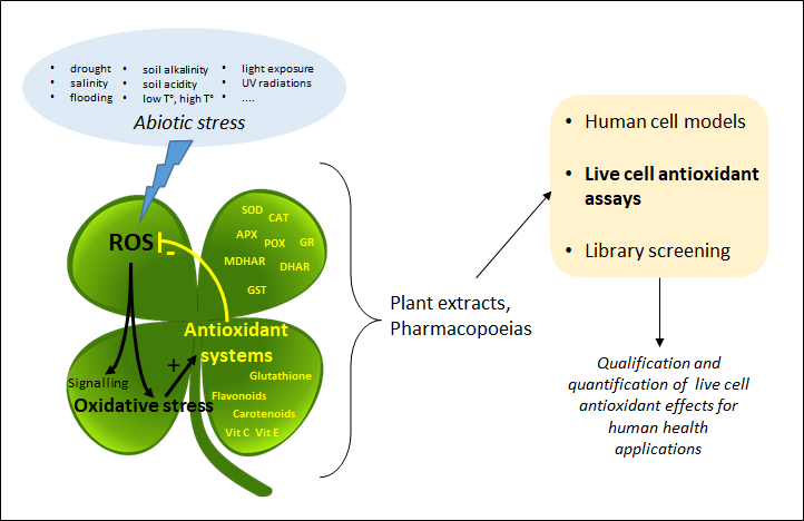
Figure 1. Strategy of valorization of plant extracts through live cell assays. Due to the strong environmental influence, plants produce oxidative stress, which forces them to equip themselves with powerful antioxidant systems. Emergence of new standardized live cell assays now allows for quantification of plant extract antioxidant power from monograph to high-throughput screening studies. SOD: superoxide dismutase; CAT: catalase; APX: ascorbate peroxidase; POX: peroxidases; GR: glutathione reductase; GPX: glutathione peroxidase; (M)DHAR: (mono)dehydroascorbate reductase; GST: glutathione-S-transferase.

2. Cell Antioxidant Assay (CAA)

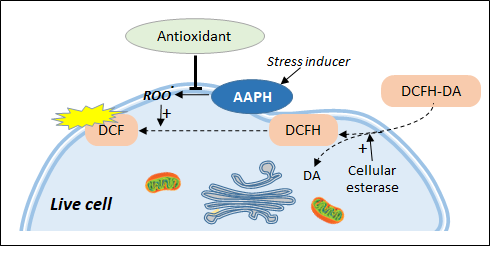
Principle: The cell antioxidant assay (CAA) was until recently the only cell-based assay commonly used for demonstrating antioxidant effects in live cells. The concept of CAA was developed after a series of studies in the 1990s [18][19][20] that led to a standardized approach allowing measurement of intracellular ROSs via the use of a 2′,7′-dichlorofluorescin diacetate (DCFH-DA) probe. DCFH-DA is a nonpolar and nonionic form of 2′,7′-dichlorofluorescin (DCFH) which can easily be transported across the cell membrane. The ester bond is then hydrolyzed by endogenous cellular esterases, bringing the probe back to its reduced (and more oxidizable) form and limiting the newly formed polar DCFH to move back in the extracellular medium (Figure 2). Oxidation of DCFH by ROS eventually leads to the fluorescent 2′,7′-dichlorofluorescein (DCF) form. The procedure was further developed in 2007 by Wolfe and Liu [21] as a live cell antioxidant assay able to analyze the effects of a panel of fruit extracts [22]. Even if DCFH provides a useful way to detect ROS production in the cytosolic compartment, the protocol for a derived antioxidant assay needs the addition of an ROS generator. AAPH (sometimes called ABAP), the free-radical initiator used by Wang and Joseph in their pioneering study [20], became the standard. AAPH is known to spontaneously decompose to form carbon-centered radicals [23] which, in the presence of molecular oxygen, initiate lipid peroxidation by attacking plasma membrane polyunsaturated fatty acids [24].

Figure 2. Live cell antioxidant assays based on chemical stress inducers. DCFH-DA is trapped in the cell in the form of DCFH which can be transformed by peroxidation products into the fluorescent DCF. Antioxidant effect is measured as the ability to inhibit the formation of AAPH-induced lipid peroxidation.

Specificity: Up to date, no action of AAPH has been demonstrated outside the plasma membrane, and CAA results need to be interpreted as the capacity of assayed samples to selectively interfere with plasma membrane-associated lipid peroxidation production [25]. This is also supported by studies on DCFH intracellular location. The traditional view is that the probe diffuses in the cytosol [26] up to the mitochondria [27] but nuclear magnetic resonance (NMR) data using liposomes as a model strongly suggest that DCFH locates within the lipid bilayer and, more precisely, between the lipid chains in the perpendicular direction to the interface [28]. The CAA assay is theoretically adaptable to any cell lines and a simpler version of the assay, called ERYCA (erythrocyte cellular antioxidant activity), has been developed on erythrocytes using AAPH as stressor and light scattering signal instead of DCFH-DA fluorescence as the readout [29]. The approach has been successfully applied to rank antioxidant effects of 34 common tropical fruits [30].
Applications to plant extracts: Among the 25 recent surveyed studies (>2018) (with 12 performed on HepG2 model), only a short list of plant extracts or compounds provided conclusive dose–response data: silybin from cypselea (Silybum marianum L.) [31] and phenolics from black rice [32] in HepG2 cells, isoflavones from black chickpea (Cicer arietinum L.) in both HepG2 and MDA-MB-231 cells, and extracts of Jerusalem artichoke (Helianthus tuberosus) tubers and leaves [33] in HaCaT and BJ fibroblast models.
CAA drawbacks: Despite the high number of studies dealing with the use of the DCFH-DA/AAPH combination to analyze antioxidant effects in live cells, many authors have pointed out severe drawbacks which strongly limit the performance of the approach. First of all, the protocol appears to be difficult to standardize. For instance, there is no consensus on the number of PBS or HBSS buffer washes inserted between extract treatment and AAPH addition, and it has been shown that these washes influence dose–response curves [21]. Despite the cutting off of the acetate group by cellular esterase activity, retention of the probe inside the cell is time-dependent, and part of the DCF fluorescence diffuses to the extracellular compartments, excluding definitive evidence that the observed antioxidant effect actually happens inside the cell. It has been shown that up to 90% of fluorescence can go back to the culture medium after only 1 h of incubation even at low DCFH-DA concentration (11 mM) [34]. Some confusion also comes from the fact that DCF can behave itself as an antioxidant or a prooxidant according to its intracellular concentration [35]. Chemical stressors also expose the cells to massive exogenous radicals, and more physiological stressors such as oleic acid have been applied with some success, but with low signal amplitude, as a surrogate to AAPH in the CAA procedure [36]. Furthermore, the diverse cell lines used in the CAA assay are cultured in diverse culture media with different compositions which are also known to influence DCF fluorescence, possibly due to spontaneous auto-oxidation of DCFH-DA to DCF [37]. Other data showed that AAPH-induced peroxyl radicals increase cytosolic calcium from both extracellular and reticulum endoplasmic compartments [38], and a recent study concluded that low concentration of calcium and magnesium ions in culture medium leads to underestimated CAA results in both HepG2 and CaCo2 cells [39]. The number of cells per well used for CAA analysis is another concern as fluorescence measurement varies with cell density [35]. Last but not least, the CAA assay cannot discriminate between the antioxidant and the cytotoxicity effect of the assayed sample and necessitates the addition of a toxicity assay (usually MTT) to specify which effect is actually observed.

3. AOP1, a New Antioxidant Live Cell Approach Based on Photoinduction
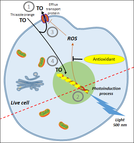
The idea of a new antioxidant live cell assay came from the abovementioned photoinduction process but with biosensors capable of emitting signals linked to the actual concentration of ROS produced by the cells. Simple photosensitizers added to the culture medium that are able to reach the plasma membrane, such as PDT agents, or enter cells have been described in the past. Among them, thiazole orange (TO) is a photosensitizer of the asymmetric cyanine subfamily known to selectively target cytosolic and nuclear 3D structured nucleic acids [40]. TO presents a very interesting property for cell biology; its fluorescence quantum yield remains very low (2 × 10⁻⁴) in the culture medium due to free rotation of its two aromatic rings around the methine bridge that links them [41]. In this situation, energy relaxation occurs on a nonradiative mode via internal conversion through an ultrafast intramolecular twisting (100 fs) at the excited state. This basically means that there is virtually no residual TO fluorescence before the photosensitizer has reached its intracellular target. TO is known to interact with nucleic acids as an intercalator and/or a minor groove binder with an increase of its fluorescence quantum yield to 0.1, denoting a 500-fold gain [42], an increase attributed to a restriction in its torsion capacity [43]. More interesting, a recent electron paramagnetic resonance (EPR) study conducted in HepG2 cells showed that TO acts as a classical photosensitizer producing both 1O2 (type II reaction) and OH (type I reaction) [41]. Lastly, TO presents another quite unique property in live cells; its fluorescence level increases during the irradiation-driven photoinduction in a process called light-up cell system (LUCS) [44]. The intimate mechanisms underlying LUCS have been partially deciphered. TO passively enters the cells but is mainly removed by efflux transport proteins (presumably of the MATE family), limiting its access to nucleic acids and resulting in a low fluorescence level. When light is applied, ROS induced by TO photoactivation alter efflux and/or other cellular functions, perturbating cell homeostasis and triggering a massive entry of TO which progressively saturates nucleic acid binding sites, resulting in a relevant increase of fluorescence level. For the first time, cell ROS level can be precisely controlled, kept at a sublethal level, and quantified by a simple fluorescence measurement [45]. This unique feature led to the development of a new promising antioxidant assay called AOP1 (Figure 3) [46]
Figure 3. AOP1 assay, live cell antioxidant assay based on photo-induced ROS production. (1) Before photoinduction, TO is massively removed from the cell by efflux transport proteins; (2) photoinduction is initiated by an energy transfer from thiazole orange (TO) to molecular oxygen at the triplet state forming singlet oxygen and subsequent free radicals (ROS); (3) ROS alter TO efflux transport and other cell functions; (4) massive entry of TO triggers an increase in fluorescence emission. Effect is measured as the ability of antioxidants to quench ROS production, keeping TO out of the cell and resulting in low fluorescence.
AOP1 protocol includes a run of moderate light applications, each leading to a moderate ROS production and a moderate increase in fluorescence. Antioxidant effect (i.e., the capacity to neutralize intracellular ROS or free radicals) is measured as the ability of extracts/samples to delay or suppress this ROS-induced increase of fluorescence [47]. The antioxidant index is calculated as the integration of measured fluorescent signal over time. AOP1 is to our knowledge the first approach able to quantitatively assess quenching of ROS or free radicals directly produced by living cells. The AOP1 assay has been already applied with success to classify 15 standard antioxidants according to their efficacy concentrations (EC50s) [46] and to assess cellular antioxidant effects of many plant extracts including a phytocomplex of bilberry (Vaccinium myrtillus) [47]. The AOP1 approach presents many benefits over the competitive CAA approach (Table 1). One concern is the availability of an appropriate light source as irradiation energy takes place in the range of 20 mJ/cm2. However, the recent emergence of high-throughput tools for optogenetics led to the development of appropriate 96-well plate illuminators that are either commercially available or produced on a DIY open-source mode [48][49].
Table 1. Features and limits of the two main live cell assays for the measurement of direct antioxidant effects.
CAA Assay AOP1 Assay
Based on the production of AAPH-induced peroxyl radicals Based on the controlled production of 1O2 and free radicals by photoinduction
Measures effects of plasma membrane-based antioxidants Measures effects of intracellular-based antioxidants
No control of ROS production Easy control of ROS production by light intensity; allows monitoring ROS production at a sublethal level (i.e., more physiological concentrations)
Interpretation limited to AAPH effects  
Does not differentiate between antioxidant and cytotoxic effects Can easily discriminate between antioxidant and cytotoxic effects
Results need to be confirmed by performing a cytotoxicity assay (e.g., MTT) No other assay needed
DCFH-DA subject to auto-oxidation Sensor not directly involved in the oxidation process
Subject to cell leakage No cell leakage
Fluorescence levels vary according to cell density No effect of cell density (measure on a ratio mode)
Needs culture medium washes that disrupt cell culture No washes required
Difficult to standardize Easy to standardize
Detection by fluorescence readers Detection by fluorescence readers + illuminator
Limited to adherent cells Works for adherent and suspension cells, and organotypic models

This entry is adapted from the peer-reviewed paper 10.3390/antiox10060944

References

  1. Soares, C.; Carvalho, M.E.A.; Azevedo, R.A.; Fidalgo, F. Plants Facing Oxidative Challenges—A Little Help from the Antioxidant Networks. Environ. Exp. Bot. 2019, 161, 4–25.
  2. Singh, R.; Singh, S.; Parihar, P.; Mishra, R.K.; Tripathi, D.K.; Singh, V.P.; Chauhan, D.K.; Prasad, S.M. Reactive Oxygen Species (ROS): Beneficial Companions of Plants’ Developmental Processes. Front. Plant. Sci. 2016, 7, 1299.
  3. Del Río, L.A. ROS and RNS in Plant Physiology: An Overview. J. Exp. Bot. 2015, 66, 2827–2837.
  4. Sun, Y.; Lu, Y.; Saredy, J.; Wang, X.; Drummer Iv, C.; Shao, Y.; Saaoud, F.; Xu, K.; Liu, M.; Yang, W.Y.; et al. ROS Systems Are a New Integrated Network for Sensing Homeostasis and Alarming Stresses in Organelle Metabolic Processes. Redox Biol. 2020, 37, 101696.
  5. Hasanuzzaman, M.; Bhuyan, M.H.M.B.; Parvin, K.; Bhuiyan, T.F.; Anee, T.I.; Nahar, K.; Hossen, M.S.; Zulfiqar, F.; Alam, M.M.; Fujita, M. Regulation of ROS Metabolism in Plants under Environmental Stress: A Review of Recent Experimental Evidence. Int. J. Mol. Sci. 2020, 21, 8695.
  6. Mittler, R. ROS Are Good. Trends Plant. Sci. 2017, 22, 11–19.
  7. Ramel, F.; Mialoundama, A.S.; Havaux, M. Nonenzymic Carotenoid Oxidation and Photooxidative Stress Signalling in Plants. J. Exp. Bot. 2013, 64, 799–805.
  8. Hernández, I.; Alegre, L.; Van Breusegem, F.; Munné-Bosch, S. How Relevant Are Flavonoids as Antioxidants in Plants? Trends Plant. Sci. 2009, 14, 125–132.
  9. Panche, A.N.; Diwan, A.D.; Chandra, S.R. Flavonoids: An Overview. J. Nutr. Sci. 2016, 5, e47.
  10. Dumanović, J.; Nepovimova, E.; Natić, M.; Kuča, K.; Jaćević, V. The Significance of Reactive Oxygen Species and Antioxidant Defense System in Plants: A Concise Overview. Front. Plant. Sci. 2020, 11, 552969.
  11. Ivanova, A.; Gerasimova, E.; Gazizullina, E. Study of Antioxidant Properties of Agents from the Perspective of Their Action Mechanisms. Molecules 2020, 25, 4251.
  12. Gulcin, İ. Antioxidants and Antioxidant Methods: An Updated Overview. Arch. Toxicol. 2020, 94, 651–715.
  13. Munteanu, I.G.; Apetrei, C. Analytical Methods Used in Determining Antioxidant Activity: A Review. Int. J. Mol. Sci. 2021, 22, 3380.
  14. Pompella, A.; Sies, H.; Wacker, R.; Brouns, F.; Grune, T.; Biesalski, H.K.; Frank, J. The Use of Total Antioxidant Capacity as Surrogate Marker for Food Quality and its Effect on Health is to be Discouraged. Nutrition 2014, 30, 791–793.
  15. Soccio, M.; Laus, M.N.; Alfarano, M.; Pastore, D. The Soybean Lipoxygenase-Fluorescein Reaction may be used to Assess Antioxidant Capacity of Phytochemicals and Serum. Anal. Methods 2016, 8, 4354–4362.
  16. Soccio, M.; Laus, M.N.; Alfarano, M.; Dalfino, G.; Panunzio, M.F.; Pastore, D. Antioxidant/Oxidant Balance as a novel approach to evaluate the effect on serum of long-term intake of plant antioxidant-rich foods. J. Funct. Foods 2018, 40, 778–784.
  17. Franco, R.; Navarro, G.; Martínez-Pinilla, E. Antioxidants versus Food Antioxidant Additives and Food Preservatives. Antioxidants 2019, 8, 542.
  18. Swann, J.D.; Acosta, D. Failure of Gentamicin to Elevate Cellular Malondialdehyde Content or Increase Generation of Intracellular Reactive Oxygen Species in Primary Cultures of Renal Cortical Epithelial Cells. Biochem. Pharm. 1990, 40, 1523–1526.
  19. Rosenkranz, A.R.; Schmaldienst, S.; Stuhlmeier, K.M.; Chen, W.; Knapp, W.; Zlabinger, G.J. A Microplate Assay for the Detection of Oxidative Products Using 2′,7′-Dichlorofluorescin-Diacetate. J. Immunol. Methods 1992, 156, 39–45.
  20. Wang, H.; Joseph, J.A. Quantifying Cellular Oxidative Stress by Dichlorofluorescein Assay Using Microplate Reader. Free Radic. Biol. Med. 1999, 27, 612–616.
  21. Wolfe, K.L.; Liu, R.H. Cellular Antioxidant Activity (CAA) Assay for Assessing Antioxidants, Foods, and Dietary Supplements. J. Agric. Food Chem. 2007, 55, 8896–8907.
  22. Wolfe, K.L.; Kang, X.; He, X.; Dong, M.; Zhang, Q.; Liu, R.H. Cellular Antioxidant Activity of Common Fruits. J. Agric. Food Chem. 2008, 56, 8418–8426.
  23. De la Haba, C.; Palacio, J.R.; Martínez, P.; Morros, A. Effect of Oxidative Stress on Plasma Membrane Fluidity of THP-1 Induced Macrophages. Biochim. Biophys. Acta BBA Biomembr. 2013, 1828, 357–364.
  24. Park, J.E.; Yang, J.-H.; Yoon, S.J.; Lee, J.-H.; Yang, E.S.; Park, J.-W. Lipid Peroxidation-Mediated Cytotoxicity and DNA Damage in U937 Cells. Biochimie 2002, 84, 1199–1205.
  25. Sunitha, D. A Review on Antioxidant Methods. Asian J. Pharm. Clin. Res. 2016, 9, 14–32.
  26. Kellett, M.E.; Greenspan, P.; Pegg, R.B. Modification of the Cellular Antioxidant Activity (CAA) Assay to Study Phenolic Antioxidants in a Caco-2 Cell Line. Food Chem. 2018, 244, 359–363.
  27. Swift, L.M.; Sarvazyan, N. Localization of Dichlorofluorescin in Cardiac Myocytes: Implications for Assessment of Oxidative Stress. Am. J. Physiol. Heart Circ. Physiol. 2000, 278, H982–H990.
  28. Afri, M.; Frimer, A.A.; Cohen, Y. Active Oxygen Chemistry within the Liposomal Bilayer. Part IV: Locating 2′,7′-Dichlorofluorescein (DCF), 2′,7′-Dichlorodihydrofluorescein (DCFH) and 2′,7′-Dichlorodihydrofluorescein Diacetate (DCFH-DA) in the Lipid Bilayer. Chem. Phys. Lipids 2004, 131, 123–133.
  29. González, E.; Vaillant, F.; Rojas, G.; Pérez, A. Novel Semiautomated Method for Assessing in Vitro Cellular Antioxidant Activity Using the Light-Scattering Properties of Human Erythrocytes. J. Agric. Food Chem. 2010, 58, 1455–1461.
  30. González, E.; Vaillant, F.; Pérez, A. In Vitro Cell-Mediated Antioxidant Protection of Human Erythrocytes by Some Common Tropical Fruits. J. Nutr. Food Sci. 2012, 2.
  31. Dobiasová, S.; Řehořová, K.; Kučerová, D.; Biedermann, D.; Káňová, K.; Petrásková, L.; Koucká, K.; Václavíková, R.; Valentová, K.; Ruml, T.; et al. Multidrug Resistance Modulation Activity of Silybin Derivatives and Their Anti-Inflammatory Potential. Antioxidants 2020, 9, 455.
  32. Gong, E.S.; Liu, C.; Li, B.; Zhou, W.; Chen, H.; Li, T.; Wu, J.; Zeng, Z.; Wang, Y.; Si, X.; et al. Phytochemical Profiles of Rice and Their Cellular Antioxidant Activity against ABAP Induced Oxidative Stress in Human Hepatocellular Carcinoma HepG2 Cells. Food Chem. 2020, 318, 126484.
  33. Nizioł-Łukaszewska, Z.; Furman-Toczek, D.; Zagórska-Dziok, M. Antioxidant Activity and Cytotoxicity of Jerusalem Artichoke Tubers and Leaves Extract on HaCaT and BJ Fibroblast Cells. Lipids Health Dis. 2018, 17, 280.
  34. Royall, J.A.; Ischiropoulos, H. Evaluation of 2′,7′-Dichlorofluorescin and Dihydrorhodamine 123 as Fluorescent Probes for Intracellular H2O2 in Cultured Endothelial Cells. Arch. Biochem. Biophys. 1993, 302, 348–355.
  35. Yazdani, M. Concerns in the Application of Fluorescent Probes DCDHF-DA, DHR 123 and DHE to Measure Reactive Oxygen Species in Vitro. Toxicol. Vitr. 2015, 30, 578–582.
  36. Zhang, D.; Xie, L.; Wei, Y.; Liu, Y.; Jia, G.; Zhou, F.; Ji, B. Development of a Cell-Based Antioxidant Activity Assay Using Dietary Fatty Acid as Oxidative Stressor. Food Chem. 2013, 141, 347–356.
  37. Chen, X.; Zhong, Z.; Xu, Z.; Chen, L.; Wang, Y. 2′,7′-Dichlorodihydrofluorescein as a Fluorescent Probe for Reactive Oxygen Species Measurement: Forty Years of Application and Controversy. Free Radic. Res. 2010, 44, 587–604.
  38. Quintanar-Escorza, M.A.; González-Martínez, M.T.; del Pilar, I.-O.M.; Calderón-Salinas, J.V. Oxidative Damage Increases Intracellular Free Calcium [Ca2+]i Concentration in Human Erythrocytes Incubated with Lead. Toxicol. In Vitro 2010, 24, 1338–1346.
  39. Zhou, J.; Gao, G.; Zhang, S.; Wang, H.; Ke, L.; Zhou, J.; Rao, P.; Wang, Q.; Li, J. Influences of Calcium and Magnesium Ions on Cellular Antioxidant Activity (CAA) Determination. Food Chem. 2020, 320, 126625.
  40. Lubitz, I.; Zikich, D.; Kotlyar, A. Specific High-Affinity Binding of Thiazole Orange to Triplex and G-Quadruplex DNA. Biochemistry 2010, 49, 3567–3574.
  41. Karunakaran, V.; Pérez Lustres, J.L.; Zhao, L.; Ernsting, N.P.; Seitz, O. Large Dynamic Stokes Shift of DNA Intercalation Dye Thiazole Orange Has Contribution from a High-Frequency Mode. J. Am. Chem. Soc. 2006, 128, 2954–2962.
  42. Kessel, D. Photodynamic Therapy: A Brief History. J. Clin. Med. 2019, 8, 1581.
  43. Nygren, J.; Svanvik, N.; Kubista, M. The Interactions between the Fluorescent Dye Thiazole Orange and DNA. Biopolymers 1998, 46, 39–51.
  44. Armitage, B.A. Cyanine Dye–Nucleic Acid Interactions. In Heterocyclic Polymethine Dyes; Strekowski, L., Ed.; Topics in Heterocyclic Chemistry; Springer: Berlin/Heidelberg, Germany, 2008; Volume 14, pp. 11–29. ISBN 978-3-540-79063-1.
  45. Derick, S.; Gironde, C.; Perio, P.; Reybier, K.; Nepveu, F.; Jauneau, A.; Furger, C. LUCS (Light-Up Cell System), a Universal High Throughput Assay for Homeostasis Evaluation in Live Cells. Sci. Rep. 2017, 7, 18069.
  46. Gironde, C.; Rigal, M.; Dufour, C.; Furger, C. AOP1, a New Live Cell Assay for the Direct and Quantitative Measure of Intracellular Antioxidant Effects. Antioxidants 2020, 9, 471.
  47. Vigliante, I.; Mannino, G.; Maffei, M.E. OxiCyan®, a Phytocomplex of Bilberry (Vaccinium Myrtillus) and Spirulina (Spirulina Platensis), Exerts Both Direct Antioxidant Activity and Modulation of ARE/Nrf2 Pathway in HepG2 Cells. J. Funct. Foods 2019, 61, 103508.
  48. Bugaj, L.J.; Lim, W.A. High-Throughput Multicolor Optogenetics in Microwell Plates. Nat. Protoc. 2019, 14, 2205–2228.
  49. Thomas, O.S.; Hörner, M.; Weber, W. A Graphical User Interface to Design High-Throughput Optogenetic Experiments with the OptoPlate-96. Nat. Protoc. 2020, 15, 2785–2787.
More
This entry is offline, you can click here to edit this entry!
ScholarVision Creations