Salt Sensation: History
Please note this is an old version of this entry, which may differ significantly from the current revision.
Subjects: Biology
Contributor:

Taste sensation and regulation are highly conserved in insects and mammals. Research conducted over recent decades has yielded major advances in our understanding of the molecular mechanisms underlying the taste sensors for a variety of taste sensations and the processes underlying the regulation of ingestion depending on our internal state. Salt (NaCl) is an essential ingested nutrient. The regulation of internal sodium concentrations for physiological processes, including neuronal activity, fluid volume, acid-base balance, and muscle contraction, are extremely important issues in animal health. Both mammals and flies detect low and high NaCl concentrations as attractive and aversive tastants, respectively. These attractive or aversive behaviors can be modulated by the internal nutrient state. However, the differential encoding of the tastes underlying low and high salt concentrations in the brain remains unclear.

  • Salt sensation
  • salt coding
  • taste perception
  • salt appetite

1. Introduction

In addition to being one of the five basic senses, the taste is also one of the two chemosensations, along with smell [1]. All life forms strive to consume integral nutrients and to steer clear of potential toxic supplements based on their unattractive taste. As an attempt to survive, numerous animals have developed chemoreceptors to determine deleterious chemicals by contact [2].
Both mammals and Drosophila melanogaster show mechanisms for coding the taste of salt. Salt is fundamental for the proper functioning of many physiological processes, including electrolyte homeostasis, muscle contraction, neuronal activity, and nutrient absorption. Although salt is key for the survival of mammals, excessive amounts of salt may cause hypertension and are therefore detrimental. Salt is perceived differently depending on the concentration and is usually attractive below 100 mM and aversive above (the aversiveness intensifies as the concentration increases above this threshold) [3][4][5]. Drosophila is an influential model for functional analysis of the neuronal circuits behind behaviors. Although cardiovascular diseases, abnormal blood pressure, stroke, and hypertension resulting in death are highlight prevalent conditions associated with elevated levels of salt consumption [6], the actual cellular and molecular mechanisms in peripheral tissues and the brain by which high/low salt concentrations are encoded or recognized remain unclear, as is the effect of salt intake on consumption behavior. This review focuses on the studies of Drosophila melanogaster to determine the regulation of salt-sensing neurons in the brain and the effect of salt on physiological functions such as feeding behavior in mammals. The invaluable information obtained in these investigations will improve our understanding of the previously mentioned human diseases. Since all higher animals share innumerable functional and anatomical features of taste transduction, studies in Drosophila and mammals may also address our questions regarding sensory coding. Here, we present a critical and reliable examination of recent literature about salt sensation and regulation in mammals and Drosophila along with professional comment on prospective advancements.

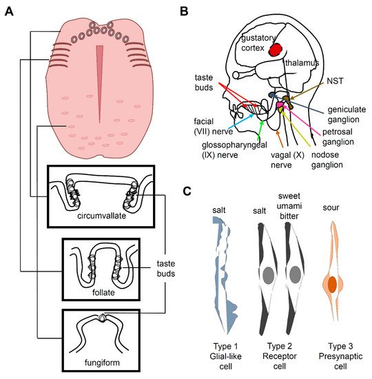
2. Salt Taste in Mammals

Taste buds, which are ovoid structures composed of 50–100 taste receptor cells (TRCs), are located in the tongue and palate epithelium. Circumvallate and foliate papillae contain dense groupings of taste buds at the back or sides of the tongue, respectively, whereas fungiform papillae contain scattered taste buds at the front of the tongue (Figure 1A) [4][7]. TRCs are neuroepithelial cells innervated by pseudounipolar neurons whose cell bodies are located at the petrosal and geniculate ganglia (Figure 1B) [7]. Three morphologically distinct types of cells (I, II, and III) are present in a taste bud and constitute five functional classes of sensory cells, each specialized to detect at least one of the five basic taste qualities, namely, sweet, umami, bitter, salty, and sour (Figure 1C). Type I TRCs are glial-like cells and were earlier proposed to transduce an attractive salty taste [8][9] Type II TRCs express T1R or T2R receptors. Sweet and umami taste receptors are heterodimers of T1R2 + T1R3 and T1R1 + T1R3, respectively [10][11][12]. Bitter taste is mediated by T2Rs [7][13][14][15]. Type III TRCs express otopetrin, a proton channel mediating sour taste [16][17][18]. Type III TRCs also express PKD2L1 and carbonic anhydrase 4 [19][20][21]. However, the cell type associated with the perception of salty taste is controversial, because a recent study reported that sodium-taste cells can be considered as a Type II cell subset [9]. The salty Type II TRCs are distinct from sweet-, umami-, and bitter-sensing Type II TRCs and are characterized by the absence of Trpm5 and the presence of ENaCα. The cells dedicated to attractive sodium taste are positive for ENaC and CALHM1/3 [8].
Figure 1. Taste perception in mammals. (A) Structure of a human tongue and the taste buds housing fungiform, folate, and circumvallate taste receptor cells (TRCs). (B) The neural pathway for human taste. The taste buds on the tongue and pharynx send gustatory information to the geniculate ganglion via the facial nerve VII, the petrosal ganglion via the glossopharyngeal nerve, and the nodose ganglion via the vagal nerve. This taste information is further transmitted to the thalamus and gustatory cortex via the nucleus of the solitary tract (NST) in the medulla oblongata. (C) Three morphologically distinct cell types (I, II, and III) are present in a taste bud and constitute five functional classes of sensory cells, each specialized to detect at least one of the five basic taste qualities. Type I TRCs are glial-like cells and earlier proposed to transduce an attractive salty taste, although it is controversial. Type II TRCs express T1R or T2R receptors and mediate the detection of sweet, umami, and bitter compounds. Distinct Type II TRCs from the sweet, umami and bitter-sensing TRCs are salt sensitive. Type III TRCs, which are presynaptic cells, express otopetrin, a proton channel for sour taste.
Most of the taste-related information is transmitted via the facial nerve (VII) and the glossopharyngeal nerve (IX) (Figure 1B). The chorda tympani is a branch of the facial nerve that transmits taste messages from the anterior region of the tongue, which contains the fungiform papillae. The taste messages from the circumvallate papillae are transmitted via the glossopharyngeal nerve to the nucleus of the solitary tract (NST) in the medulla oblongata, while the foliate papillae send information via both VII and IX cranial nerves [22][23][24]. When the superficial petrosal and glossopharyngeal nerves are abolished, the chorda tympani nerve occupies the space in the terminal field, which indicates that these two nerves compete or prune axon terminals to occupy the terminal space during development [25]. The taste information in the NST passes to the thalamus, which projects to the cerebral cortex. The taste message is stored in the primary gustatory cortex in the insula and transported to the somatosensory cortex of the postcentral gyrus specifically dedicated to the tongue. This message is also carried to the prefrontal cortex, which coordinates taste association and perception of flavor.
Salt responses are recorded from the chorda tympani nerve [18]. Depending on salt concentration, animals have two distinctive behaviors that are well reported in rodents: attraction (<100 mM) and aversion (>300 mM). At low concentrations, mice consume salt, but their behavior and neural responses are largely blocked by amiloride [26]. In contrast, at high concentrations of salt, mice exhibit innate aversion, and these responses are unaffected by amiloride. Thus, a low concentration of salt is sensitive and a high concentration of salt is insensitive to the diuretic amiloride [27]. Because epithelial Na+ channels (ENaCs) are specifically inhibited by amiloride, they have been proposed to participate in low-concentration salt taste [27][28]. ENaCs are non-voltage-gated and constitutively active channels that are highly selective of Na+ and Li+ over other monovalent cations such as K+ [27]. An ENaC is composed of three subunits [29], all of which are expressed in a small number of unique taste cells, with some being uncategorized TRCs [3]. In humans, the salt taste is amiloride-insensitive [30]. In addition to α, β, and γ ENaC subunits, humans express an additional δ ENaC subunit [31]. Heterologous expression of fully functional human α ENaC and δ ENaC was achieved in Xenopus oocytes [31][32][33]. This may provide evidence that ENaC functions in salt sensation. Therefore, further studies are required to identify evolutionary changes in α, β, γ ENaCs in humans. ENaCs also play a major role in body fluid and blood pressure regulation [34]. The δ-ENaC is also expressed in the brain, pancreas, testis, and ovary. Therefore, it is interesting to find any other roles of ENaC in different organs. Amiloride-insensitive high-salt transduction constitutes ~80% of the high-salt responses recorded in the chorda tympani nerve in mice [35]. Furthermore, amiloride-insensitive high-salt responses are detected in type II TRCs, but not in type III TRCs in the fungiform papillae [36]. In type II TRCs, the salt response is highly dependent on the presence of Cl-, which is called “anion effect,” and involves the use of internal Ca2+ stores, whereas type III TRCs utilize voltage-gated Ca2+ channels activated by external Ca2+. Cl, not Na+, plays a major role in the amiloride-insensitive high-salt responses [36], but the contribution of the cation remains unclear. In the circumvallate papilla, the amiloride-insensitive high salt response is mediated in type III TRCs through either an anion-selective or osmolality-sensitive channel [30][37]. Recent studies in the field of salt sensation have shown substantial progress in our understanding of the effects of low and high salt concentrations on peripheral responses in the tongue. Therefore, future studies in this regard should be focused on determining how excessive salt intake and hypertension can be avoided using the salt taste sensation.

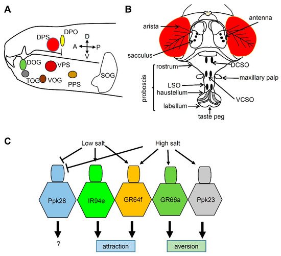
3. Salt Taste in Drosophila

The taste system in adult insects spreads throughout the body and includes the mouth, legs, wings, and ovipositor, where taste sensilla are located, although it is mainly distributed in the head region during the larval stage (Figure 2A) [38][39]. The mouth part is composed of the external organ, the labellum, and the internal organ, the pharynx (Figure 2B). The 31 taste sensilla in the labellum are classified into long (L), intermediate (I), and short (S) bristles, depending on their length. Each sensillum contains 2–4 gustatory receptor neurons (GRNs).
Figure 2. Chemosensory organs and taste sensation in Drosophila melanogaster. (A) The larval anterior part showing the chemosensory organs. DOG: dorsal organ ganglion, DPS: dorsal pharyngeal sensilla, DPO: dorsal pharyngeal organ, TOG: terminal organ ganglion, VOG: ventral organ ganglion, VPS: ventral pharyngeal sensilla, PPS: posterior pharyngeal sensilla. SOG (suboesophageal ganglion) indicates the first gustatory center in the brain. The orientation of the larvae is indicated by arrows. A: anterior, P: posterior, D: dorsal, V: ventral. (B) The adult fly head showing gustatory organs (labellum, taste peg, labial sense organ [LSO], dorsal cibarial sense organ [DCSO], and ventral cibarial sense organ [VCSO]) and olfactory organs (antenna and maxillary palp). Arista and sacculus sense temperature and humidity, respectively. (C) Fly gustatory receptor neurons (GRNs) can be divided into five classes based on the reporter expression. Low salt activates IR94e- and GR64f-positive GRNs, but inhibits water-sensing Ppk28 GRNs by osmolarity. High salt activates three types of GRNs, namely, GR64f-, GR66a-, and Ppk23-positive GRNs, but inhibits water-sensing Ppk28 GRNs.
Flies can sense five basic tastes—sweet, umami, bitter, salty, and sour—by utilizing chemoreceptors such as gustatory receptors (GRs), ionotropic receptors (IRs), transient receptor potential channels (TRPs), and pickpockets (PPKs) [40]. Flies enjoy ingesting low concentrations of salt but avoid high concentrations as humans do [41]. Among chemoreceptors, PPKs and IRs seem to meditate salt response in Drosophila during the larval and adult stages, respectively. Flies have 31 family members called PPKs, which are homologs of the amiloride-sensitive degenerin/epithelial Na+ channels (DEG/ENaC) [42]. Two PPKs, PPK11 and PPK19, detect Na+ and K+ during the larval stage [43][44]. However, it is not clear whether PPK11 and PPK19 are required for sensing salts during the adult stage. IR76b is required for attractive behavior to low salt concentrations [41], and Ir76b mutants have no electrophysiological response to low salt concentrations. IR25a and IR76b are also reported to play widespread roles in sensing low-salt, high-salt, and calcium levels, suggesting that IRs are major sensors that detect salt in insects [43][45][46]. Many genetic tools are available to express artificial reporters in flies by using the GAL4/UAS system, including GRN-specific reporters such as Gr64f-GAL4 (sugar-sensing) [47], Gr66a-GAL4 (bitter-sensing) [48], ppk28-GAL4 (water-sensing, osmolarity sensitive) [49], ppk23-GAL4 (calcium-sensing) [46], and Ir94e-GAL4 (low salt-sensing) [43]. The cellular basis of salt-responsive behaviors was studied by expressing the Kir2.1 potassium channel in specific GRNs [43]. This study suggested that Gr64f-GAL4- and Ir94e-GAL4-expressing GRNs have low salt tuning properties and cause salt attraction, whereas Gr66a-GAL4- and ppk23-GAL4-expressing GRNs (especially the glutaminergic neurons of ppk23+ GRNs) have high salt properties and mediate salt aversion (Figure 2C). However, the authors of that study did not test whether these GRNs mediate salt response during the larval stage. Furthermore, Gr2a-positive GRNs in the pharynx are involved in the aversion against high salt, although the Gr2a mutant shows normal high-salt aversion [50]. This indicates that complex coding mechanisms to sense salt are separately mediated by the labellum and pharynx. Future studies should investigate the full repertoire of low- and high-salt sensors.

This entry is adapted from the peer-reviewed paper 10.3390/metabo11030175

References

  1. Matthews, R.W. The Insects: Structure and Function; Oxford University Press: Oxford, UK, 1971.
  2. Joseph, R.M.; Carlson, J.R. Drosophila Chemoreceptors: A Molecular Interface between the Chemical World and the Brain. Trends Genet. 2015, 31, 683–695.
  3. Chandrashekar, J.; Kuhn, C.; Oka, Y.; Yarmolinsky, D.A.; Hummler, E.; Ryba, N.J.; Zuker, C.S. The cells and peripheral representation of sodium taste in mice. Nature 2010, 464, 297–301.
  4. Lindemann, B. Receptors and transduction in taste. Nature 2001, 413, 219–225.
  5. Oka, Y.; Butnaru, M.; von Buchholtz, L.; Ryba, N.J.; Zuker, C.S. High salt recruits aversive taste pathways. Nature 2013, 494, 472–475.
  6. Kaushik, S.; Kumar, R.; Kain, P. Salt an Essential Nutrient: Advances in Understanding Salt Taste Detection Using Drosophila as a Model System. J. Exp. Neurosci. 2018, 12, 1179069518806894.
  7. Chandrashekar, J.; Hoon, M.A.; Ryba, N.J.; Zuker, C.S. The receptors and cells for mammalian taste. Nature 2006, 444, 288–294.
  8. Nomura, K.; Nakanishi, M.; Ishidate, F.; Iwata, K.; Taruno, A. All-electrical Ca2+-independent signal transduction mediates attractive sodium taste in taste buds. Neuron 2020, 106, 816–829.
  9. Ohmoto, M.; Jyotaki, M.; Foskett, J.K.; Matsumoto, I. Sodium–Taste Cells Require Skn-1a for Generation and Share Molecular Features with Sweet, Umami, and Bitter Taste Cells. Eneuro 2020, 7.
  10. Zhao, G.Q.; Zhang, Y.; Hoon, M.A.; Chandrashekar, J.; Erlenbach, I.; Ryba, N.J.; Zuker, C.S. The receptors for mammalian sweet and umami taste. Cell 2003, 115, 255–266.
  11. Xu, H.; Staszewski, L.; Tang, H.; Adler, E.; Zoller, M.; Li, X. Different functional roles of T1R subunits in the heteromeric taste receptors. Proc. Natl. Acad. Sci. USA 2004, 101, 14258–14263.
  12. Nelson, G.; Chandrashekar, J.; Hoon, M.A.; Feng, L.; Zhao, G.; Ryba, N.J.; Zuker, C.S. An amino-acid taste receptor. Nature 2002, 416, 199–202.
  13. Vegezzi, G.; Anselmi, L.; Huynh, J.; Barocelli, E.; Rozengurt, E.; Raybould, H.; Sternini, C. Diet-induced regulation of bitter taste receptor subtypes in the mouse gastrointestinal tract. PLoS ONE 2014, 9, e107732.
  14. Toda, Y.; Nakagita, T.; Hayakawa, T.; Okada, S.; Narukawa, M.; Imai, H.; Ishimaru, Y.; Misaka, T. Two distinct determinants of ligand specificity in T1R1/T1R3 (the umami taste receptor). J. Biol. Chem. 2013, 288, 36863–36877.
  15. Avau, B.; Rotondo, A.; Thijs, T.; Andrews, C.N.; Janssen, P.; Tack, J.; Depoortere, I. Targeting extra-oral bitter taste receptors modulates gastrointestinal motility with effects on satiation. Sci. Rep. 2015, 5, 1–12.
  16. Liman, E.R.; Zhang, Y.V.; Montell, C. Peripheral Coding of Taste. Neuron 2014, 81, 984–1000.
  17. Roper, S.D.; Chaudhari, N. Taste buds: Cells, signals and synapses. Nat. Rev. Neurosci. 2017, 18, 485–497.
  18. Hill, D.L.; Mistretta, C.M.; Bradley, R.M. Developmental changes in taste response characteristics of rat single chorda tympani fibers. J. Neurosci. 1982, 2, 782–790.
  19. LopezJimenez, N.D.; Cavenagh, M.M.; Sainz, E.; Cruz-Ithier, M.A.; Battey, J.F.; Sullivan, S.L. Two members of the TRPP family of ion channels, Pkd1l3 and Pkd2l1, are co-expressed in a subset of taste receptor cells. J. Neurochem. 2006, 98, 68–77.
  20. Kataoka, S.; Yang, R.; Ishimaru, Y.; Matsunami, H.; Sévigny, J.; Kinnamon, J.C.; Finger, T.E. The candidate sour taste receptor, PKD2L1, is expressed by type III taste cells in the mouse. Chem. Senses 2008, 33, 243–254.
  21. Lossow, K.; Hermans-Borgmeyer, I.; Behrens, M.; Meyerhof, W. Genetic labeling of Car4-expressing cells reveals subpopulations of type III taste cells. Chem. Senses 2017, 42, 747–758.
  22. AlJulaih, G.H.; Lasrado, S. Anatomy, Head and Neck, Tongue Taste Buds; Statpearls: Treasure Island, FL, USA, 2019.
  23. Breslin, P.A.; Spector, A.C. Mammalian taste perception. Curr. Biol. 2008, 18, R148–R155.
  24. Adler, E.; Hoon, M.A.; Mueller, K.L.; Chandrashekar, J.; Ryba, N.J.; Zuker, C.S. A novel family of mammalian taste receptors. Cell 2000, 100, 693–702.
  25. Corson, S.L.; Hill, D.L. Chorda Tympani Nerve Terminal Field Maturation and Maintenance Is Severely Altered Following Changes to Gustatory Nerve Input to the Nucleus of the Solitary Tract. J. Neurosci. 2011, 31, 7591–7603.
  26. Bachmanov, A.A.; Beauchamp, G.K.; Tordoff, M.G. Voluntary consumption of NaCl, KCl, CaCl 2, and NH 4 Cl solutions by 28 mouse strains. Behav. Genet. 2002, 32, 445–457.
  27. Brand, J.G.; Teeter, J.H.; Silver, W.L. Inhibition by amiloride of chorda tympani responses evoked by monovalent salts. Brain Res. 1985, 334, 207–214.
  28. Heck, G.L.; Mierson, S.; DeSimone, J.A. Salt taste transduction occurs through an amiloride-sensitive sodium transport pathway. Science 1984, 223, 403–405.
  29. Canessa, C.M.; Schild, L.; Buell, G.; Thorens, B.; Gautschi, I.; Horisberger, J.-D.; Rossier, B.C. Amiloride-sensitive epithelial Na+ channel is made of three homologous subunits. Nature 1994, 367, 463–467.
  30. Lewandowski, B.C.; Sukumaran, S.K.; Margolskee, R.F.; Bachmanov, A.A. Amiloride-Insensitive Salt Taste Is Mediated by Two Populations of Type III Taste Cells with Distinct Transduction Mechanisms. J. Neurosci. 2016, 36, 1942–1953.
  31. Grant, G.J.; Coca, C.; Zhao, X.-M.; Helms, M.N. Oxidized Glutathione Increases Delta-Subunit Expressing Epithelial Sodium Channel Activity in Xenopus laevis Oocytes. Emed. Res. 2020, 2, 100008.
  32. Wichmann, L.; Vowinkel, K.S.; Perniss, A.; Manzini, I.; Althaus, M. Incorporation of the δ-subunit into the epithelial sodium channel (ENaC) generates protease-resistant ENaCs in Xenopus laevis. J. Biol. Chem. 2018, 293, 6647–6658.
  33. Baldin, J.-P.; Barth, D.; Fronius, M. Epithelial na+ channel (ENaC) Formed by one or two subunits forms functional channels that respond to shear force. Front. Physiol. 2020, 11, 141.
  34. Pitzer, A.L.; Van Beusecum, J.P.; Kleyman, T.R.; Kirabo, A. ENaC in Salt-Sensitive Hypertension: Kidney and Beyond. Curr. Hypertens. Rep. 2020, 22, 1–10.
  35. Lu, B.; Breza, J.M.; Contreras, R.J. Temperature Influences Chorda Tympani Nerve Responses to Sweet, Salty, Sour, Umami, and Bitter stimuli in mice. Chem. Senses 2016, 41, 727–736.
  36. Roebber, J.K.; Roper, S.D.; Chaudhari, N. The Role of the Anion in Salt (NaCl) Detection by Mouse Taste Buds. J. Neurosci. 2019, 39, 6224–6232.
  37. Ninomiya, Y. Reinnervation of cross-regenerated gustatory nerve fibers into amiloride-sensitive and amiloride-insensitive taste receptor cells. Proc. Natl. Acad. Sci. USA 1998, 95, 5347–5350.
  38. Stocker, R.F. The organization of the chemosensory system in Drosophila melanogaster: A rewiew. Cell Tissue Res. 1994, 275, 3–26.
  39. Lee, Y.; Poudel, S. Taste sensation in Drosophila melanogaster. Hanyang Med. Rev. 2014, 34, 130–136.
  40. Rimal, S.; Lee, Y. The multidimensional ionotropic receptors of Drosophila melanogaster. Insect Mol. Biol. 2018, 27, 1–7.
  41. Zhang, Y.V.; Ni, J.; Montell, C. The Molecular Basis for Attractive Salt-Taste Coding in Drosophila. Science 2013, 340, 1334–1338.
  42. Zelle, K.M.; Lu, B.; Pyfrom, S.C.; Ben-Shahar, Y. The Genetic Architecture of Degenerin/Epithelial Sodium Channels in Drosophila. G3 Genes Genomes Genet. 2013, 3, 441–450.
  43. Jaeger, A.H.; Stanley, M.; Weiss, Z.F.; Musso, P.-Y.; Chan, R.C.; Zhang, H.; Feldman-Kiss, D.; Gordon, M.D. A complex peripheral code for salt taste in Drosophila. Elife 2018, 7, e37167.
  44. Liu, L.; Johnson, W.A.; Welsh, M.J. Drosophila DEG/ENaC pickpocket genes are expressed in the tracheal system, where they may be involved in liquid clearance. Proc. Natl. Acad. Sci. USA 2003, 100, 2128–2133.
  45. Lee, M.J.; Sung, H.Y.; Jo, H.; Kim, H.-W.; Choi, M.S.; Kwon, J.Y.; Kang, K. Ionotropic Receptor 76b Is Required for Gustatory Aversion to excessive Na+ in Drosophila. Mol. Cells 2017, 40, 787.
  46. Lee, Y.; Poudel, S.; Kim, Y.; Thakur, D.; Montell, C. Calcium Taste Avoidance in Drosophila. Neuron 2018, 97, 67–74.e64.
  47. Jiao, Y.; Moon, S.J.; Wang, X.; Ren, Q.; Montell, C. Gr64f Is Required in Combination with Other Gustatory Receptors for Sugar Detection in Drosophila. Curr. Biol. 2008, 18, 1797–1801.
  48. Moon, S.J.; Köttgen, M.; Jiao, Y.; Xu, H.; Montell, C. A Taste Receptor Required for the Caffeine Response In Vivo. Curr. Biol. 2006, 16, 1812–1817.
  49. Cameron, P.; Hiroi, M.; Ngai, J.; Scott, K. The molecular basis for water taste in Drosophila. Nature 2010, 465, 91–95.
  50. Kim, H.; Jeong, Y.T.; Choi, M.S.; Choi, J.; Moon, S.J.; Kwon, J.Y. Involvement of a Gr2a-Expressing Drosophila Pharyngeal Gustatory Receptor Neuron in Regulation of Aversion to High-Salt Foods. Mol. Cells 2017, 40, 331–338.
More
This entry is offline, you can click here to edit this entry!