Silver Nanoparticles Green Chemistry Synthesis: History
Please note this is an old version of this entry, which may differ significantly from the current revision.
Subjects: Oncology
Contributor:

Nanobiotechnology has grown rapidly and become an integral part of modern disease diagnosis and treatment. Biosynthesized silver nanoparticles (AgNPs) are a class of eco-friendly, cost-effective and biocompatible agents that have attracted attention for their possible biomedical and bioengineering applications. Like many other inorganic and organic nanoparticles, such as AuNPs, iron oxide and quantum dots, AgNPs have also been widely studied as components of advanced anticancer agents in order to better manage cancer in the clinic. AgNPs are typically produced by the action of reducing reagents on silver ions. In addition to numerous laboratory-based methods for reduction of silver ions, living organisms and natural products can be effective and superior source for synthesis of AgNPs precursors. Currently, plants, bacteria and fungi can afford biogenic AgNPs precursors with diverse geometries and surface properties.

  • silver nanoparticles
  • green chemistry
  • cancer
  • anti-cancer effect

Note: The following contents are extract from your paper. The entry will be online only after author check and submit it.

1. Introduction

Cancer is one of the leading causes of mortality, resulting in one in six deaths (or 9.6 million people) in 2018 alone; however, 70% of deaths from cancer occur in middle- and low-income countries [1]. Surgery, chemotherapy, radiation therapy and hormone therapy are the most common approaches for cancer treatment and management. In recent years, therapeutic and diagnostic approaches based on nanotechnology have shown potential to improve cancer therapy [2]. Cancer nanotechnology unfolded a newer horizon of interdisciplinary research across chemistry, medicine, engineering and biology focusing on the major advancement in cancer detection, diagnosis and treatment [3]. In recent years, nanoparticles (NPs) have gained an immense scientific attraction due to their large surface area to volume ratio and high reactivity with unmatchable properties [4]. Recently, nanotechnology-based anticancer drugs, such as Abraxane® (Celgene, Summit, NJ, USA), Doxil® (Johnson & Johnson, New Brunswick, NJ, USA) and Myocet (Perrigo, Dublin, Ireland), have been approved by the US Food and Drug Administration for clinical use [5].
Particles with at least one dimension smaller than 100 nm are defined as NPs. Their physical (e.g., plasmon resonance and fluorescent enhancement) and chemical (e.g., catalytic activity enhancement) properties arise from their surface geometry, which determines the area/volume ratio. When NPs diameter decreases for a spherical particle, the surface area increases proportionately to the square of diameter and there is a resulting increase of surface activity compared to bulk materials with larger dimensions. In some cases, a decrease in size combined with an increase in the area can improve material biocompatibility [6]. Although nanotechnology is one of the most promising research fields and is currently viewed as a frontier in medicine, only a few nanoproducts are currently considered for bio-application due to their potential toxicity and unknown safety. Silver, a precious metal, has been widely used in medicine since ancient times. Hippocrates, the father of modern medicine, believed that silver could treat disease and promote wound healing [7]. It has fascinating material properties, which are cost-effective as well as abundant in nature compared to other precious metals. Previous studies revealed that the physical, optical and catalytic properties of silver nanoparticles (AgNPs) are strongly influenced by their size, distribution, morphologic shape and surface properties [8]. Hereafter, AgNPs have received considerable attention from scientists for their unique properties [9].
Metallic NPs such as silver, gold and platinum NPs have been widely tested in humans. For medicinal applications, NPs synthesis must be biocompatible and either non-toxic or low-toxicity protocols should be used. The most common method which has been used to produce AgNPs is chemical synthesis, recruiting reagents whose function is to reduce the silver ions and stabilize the nanoparticles. However, these reagents are toxic and may have potential health hazards [10]. In addition, these production methods are usually expensive and labor-intensive. To address this issue, approaches for AgNPs synthesis through green chemistry (such as the use of biologic sources) have shown great promise recently. During the past decade, it has been demonstrated that many biologic systems, including plants, algae, bacteria, yeast and fungi can transform inorganic metal ions into metal nanoparticles via the reductive capacities of the proteins and metabolites present in these organisms. Biologic methods of AgNPs synthesis have been reported using plants [11,12], bacteria [13], fungi [14,15], seaweed [16], algae [17] and lichen [18]. These eco-friendly methods can present alternatives methods to chemical and physical syntheses of AgNPs. Physicochemical properties of AgNPs can have a significant impact on their biologic activity, therefore particle characterization is normally completed after synthesis [19]. In addition, AgNPs synthesized using biologic approaches typically retain a homogenous chemical composition with few defects [20]. These AgNPs show promising activity against a range of cancer cell lines. Such eco-friendly methods provide alternatives to the traditional chemical and physical synthesis of AgNPs for use in anti-cancer treatments.
AgNPs can cause cytotoxicity of the cancer cells by altering their morphology and reducing the viability along with oxidative stress in different cancer cells [21,22]. In addition, AgNPs play a pivotal role in tumor control via their cytotoxic effects [23]. The purpose of this review is to provide an overview of the main published studies concerning the use of biologic synthesis of silver nanoparticles and the applications of these materials in different cancer cell lines. Several biologic synthesis mechanisms of AgNPs are discussed together with the importance of biologically synthesized AgNPs on cancer cell lines.

2. Properties of AgNPs

Unique physicochemical properties of AgNPs provides them the ease of entrance into the cells and gives them the ability to efficiently interact with biomolecules inside the cells or on the cell surfaces [24]. Some of the crucial properties of the AgNPs are described below.

2.1. Shape and Size

While picking a drug material for the treatment of certain disease types including cancer, hemocompatibility of that material should be kept in mind [25]. Kwon et al. demonstrated the minimization of the damage to membrane including hemolysis, potassium efflux, protein leakage and alterations in cell shape and membrane fragility by the use of AgNPs of specific shape and size [26]. Therefore, AgNPs have been evidenced to have great potential in anti-cancer activity since they show selective participation in the interruption of mitochondrial respiratory chain that leads to the production of reactive oxygen species (ROS) and disruption of adenosine triphosphate (ATP) synthesis, thus resulting in nucleic acid damage [27]. Various studies have established that temperature, pH of the solution, precursor concentration, the molar ratio of capping agent to that of precursor, the types of reducing agents, the strength of chemical interaction between the precursor and different crystallographic planes of AgNPs and the synthesis method determine the size and shape of the AgNPs [28]. Depending on the synthesis methods, a range of particle sizes and shapes (e.g., cubes, prisms, spheres, rods, wires, plates, etc.) can be obtained [29]. For instance, in the synthesis of AgNPs from the bacteria Xanthomonas oryzae, spherical as well as triangular and rod-shaped particles with an average size of 14.86 nm were obtained depending on the experimental conditions [30], whereas bacterial synthesis from Pseudomonas stutzeri AG259 resulted in triangular and hexagonal particles with an average size of 200 nm [31]. In the fungal synthesis from Fusarium acuminatum, spherical AgNPs in the range of 5–40 nm (average size 13 nm) were reported [32]. On the other hand, AgNPs synthesis from the fungus Trichoderma viride, spherical and sporadically rod-like particles in the range of 10–40 nm at 27 °C. Decreasing the temperature to 10 °C increased the particle size to 80–100 nm [15]. Studies have established that low reaction temperatures can facilitate the formation of two-dimensional nanostructures [33]. To underscore the range of sizes and shapes of AgNPs which plant-based green synthesis can yield, the study on Nelumbo nucifera can be used as an example. Synthesis using the leaf extract of this plant resulted in AgNPs with spherical, triangular, truncated triangular and decahedral morphologies. Sizes of the particles ranged between 25 and 80 mm averaging 45 mm [34]. Vilchis-Nestor et al. reported more spherical and larger AgNPs under ambient conditions with the increase in the concentration of the Camellia sinensis (green tea) extracts [35].

2.2. Optical Properties

AgNPs efficiently interact with light through a phenomenon known as surface plasmon resonance (SPR) more efficiently than any known organic or inorganic chromophores. This strong interaction is a result of both the restriction of a large density of conducting electrons to smaller dimensions compared to the mean free path and the unique frequency dependence of the dielectric function in metallic silver. The combination of these two factors gives rise to SPR related properties. The size and shape of NPs and the dielectric function of the surrounding medium determines the frequency and resonance strength [36]. The light-interaction cross-sections of AgNPs is dependent on the photon electric field which can extend up to 10 times larger than the geometric cross-sections of the AgNPs. As such, these structures are capable of interaction with light photons that are not physically incident upon them [37,38]. Because of the unique optical characteristics of the AgNPs, non-invasive techniques like dark-field microscopy or surface-enhanced Raman Scattering can be utilized for the inspection of their tracking and cellular uptake [39]. Certain optical characteristics of AgNPs can be modified to obtain interesting results. For example, the absorption spectrum of AgNPs is tunable through careful optimization of the synthesis conditions to the near-infrared region. This eliminates the interference from tissue auto-fluorescence, resulting in NPs which have great promise in tumor-targeting and deep tissue-imaging [40].

2.3. Electrical Properties

One of the important electrical properties of AgNPs in a colloidal suspension is zeta potential which is influenced by the size of the NPs. AgNPs possess a large absolute value of zeta potential corresponding to a high surface electrical charge which can result in a strong repulsive force among particles to resist agglomeration [41]. Agglomeration needs to be suppressed at any cost to prevent the growth of the particles from nanoscale to microscale. It has been established that a high zeta potential value of around −25 mV ensures a large enough energy barrier for the stabilization of the nanosuspension. For example, Sankar et al. reported a zeta potential of about −26 ± 0.77 mV for the biosynthesis of AgNPs with an average size of 136 ± 10.09 nm from Origanum vulgare [42].

3. Biologic Synthesis of AgNPs

Both “top-down” and “bottom-up” approaches can be adopted for NPs synthesis, including AgNPs [43]. A top-down approach involves reducing particle size of an appropriate starting material until NPs are obtained. Different physical and chemical processes like ball milling, chemical etching, laser ablation and sputtering are known top-down approaches [44]. However, a major drawback of top-down approaches is the occurrence of surface defects on the NPs [45]. Conversely, a bottom-up approach produces NPs by assembling smaller entities like atoms, molecules or particles to assemble the NPs [46]. Bottom-up methods are typically chemical or biologic in nature (e.g., sol-gel processes, spray pyrolysis, electrochemical precipitation and laser pyrolysis) [47].
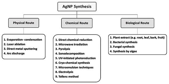
The downside of most AgNPs synthesis protocols involving chemical and physical methods include associated high cost and the use of toxic and potentially hazardous materials which may pose environmental and biologic threats (Figure 1). NPs produced through chemical synthesis are often not suitable for medical applications because of toxic substances absorbed onto the NPs surfaces. Commercial AgNPs have to be handled and must be cost-effective for practical purposes. Therefore, alternative synthetic methods are required that are environmentally sound and economically feasible. The pursuit of such methods has led to the investigation of biologic processes for synthesis [48]. In biologic synthesis of AgNPs, molecules produced by living organisms (e.g., bacteria, fungi or plants), act as reducing and stabilizing agents [49]. The microbial enzymes or the plant phytochemicals possessing antioxidant or reducing properties are mainly responsible for NP biologic synthesis [50].
Figure 1. Different routes of AgNPs synthesis.

3.1. Bacterial Synthesis of AgNPs

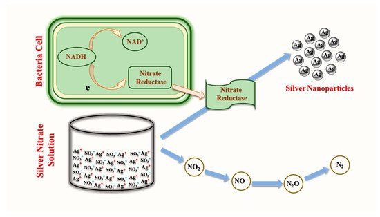
Biosynthesis of NPs may occur extracellularly or intracellularly. A widely utilized extracellular mechanism for AgNPs synthesis via bacteria employs NADH-dependent nitrate reductase-mediated reduction of silver ion (Ag+) by several bacteria species, such as Streptomyces sp. LK3 [51] and Bacillus licheniformis [52]. During reduction, nitrate ions (NO3) in silver nitrate (AgNO3) are reduced to nitrite (NO2) by accepting two protons then releasing two electrons and water. Electrons released in this reaction are transferred to the Ag+ ion to form elemental silver (Ag0) [53]. This mechanism is possibly dependent on electron transport pathways and enzymatic metal reduction processes (Figure 2).
Figure 2. Extracellular enzymatic bacterial synthesis of AgNPs (modified from [54]).
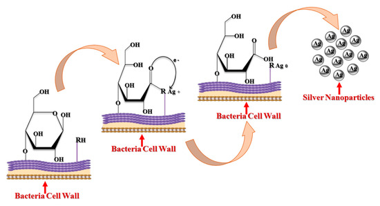
However, enzymes are not always necessary for bacterial synthesis of AgNPs. For example, non-enzymatic intracellular synthesis was reported in Lactobacillus A09, where Ag+ reduction occurred on the bacterial cell surface [53]. Gram-positive bacterial cell walls, such as those of Lactobacillus A09, contain numerous anionic surface groups. These groups can provide silver ion biosorption sites [55]. The pH of the surrounding medium decreased slightly after adsorption of Ag+ on Lactobacillus A09, suggesting competition between proton and silver ion binding to negatively charged sites [56]. Potentially an increase in pH can open bacteria cell wall monosaccharide rings and oxidize these moieties to open-chain aldehydes. At the same time, dissociation of protons from protonated anionic functional groups (–RH) creates negatively charged Ag+ adsorption sites on the cell surface. The two electron released while forming the aldehyde group from an alcohol can reduce Ag+ ions to elemental Ag0 [57]. The steps involved in the bacterial synthesis of AgNPs are mediated through glucose ring-opening (Figure 3).
Figure 3. Intracellular non-enzymatic bacterial synthesis of AgNPs (modified from [57]).

3.2. Fungal Synthesis of AgNPs

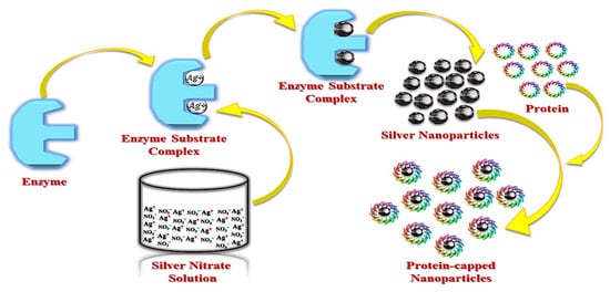
Due to their inherent ability to efficiently secrete protein, fungi can produce substantially larger amounts of AgNPs than bacteria [58]. However, the exact mechanism by which fungi synthesize AgNPs is not completely understood. Mukherjee et al. hypothesized that the following steps may occur during synthesis in Verticillium model system: Ag+ ions interact electrostatically with negatively charged carboxylate groups in the mycelial cell wall and are trapped at the cell surface. Subsequently, trapped ions are reduced by specific fungal enzymes [46]. Enzymes involved in fungal NP synthesis are generally reductases [59]. For example, during synthesis of AgNPs by Aspergillus flavus, a 32-kDa reductase protein secreted by the fungal isolate, reduces Ag+ ions [60]. A schematic representation of AgNPs synthesis by fungi is presented in Figure 4.
Figure 4. Fungal synthesis of AgNPs (modified from [60]).

3.3. Plant Synthesis of AgNPs

Plant extracts have been used for AgNPs synthesis and can be superior to bacterial and fungal synthetic systems due to their availability, low toxicity and safety. A wide range of extracted phytochemicals can rapidly reduce silver ions. Reduction can be rapid compared to microbial fermentation [50], as this approach can avoid lengthy sterile cell-culture under controlled conditions [61]. Furthermore, plant extracts can act as both reducing and stabilizing agent during AgNPs synthesis. Approaches of reduction with plant extracts can be applied to other metallic NPs [62]. The plant source determines the properties of the AgNPs [63]. The concentration of individual phytochemicals and combinations of phytochemicals present vary considerably in extracts depending on the plant source. Therefore, AgNPs’ properties can be selected by controlling the extract composition [64]. The biosynthesis of AgNPs by plant extracts commonly involves mixing of an aqueous solution of silver nitrate with an aqueous plant extract. Typical reactions take place at room temperature and requires only a few minutes to complete [47].
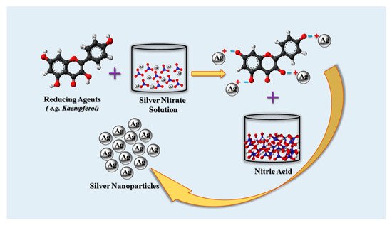
Numerous phytochemicals have been identified that are capable of producing AgNPs. As examples, flavonoids, terpenoids, terpenes, flavones, phenolics, polysaccharides, saponins, tannins and alkaloids have all been used for AgNPs synthesis [47,65]. Phytochemical functional groups, such as hydroxyl, aldehyde, ketone, carboxyl and amino groups are capable of reducing Ag+ ions [50,66]. Due to the diversity of phytochemicals the exact mechanism of AgNPs synthesis varies. However, the chief mechanism is the reduction of Ag+ ions by specific functional groups. For example, Punica granatum peel extract contains the flavonoids- kaempferol and naringin along with their glycosides. All of these compounds have hydroxyl (–OH) groups that cause the reduction of the Ag+ ions, resulting in the formation of AgNPs [67]. A typical reduction is possible by the use of kaempferol as a reducing agent (Figure 5).
Figure 5. Synthesis of AgNPs by reaction of AgNO3 with phytochemicals (concept of this representation was based on [67]).
Interestingly, not all phytochemicals present in plant extracts capable of reducing other compounds remain active during Ag+ ions reduction and only certain phytochemicals are involved in this reduction. For example, phytochemical screening revealed the presence of phenols, glycosides, flavonoids, reducing sugars, resins, tannins, terpenoids, steroids, alkaloids and saponins in Anagallis tenella leaf extract (Table 1).
Table 1. Active phytochemical fractions associated with AgNPs synthesis in specific plants.
Plant Phytochemical Reference
Alternanthera tenella Flavonoids [65]
Cocos nucifera Carbohydrates, alkaloids, terpenoids, tannins, saponins, phenolics and reducing sugars [68]
Lemongrass Reducing sugars [69]
Ocimum sanctum Caffeine [70]
Chrysanthemum indicum Tannins, flavonoids and glycosides [71]
Dalbergia spinose Reducing sugars and flavonoids [72]
Cinnamomum camphora Phenolics, terpenoids, polysaccharides and flavones [73]
Eucalyptus hybrid Flavonoids and terpenoids [74]
However, it was discovered that only flavonoids were involved in AgNPs synthesis [65]. Active phytochemicals associated with AgNPs synthesis in other plants are listed in Table 1. As stated earlier, the wide variety of phytochemicals present in different plants that contribute to AgNPs synthesis complicates this field of study. Each plant has a unique spectrum of phytochemicals that act as organic reducing agents [72].

This entry is adapted from the peer-reviewed paper 10.3390/cancers12040855

This entry is offline, you can click here to edit this entry!
ScholarVision Creations