1. Introduction
Prostate cancer, the most prevalent cancer in men worldwide, is generally considered a relatively slow-progressing cancer type, ranked fifth in mortality amongst other malignancies [
1,
2]. This is mainly reflective of the course of prostatic adenocarcinoma, which is by far the most common histologic type of the disease. It is typically androgen-dependent, with androgen deprivation being the backbone of therapy for decades. Despite the almost invariable emergence of castration resistance, most castration-resistant prostatic adenocarcinomas remain dependent on androgen signaling, with novel androgen signaling inhibitors like abiraterone acetate, enzalutamide, apalutamide and darolutamide demonstrating efficacy [
3,
4,
5,
6,
7,
8,
9].
Neuroendocrine (NE) tumor differentiation, mainly in the form of pure or mixed small-cell prostate cancer (SCPC), is a histologic subtype with a distinct phenotype, characterized by an aggressive clinical course, lack of responsiveness to hormonal therapies and an overall poor prognosis [
10]. While de novo SCPC is rare (<2%), treatment-emergent small-cell differentiation is present in up to 20% of patients with castration-resistant prostate cancer (CRPC) [
11,
12,
13]. Interestingly, there is a subset of CRPC without histologic evidence of NE differentiation that exhibits similar clinical behavior with SCPC, likely indicative of a shared underlying biology [
14,
15]. This aggressive-variant prostate cancer (AVPC) also shares the responsiveness of SCPC to platinum-based chemotherapy, with the clinical benefit being, however, short-lived and the majority of patients dying within two years from diagnosis [
16,
17,
18,
19,
20,
21]. With the lack of optimal therapies, the mortality rate of these patients has sadly remained unchanged during the last decades, while the incidence of ‘acquired’ NE tumor differentiation and AVPC is rising [
22]. Systematic research efforts are imperative in order to enhance our understanding of the biology of this disease entity and improve treatment outcomes for our patients through early detection as well as personalized, effective and safe therapeutic strategies. This review paper presents an overview of our current knowledge on NEPC/AVPC with a focus on their clinical characteristics and management, including available as well as experimental treatments.
2. Pathologic Classification of Prostate Cancer with Neuroendocrine Differentiation
NE cells are physiologically scattered within the epithelium of the glands in all anatomic zones of the prostate and are more commonly present in the prostatic gland compared to other organs of the genitourinary tract. By light microscopy, they can be seen in the basal layer between the secretory cells and contain a variety of peptide hormones, such as chromogranin A (CgA), neuron-specific enolase (NSE), serotonin, histamine, calcitonin and neuropeptide Y, vasoactive intestinal peptide, bombesin, and parathyroid hormone-related protein [
23,
24,
25]. NE cells are typically characterized by lack of androgen receptor (AR) and positivity for either synaptophysin, chromogranin, or CD56 in immunohistochemistry [
25].
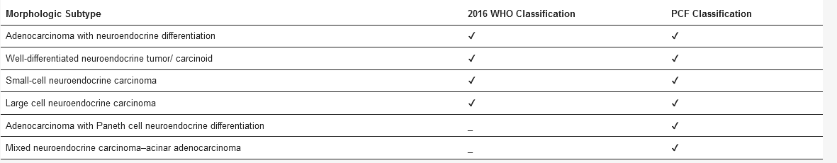
In the last decade, two similar histomorphologic classifications of prostate cancer with NE differentiation have been developed by the World Health Organization (WHO) and the Prostate Cancer Foundation (PCF), in an attempt to systematically describe this heterogeneous prostate cancer subtype [
25,
26]. ()
Table 1. Classifications of prostate cancer with neuroendocrine (NE) differentiation.
Most prostatic adenocarcinomas contain sparse benign NE cells as a part of the epithelium, but only 5–10% of these tumors will have the characteristic large, multifocal groups of NE cells and fall into the adenocarcinoma with NE differentiation category [
27]. Small infiltrating glands with prominent-marginated nucleoli, hyperchromasia, and intraluminal amorphous secretion or blue-tinged mucin as well as isolated tumor cells with eosinophilic granules are frequently present. Immunohistochemical detection of focal areas with at least one NE marker such as CgA, synaptophysin or CD56 may assist in the final diagnostic confirmation of NE differentiation. However, it is controversial whether this subtype is associated with worse oncologic outcomes. Hence, the use of immunohistochemical staining for NE markers in morphologically typical adenocarcinoma of the prostate with the aim to identify NE differentiation is not routinely recommended [
25,
28].
Well-differentiated NE tumors of the prostate, previously termed carcinoids, morphologically resemble similar tumors of other sites like the gastrointestinal tract and should be distinguished from the clinically aggressive NEPC category, as they tend to have a favorable prognosis [
25,
29,
30,
31]. The vast majority of prostatic carcinoids are prostatic adenocarcinomas with “carcinoid-like” morphology, characterized by insular, trabecular, glandular, or mixed architectural pattern and round nuclei with moderately clumped “salt and pepper” chromatin [
32]. Pure carcinoids of the prostate are extremely rare. For the diagnosis to be made, the tumor, beyond morphology, must also not be closely associated with concomitant adenocarcinoma of the prostate, it must be positive for NE markers and negative for PSA [
25].
De novo SCPC is an exceptionally rare (<2%), but very aggressive and fatal primary cancer [
11]. Approximately half of these tumors coexist with typical adenocarcinomas, while the remaining cases present as pure small-cell carcinomas [
25]. Histologic characteristics include small, undifferentiated cells with high mitotic activity in the absence of glandular structures, while high nuclear to cytoplasmic ratio, indistinct cell borders and lack of prominent nucleoli are frequent findings. These tumor cells are histologically identical to small-cell lung cancer with a 90% presence of NE markers [
33,
34]. To distinguish primary SCPC from small-cell carcinomas of other sites, presence of fusion of ETS-related gene with transmembrane protease, serine 2 (TMPRSS2-ERG fusion) is strongly suggestive of a primary prostatic tumor [
35,
36]. The differentiation of this histologic subtype from adenocarcinoma with NE differentiation and carcinoids is crucial, as it is typically nonresponsive to androgen signaling targeting therapies and requires different treatment. The prognosis is dismal, with a median overall survival of less than one year [
16,
21].
Large-cell prostatic carcinoma (LCPC) is an extremely rare, aggressive malignancy, with mainly case reports available in the literature [
25,
37]. It is a high-grade tumor that shows NE differentiation by immunohistochemistry. Morphologically, it consists of cells in large nests with peripheral palisading and geographic necrosis without glandular structures. The cells are characterized by prominent nucleoli, clumpy chromatin, and abundant cytoplasm. Most of the cases described to date have been mixed LCPC with adenocarcinomas [
37]. The outcome is poor, with a small case series reporting a mean survival of seven months (range 3–12) after completion of platinum-based chemotherapy following the detection of LCPC [
38].
The mixed NE carcinoma-acinar adenocarcinoma included in the PCF meeting classification, is not considered a distinct entity per the 2016 WHO classification [
39]. Adenocarcinoma with Paneth cell NE differentiation, which is also included only in the PCF meeting classification, is an entity with incompletely understood clinical significance, with the limited available data pointing to an overall favorable prognosis [
40].
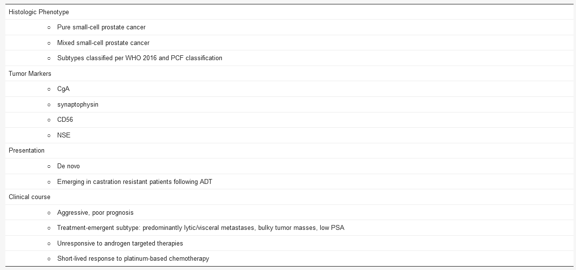
A summary of the characteristics of prostate cancer with neuroendocrine differentiation is provided in below.
Table 2. Characteristics of prostate cancer with NE differentiation.
3. Aggressive Variants of Castration Resistant Prostate Cancer
Although small cell/NE differentiation may present de novo in previously untreated patients, it is relatively rare (<2%) [
12]. More commonly, NE differentiation develops in castration-resistant patients following androgen deprivation therapy [
41]. This phenomenon is relatively common in advanced disease stages, presenting in approximately one fifth of patients with metastatic castration resistant prostate cancer (mCRPC) [
13]. Even though the mechanisms of emergence are still under investigation, a recent study points to a model of divergent clonal evolution of CRPC-adenocarcinoma to CRPC with NE differentiation, with adaptation from an AR-driven state to an AR-independent state [
42]. An alternative school of thought supports that treatment pressure with androgen signaling inhibitors may enable prostate cancer lineage plasticity and adenocarcinoma trans-differentiation by mechanisms including the acquisition of transcription factors (SOX2,11) and the loss of TP53 and phosphatase and tensin homolog (PTEN) [
43,
44]. As this phenomenon is limited to a subset of adenocarcinomas, it is crucial to identify baseline features associated with its advent.
Clinically, treatment-emergent NE/small cell differentiation has been associated with distinct manifestations, including predominantly visceral or lytic bone metastases and bulky tumor masses, frequently in the setting of low PSA level with high-volume tumor burden. Early emergence of castration resistance has also been described [
16,
19,
45,
46]. These tumors are typically not responsive to hormonal therapy, while they are sensitive to cytotoxic chemotherapy [
47,
48]. Responses are however short-lived and overall survival is reduced. Whether outcomes for pure and mixed tumors differ is not definitely answered yet, with some authors suggesting similar clinical behavior [
13,
16,
49], while others report shorter overall survival (OS) for pure SCPC [
46].
This aggressive variant has been reported more frequently, likely as a result of increasing awareness amongst clinicians and longer survival. Several terminologies have been historically used to describe this CRPC subset, with commonly used terminologies, however, being often ill-defined and having deficiencies [
14]. For instance, “NEPC” is an ambiguous term, as it implies the presence of histologic NE differentiation or other NE markers, even though this is not the case in many patients. Also, specific pathology features of NE differentiation are not necessarily associated with an aggressive clinical course (e.g., Paneth cell differentiation). The term “anaplastic” is likely misleading, as this is also an established term designated to pleomorphic cytology by surgical pathologists. The terminology “AR-negative prostate cancer” is considered too limiting, while “therapy-related NE prostate cancer” is being discouraged, as it may drive clinicians to withhold potentially effective hormonal therapies. While the term “AVPC” might be overall less confusing as it does not imply any histologic correlate and is more reflective of the clinical phenotype of this disease, it may potentially be more contaminated. [
14]
For the purpose of systematically studying this disease variant, a set of criteria were proposed to define it [
19]. CRPC characterized by one or more of the following was determined to be AVPC:
-
histologic evidence of SCPC (pure or mixed);
-
presence of only visceral metastases;
-
predominantly lytic bone lesions;
-
bulky (≥5 cm) lymphadenopathy or large (≥5 cm) high-grade (Gleason ≥ 8) tumor mass in prostate/pelvis;
-
low PSA at presentation with extensive bone metastatic disease;
-
presence of NE markers at histology (CgA and synaptophysin) or serum (CgA and gastrin-releasing peptide) combined with either elevated lactate dehydrogenase (LDH), malignant hypercalcemia or elevated serum carcinoembryonic antigen (CEA);
-
progression to CRPC in six months or less after initiation of hormonal therapy.
Of note, the presence of SCPC, either pure or mixed, is considered AVPC regardless of hormonal status [
19].
In this review, we are going to use the term AVPC according to the principles of the above definition to describe this clinically aggressive-variant disease with or without small-cell histology. AVPC was shown to share the responsiveness of SCPC to platinum based chemotherapy, indicating a likely shared underlying biology [
19]. Even though AVPC is a morphologically heterogeneous group of tumors [
14,
50], it may share molecular characteristics with SCPC. Strikingly, joint alterations in two or more of RB1, TP53 and/or PTEN were shown to correlate with this aggressive clinical phenotype similarly to SCPC, consistent with preclinical models that support the role of combined tumor suppressor alterations in prostate cancer progression and development of resistance to novel hormonal agents [
15,
43,
44,
51]. Apart from alterations in tumor suppressors, further features may include AR loss, induction of neuroendocrine/neural as well as mitotic programs, and genomic instability [
12,
14,
15].