Breast Reconstruction: History
Please note this is an old version of this entry, which may differ significantly from the current revision.
Contributor: , , , , , , , , , , ,

Breast cancer accounted for 685,000 deaths globally in 2020, and half of all cases occur in women with no specific risk factor besides gender and age group. The number of mastectomies performed for younger women increased, raising the need for adequate breast reconstructive surgery. Advances in oncological treatment have made it possible to limit the extent of what represents radical surgery for breast cancer, a marked trend toward mastectomies in breast-conserving surgery-eligible patients are seen. Prophylactic mastectomies have also registered an upward trend. This trend together with new uses for breast reconstruction like chest feminization in transgender patients has increased the need for breast reconstruction surgery

  • breast reconstruction
  • reconstruction following mastectomy
  • prophylactic mastectomy
  • chest feminization
  • transgender
  • implant reconstruction of breast
  • immediate reconstruction
  • delayed reconstruction
  • two-stage breast reconstruction

1. Introduction

Breast cancer accounted for 685,000 deaths globally in 2020, and half of all cases occur in women with no specific risk factor besides gender and age group. During the last four decades, we have seen a 40% reduction in age-standardized breast cancer mortality [1] and have also witnessed a reduction in the medium age at diagnosis, which in turn means that the number of mastectomies performed for younger women increased, raising the need for adequate breast reconstructive surgery. Advances in oncological treatment have made it possible to limit the extent of what represents radical surgery for breast cancer, yet in the past decade, we have seen a marked trend toward mastectomies in breast-conserving surgery-eligible patients [2]. Prophylactic mastectomies have also registered an upward trend [3][4]. This trend together with new uses for breast reconstruction like chest feminization in transgender patients [5] has increased the need for breast reconstruction surgery.
Breast cancer is the most commonly diagnosed neoplasm in the female population in the world [6]. It is the leading cause of cancer-related death in women in most countries of the world, except in developed countries, where it ranks second after lung tumors. However, mortality has been steadily declining for over 30 years, with an average 5-year survival of 86% and 75% at 10 years [7]. This trend is attributed both to the increase in the effectiveness of oncological treatments and to early screenings and screening programs similar to those for other neoplastic diseases [8][9].
Breast reconstruction is an important component of breast cancer treatment. With the increase in life expectancy, it has become essential to ensure a good quality of life for patients, forcing a continuous evolution of surgical techniques. Breast reconstruction is necessary not only after performing a modified radical mastectomy, but also after conservative interventions on the breast that have not been accompanied by an optimal aesthetic effect. The need to complete the surgical treatment of breast cancer with breast reconstruction derives from the beneficial impact at the psychological level, respectively, for the body image, sexuality, and general quality of life of patients [10]. In recent years, the ever-increasing number of patients opting for prophylactic mastectomies due to a genetic predisposition for developing breast cancer or a family history of cancer [3][4][11][12][13] has given birth to a new type of integrated treatment plan in oncology. Changes in guidelines, prompting the genetic testing of BRCA mutations and the availability of those tests even in the absence of an oncologist’s recommendation, have determined an increase in the number of women getting tested and then opting for a contralateral or bilateral prophylactic mastectomy. A good example for this trend is the Angelina Jolie effect on the Western population; following the known actress’s double prophylactic mastectomy, there was a noticeable increase in the number of healthy women requesting this procedure and having it performed.

2. Breast Reconstruction

2.1. Timing of Breast Reconstruction—Immediate or Delayed
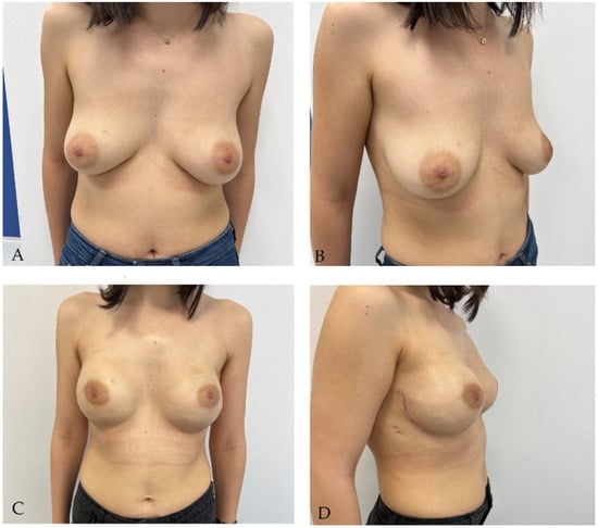
Breast reconstruction is classified by the type and time of surgery. Immediate reconstruction takes place at the same time as the mastectomy, and secondary (or delayed) reconstruction is performed from a few months to several years after the mastectomy. Currently, it is performed at least three months after the end of radiotherapy and generally at about a year after the mastectomy [14]. The two main types of reconstruction are with implants or autologous tissue; they can also be used together in mixed procedures.
Immediate breast reconstruction (Figure 1) has certain benefits over the secondary one, especially in terms of patient satisfaction, quality of life, and psychological status post-mastectomy [15]. These patients are relatively more protected from the psychological effects of mastectomies, and studies have shown a stable evolution of quality of life and satisfaction of this group compared to patients receiving delayed reconstruction [16][17]. In the latter case, the quality of life is significantly improved with the reconstructive procedures, with the results ultimately being equalized in the long run [18]. Also, after the immediate reconstruction, a more natural and aesthetic result is obtained, with the intervention usually being associated with a skin-sparing mastectomy which respects the inframammary groove and keeps the skin intact, proven safe from the oncological point of view by a series of studies, provided the correct selection of patients [19][20][21][22]. An important factor for selection is the appreciation of the thickness of the skin flap, this being correlated with the aesthetic results and with the possible postoperative complications [23]. Patients undergo fewer major surgeries and require fewer days of hospitalization, recovering faster postoperatively. From an oncological point of view, immediate reconstruction is considered safe and has been shown to not increase the risk of local recurrence compared to that of mastectomies without reconstruction [24][25]. At the same time, this technique does not change the effectiveness of adjuvant radiotherapy [26].
Figure 1. Patient with stage Ia invasive ductal carcinoma of left breast and BRCA positive status. She underwent bilateral subcutaneous mastectomy with left sentinel lymph node identification using Indocyanine green followed by immediate bilateral reconstruction with 350 cc round implants: (A,B) aspect before reconstructive surgery; (C,D) aspect at 3 months after reconstructive surgery.
Despite the many benefits of immediate reconstruction, many surgeons choose to postpone the operation for another time (Figure 2), often for reasons of oncological safety. Definite diagnoses of malignancies of radiologically detected breast tumors are made more and more frequently by guided biopsies, and the real extension of the tumor tissue can be evaluated macroscopically only intraoperatively, and microscopically only when examining the mastectomy piece. Thus, the subsequent therapeutic attitude is often decided intraoperatively [27]. However, in order to evaluate the quality and thickness of the skin flap intended for immediate breast reconstruction, mammography, breast ultrasound, and magnetic resonance imaging (MRI) can be used preoperatively to complete the clinical examination. The results of these investigations guide the decision on surgery for immediate breast reconstruction and have been shown to be true to intraoperative findings. The thickness of the flap is important in choosing the type of implant used, but also for avoiding postoperative complications such as skin necrosis [23].
Figure 2. Patient with right radical mastectomy for breast cancer followed by radiotherapy; she underwent right breast delayed reconstruction using latissimus dorsi pediculated flap and a 225 cc round implant. (A) aspect before reconstructive surgery; (B) aspect at 3 months after reconstructive surgery.
The indications of radiotherapy, typically applied to patients at high risk of recurrence (>4 positive lymph nodes or positive resection margins), tend to increase, with studies proving its usefulness in patients with 1–3 positive lymph nodes [28][29][30]. Although radiotherapy does not contraindicate immediate reconstruction, the higher rate of complications, especially in implant-only reconstructions, is a second reason why in these patients, either a two-stage reconstruction or a delayed reconstruction is chosen [31]. After reconstructive surgery, radiation therapy may affect the aspect of the operated breast, including altered skin color and rigidity. It can also lead to capsular contraction which mandates the removal of implant. Patients undergoing radiation therapy after reconstructive surgery need to be advised about the possibility of additional corrective surgery [32].
Another contraindication of immediate reconstruction is any modifications of the flaps (tegument and subcutaneous tissue), namely, the presence of necrosis, inflammation, or signs of dermal neoplastic dissemination resulting in a large skin defect after the mastectomy.

2.2. Two-Stage Breast Reconstruction

In 2002, the technique of two-stage breast reconstruction was initially described for delayed reconstruction. Later, the technique was especially used to improve the results in cases associated with radiotherapy [33]. The ionizing radiation used on either the chest wall or the axilla irreversibly alters the tissues in the irradiated field, regardless of their nature. In the short term, erythema and scaling of the skin can appear, and in the long-term, severe fibrosis, telangiectasia, hyperpigmentation, and tissue atrophy [34]. Under these conditions, many surgeons prefer to place a tissue expander at the time of the mastectomy, preserved during radiotherapy, which aims to maintain both the shape and the skin needed for the final reconstruction [35]. The expander can be filled at the time of the intervention, or progressively, depending on the condition of the flaps and the center where the intervention is carried out [36]. It can be partially emptied before radiotherapy sessions in order to favor the alignment of the irradiation fields, but this step is not always necessary [37]. Subsequently—it is recommended no later than 3 months after the completion of radiotherapy—the second stage of reconstruction is performed, usually with autologous tissue. For patients who do not require adjuvant radiotherapy, the recommendation is that the second stage of reconstruction be performed no later than two weeks after the mastectomy [31].

2.3. Breast Reconstruction with Implant

Regarding the type of intervention, at present, it is estimated that 80% of breast reconstructions are performed with an implant [35]. This type of intervention is shorter and easier from a technical point of view, and postoperative recovery is faster.
In the long run, however, complications are more common than in cases of breast augmentation (30% at 5 years compared to 12% at 5 years) and are accentuated by the history of radiation therapy [36][37]. The main complications are capsular contracture, implant rupture, hematoma, and infections [38]. Implant reconstruction is associated with aesthetic complications like asymmetry, chest wall deformity, mispositioning or displacement, ptosis, wrinkling or rippling (wrinkling of the implant that can be felt or seen through the skin), skin rush, redness and bruising, and inflammation. The implant can suffer deflation, rupture, or extrusion. Many of these complications will require additional surgeries, a possibility that the patient should be informed about. Seroma, hematoma, delayed wound healing, infection, and necrosis of the skin/flap can also occur after implant breast reconstruction. These complications will require additional treatment and will most often delay adjuvant therapies with effects on the overall oncologic outcome. Following infection, hematoma formation, and seroma formation, capsular contraction can occur. Grade III and IV capsular contraction (hardening of the breast around the implant, causing painful tightening of the tissues) will require corrective surgery, but could occur again after the procedure. Implants are associated also with more exotic complications including other cancers; there have been reports of Breast Implant-Associated Anaplastic Large Cell Lymphoma (BIA-ALCL), squamous cell carcinoma, and mesenchymal tumors after breast reconstruction with implants.
However, implant reconstructions remain preferable for many surgeons because they avoid the complications of the donor areas and generate lower costs, and in the absence of radiotherapy or in a two-stage reconstruction, they are a simple solution with good aesthetic results. Acellular dermal matrices (ADMs) are used either as a first intervention to support the implant in the lower pole, not covered by the pectoralis major, or in reinterventions [39]. These biological materials are made from human, bovine, or porcine dermis processed to remove all cellular components—which can generate an immune response—and keeping the extracellular matrix containing mainly collagen (85%) along with proteoglycans, glycosaminoglycans, and elastin, arranged in a network, in the meshes of which the host cells are arranged [40]. This integration of the matrix provides good support for the breast prosthesis and a high-quality capsule, resulting in a natural appearance of the final reconstructed breast. The high costs are the main disadvantage and make dermal substitutes inaccessible on a large scale. With the increase in accessibility, it is estimated that the approach to implant reconstructions in a single stage will change significantly.

2.4. Autologous Breast Reconstruction Techniques

Autologous techniques are considered by many authors to be the gold standard in breast reconstruction. They consist in restoring the contour and volume of the mammary gland with the help of either rotating, pediculated flaps, which retain their vascular source, or micro-surgically freely transferred flaps from other areas of the body, most often from the abdomen. The intervention can be performed immediately or delayed, like the implant reconstruction. Moreover, if the volume provided by the flap is not sufficient, other techniques such as the free transfer of autologous fat (lipofilling) or placement of an implant may be associated [41].
  • The advantages of flap reconstruction.
Flap reconstruction offers several advantages, including improving the quality of irradiated tissue by bringing healthy tissue into a scar area; the final appearance after reconstruction is a natural one that mimics, in time, the physiological ptosis of the contralateral breast, does not require reinterventions for replacement after a period of time, can be used in patients who do not want or do not tolerate an implant, and is the recommended type of reconstruction for the radio-treated patients [42].
b.
Types of transferred free flaps.
First described in 1989 by Koshima and Soeda [43], the freely transferred flap based on the inferior epigastric artery (DIEP) has long been the preferred alternative in autologous breast reconstruction in specialized centers [43]. Other free flaps that are described but rarely used in practice are the TRAM (transverse rectus abdominis myocutaneous flap), more commonly used in its pediculated version; TUG (transverse upper gracilis flap); SGAP (upper gluteal artery perforator flap); or IGAP (lower gluteal artery flap). Lower-limb flaps are indicated in selected cases, in the absence of a suitable abdominal donor area or in patients with previous interventions at this level [44].
c.
Latissimus dorsi pediculated flap.
First described by Tansini for covering chest wall defects in 1906, the latissimus dorsi pediculated flap began to be used in breast reconstruction after almost 70 years [45]. Until the middle of the twentieth century, the radical mastectomy technique described by Halsted recommended either grafting or secondhand healing of the resulting defect, strongly contraindicating any form of reconstruction, as it was considered to “hide possible recurrences and promote the spread of tumor cells” [46].
The evolution of oncological treatments, a better understanding of the pathology, and the increase of patients’ life expectancy, together with the appearance of breast implants, changed the approach of these cases. Schneider and Botswick described in 1977 and 1978, respectively, the latissimus dorsi flap accompanied by the implant in restoring the physiological contour and ptosis of the breast after a mastectomy [47][48]. Subsequently, Papp and McCraw modified the flap, including subcutaneous adipose tissue overlying the muscle in order to achieve implant-free reconstruction [49].
Although it is no longer the gold standard in autologous reconstruction, the reliability and predictability of its anatomy still make it preferred by many surgeons for delayed reconstructions and also the preferred rescue option in the event of free-transfer flaps failure [50]. Currently, its primary uses are in patients who do not have sufficient reserves for a free flap; those with a personal history of abdominal interventions; or those with significant comorbidities such as obesity and diabetes or in smoking patients [51].
The most common complications are seromas in the donor area, usually easy to treat without further intervention. Associated with alloplastic procedures, capsular contracture has been described more frequently in association with implants and less frequently in two-stage reconstructions, when the implant is preceded by an expander. Rare complications are contour defects in the donor area, limited shoulder mobility, and decreased muscle strength in the arm and the scapula alatae [52].
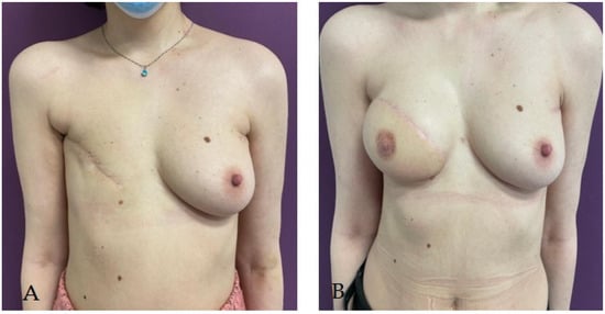
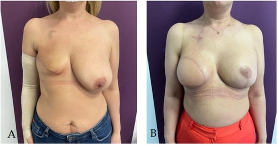
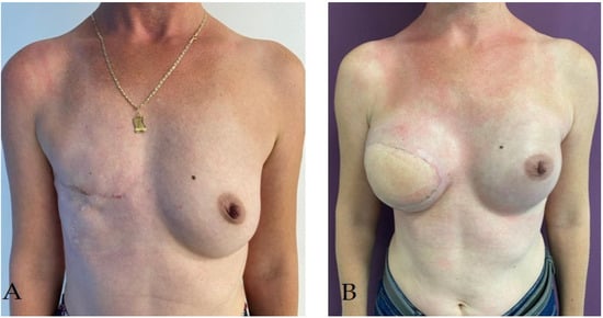
In Figure 3, Figure 4 and Figure 5, the researchers present various immediate or delayed reconstructive techniques using the autologous or mixed procedures the researchers employed for the patients.
Figure 3. Patient with right radical mastectomy for breast cancer followed by radiotherapy; she underwent right breast delayed reconstruction using latissimus dorsi pediculated flap and a 320 cc round implant simultaneous with prophylactic left subcutaneous mastectomy (due to BRCA-positive status) with immediate reconstruction using a pediculated inferior dermoadipous flap and a 350 cc round implant: (A) aspect before reconstructive surgery; (B) aspect at 3 months after reconstructive surgery.
Figure 4. Bilateral prophylactic mastectomy in patient with BRCA-positive status and heavy family history of breast cancer. Immediate reconstruction using 325 cc round implant and pediculated inferior dermoadipous flap followed by nipple–areola complex graft: (A) aspect before reconstructive surgery; (B) aspect at 3 months after reconstructive surgery; (C) final aspect at 32 months after reconstructive surgery.
Figure 5. Patient with right radical mastectomy for breast cancer; she underwent right breast delayed reconstruction using latissimus dorsi pediculated flap and a 275 cc round implant simultaneous with prophylactic left subcutaneous mastectomy (due to BRCA-positive status) with immediate reconstruction using a 325 cc round implant: (A) aspect before reconstructive surgery; (B) aspect at 3 months after reconstructive surgery.

This entry is adapted from the peer-reviewed paper 10.3390/life14010138

References

  1. WHO. Statistics on Breast Cancer. Available online: https://www.who.int/news-room/fact-sheets/detail/breast-cancer (accessed on 22 July 2023).
  2. Kummerow, K.L.; Du, L.; Penson, D.F.; Shyr, Y.; Hooks, M.A. Nationwide Trends in Mastectomy for Early-Stage Breast Cancer. JAMA Surg. 2015, 150, 9–16.
  3. Tuttle, T.M.; Habermann, E.B.; Grund, E.H.; Morris, T.J.; Virnig, B.A. Increasing Use of Contralateral Prophylactic Mastectomy for Breast Cancer Patients: A Trend toward More Aggressive Surgical Treatment. J. Clin. Oncol. 2007, 25, 5203–5209.
  4. Soran, A.; Kamali Polat, A.; Johnson, R.; McGuire, K.P. Increasing Trend of Contralateral Prophylactic Mastectomy: What Are the Factors behind This Phenomenon? Surgeon 2014, 12, 316–322.
  5. Patel, H.; Samaha, Y.; Ives, G.; Lee, T.-Y.; Cui, X.; Ray, E. Chest Feminization in Male-to-Female Transgender Patients: A Review of Options. Transgender Health 2021, 6, 244–255.
  6. Ferlay, J.; Steliarova-Foucher, E.; Lortet-Tieulent, J.; Rosso, S.; Coebergh, J.W.W.; Comber, H.; Forman, D.; Bray, F. Cancer Incidence and Mortality Patterns in Europe: Estimates for 40 Countries in 2012. Eur. J. Cancer 2013, 49, 1374–1403.
  7. Sancho-Garnier, H.; Colonna, M. Epidémiologie Des Cancers Du Sein Breast Cancer Epidemiology. Presse Medicale 2019, 48, 1076–1084.
  8. Simion, L.; Rotaru, V.; Cirimbei, C.; Gales, L.; Stefan, D.-C.; Ionescu, S.-O.; Luca, D.; Doran, H.; Chitoran, E. Inequities in Screening and HPV Vaccination Programs and Their Impact on Cervical Cancer Statistics in Romania. Diagnostics 2023, 13, 2776.
  9. Simion, L.; Chitoran, E.; Cirimbei, C.; Stefan, D.-C.; Neicu, A.; Tanase, B.; Ionescu, S.O.; Luca, D.C.; Gales, L.; Gheorghe, A.S.; et al. A Decade of Therapeutic Challenges in Synchronous Gynecological Cancers from the Bucharest Oncological Institute. Diagnostics 2023, 13, 2069.
  10. UpToDate. Overview of Breast Reconstruction. Available online: https://www.uptodate.com/contents/overview-of-breast-reconstruction?search=Overview%20of%20breast%20reconstruction&source=search_result&selectedTitle=1~150&usage_type=default&display_rank=1 (accessed on 21 July 2023).
  11. Angelos, P.; Bedrosian, I.; Euhus, D.M.; Herrmann, V.M.; Katz, S.J.; Pusic, A. Prophylactic Mastectomy: Challenging Considerations for the Surgeon. Ann. Surg. Oncol. 2015, 22, 3208–3212.
  12. Dragun, A.E.; Pan, J.; Riley, E.C.; Kruse, B.; Wilson, M.R.; Rai, S.; Jain, D. Increasing Use of Elective Mastectomy and Contralateral Prophylactic Surgery among Breast Conservation Candidates: A 14-Year Report from a Comprehensive Cancer Center. Am. J. Clin. Oncol. 2013, 36, 375–380.
  13. Metcalfe, K.A.; Eisen, A.; Poll, A.; Candib, A.; McCready, D.; Cil, T.; Wright, F.; Demsky, R.; Mancuso, T.; Sun, P.; et al. Frequency of Contralateral Prophylactic Mastectomy in Breast Cancer Patients with a Negative BRCA1 and BRCA2 Rapid Genetic Test Result. Ann. Surg. Oncol. 2021, 28, 4967–4973.
  14. Mathelin, C.; Bruant-Rodier, C. Indications for Breast Reconstruction after Mastectomy According to the Oncological Situation. Ann. Chir. Plast. Esthet. 2018, 63, 580–584.
  15. Atisha, D.; Alderman, A.K.; Lowery, J.C.; Kuhn, L.E.; Davis, J.; Wilkins, E.G. Prospective Analysis of Long-Term Psychosocial Outcomes in Breast Reconstruction: Two-Year Postoperative Results from the Michigan Breast Reconstruction Outcomes Study. Ann. Surg. 2008, 247, 1019–1028.
  16. Platt, J.; Zhong, T. Patient-Centered Breast Reconstruction Based on Health-Related Quality-of-Life Evidence. Clin. Plast. Surg. 2018, 45, 137–143.
  17. Zhong, T.; McCarthy, C.; Min, S.; Zhang, J.; Beber, B.; Pusic, A.L.; Hofer, S.O.P. Patient Satisfaction and Health-Related Quality of Life after Autologous Tissue Breast Reconstruction: A Prospective Analysis of Early Postoperative Outcomes. Cancer 2012, 118, 1701–1709.
  18. Zhong, T.; Hu, J.; Bagher, S.; Vo, A.; O’Neill, A.C.; Butler, K.; Novak, C.B.; Hofer, S.O.P.; Metcalfe, K.A. A Comparison of Psychological Response, Body Image, Sexuality, and Quality of Life between Immediate and Delayed Autologous Tissue Breast Reconstruction: A Prospective Long-Term Outcome Study. Plast. Reconstr. Surg. 2016, 138, 772–780.
  19. Simmons, R.M.; Fish, S.K.; Gayle, L.; La Trenta, G.S.; Swistel, A.; Christos, P.; Osborne, M.P. Local and Distant Recurrence Rates in Skin-Sparing Mastectomies Compared With Non-Skin-Sparing Mastectomies. Ann. Surg. Oncol. 1999, 6, 676–681.
  20. Toth, B.A.; Lappert, P. Modified Skin Incisions for Mastectomy: The Need for Plastic Surgical Input in Preoperative Planning. Plast. Reconstr. Surg. 1991, 87, 1048–1053.
  21. Kroll, S.; Khoo, A.; Singletary, E.; Ames, F.; Wang, B.G.; Reece, G.; Miller, M.; Evans, G.; Robb, G. Local Recurrence Risk after Skin-Sparing and Conventional Mastectomy: A 6-Year Follow-Up. Plast. Reconstr. Surg. 1999, 104, 421–425.
  22. Galimberti, V.; Vicini, E.; Corso, G.; Morigi, C.; Fontana, S.; Sacchini, V.; Veronesi, P. Nipple-Sparing and Skin-Sparing Mastectomy: Review of Aims, Oncological Safety and Contraindications. Breast 2017, 34, S82–S84.
  23. Radu, M.; Bordea, C.; Noditi, A.; Blidaru, A. Assessment of Mastectomy Skin Flaps for Immediate Implant-Based Breast Reconstruction. J. Med. Life 2018, 11, 137–145.
  24. Huang, C.J.; Hou, M.F.; Lin, S.D.; Chuang, H.Y.; Huang, M.Y.; Fu, O.Y.; Lian, S.L. Comparison of Local Recurrence and Distant Metastases between Breast Cancer Patients after Postmastectomy Radiotherapy with and without Immediate TRAM Flap Reconstruction. Plast. Reconstr. Surg. 2006, 118, 1079–1086.
  25. Gieni, M.; Avram, R.; Dickson, L.; Farrokhyar, F.; Lovrics, P.; Faidi, S.; Sne, N. Local Breast Cancer Recurrence after Mastectomy and Immediate Breast Reconstruction for Invasive Cancer: A Meta-Analysis. Breast 2012, 21, 230–236.
  26. Strålman, K.; Mollerup, C.L.; Kristoffersen, U.S.; Elberg, J.J. Long-Term Outcome after Mastectomy with Immediate Breast Reconstruction. Acta Oncol. 2008, 47, 704–708.
  27. Kronowitz, S.J. Delayed-Immediate Breast Reconstruction: Technical and Timing Considerations. Plast. Reconstr. Surg. 2010, 125, 463–474.
  28. Gradishar, W.; Salerno, K.E. NCCN guidelines update: Breast cancer. J. Natl. Compr. Cancer Netw. 2016, 14, 641–644.
  29. Harris, E.E.R.; Freilich, J.; Lin, H.Y.; Chuong, M.; Acs, G. The Impact of the Size of Nodal Metastases on Recurrence Risk in Breast Cancer Patients with 1-3 Positive Axillary Nodes after Mastectomy. Int. J. Radiat. Oncol. Biol. Phys. 2013, 85, 609–614.
  30. McBride, A.; Allen, P.; Woodward, W.; Kim, M.; Kuerer, H.M.; Drinka, E.K.; Sahin, A.; Strom, E.A.; Buzdar, A.; Valero, V.; et al. Locoregional Recurrence Risk for Patients with T1,2 Breast Cancer with 1-3 Positive Lymph Nodes Treated with Mastectomy and Systemic Treatment. Int. J. Radiat. Oncol. Biol. Phys. 2014, 89, 392–398.
  31. Kronowitz, S.J.; Hunt, K.K.; Kuerer, H.M.; Babiera, G.; McNeese, M.D.; Buchholz, T.A.; Strom, E.A.; Robb, G.L. Delayed-Immediate Breast Reconstruction. Plast. Reconstr. Surg. 2004, 113, 1617–1628.
  32. Doherty, C.; Mcclure, J.A.; Baxter, N.N.; Brackstone, M. Complications from Postmastectomy Radiation Therapy in Patients Undergoing Immediate Breast Reconstruction: A Population-Based Study. Adv. Radiat. Oncol. 2023, 8, 101104.
  33. See, M.S.F.; Farhadi, J. Radiation Therapy and Immediate Breast Reconstruction: Novel Approaches and Evidence Base for Radiation Effects on the Reconstructed Breast. Clin. Plast. Surg. 2018, 45, 13–24.
  34. Bellini, E.; Pesce, M.; Santi, P.L.; Raposio, E. Two-Stage Tissue-Expander Breast Reconstruction: A Focus on the Surgical Technique. Biomed. Res. Int. 2017, 2017, 1791546.
  35. Cemal, Y.; Albornoz, C.R.; Disa, J.J.; Mccarthy, C.M.; Mehrara, B.J.; Pusic, A.L.; Cordeiro, P.G.; Matros, E. A Paradigm Shift in U.S. Breast Reconstruction: Part 2. the Influence of Changing Mastectomy Patterns on Reconstructive Rate and Method. Plast. Reconstr. Surg. 2013, 131, 320e–326e.
  36. Sherine, E.G.; Woods, J.E.; O’Fallon, M.; Beard, M.; Kurland, L.T.; Melton, J.L. Complications Leading to Surgery after Breast Implantation. N. Engl. J. Med. 1997, 336, 677–682.
  37. Percec, I.; Bucky, L.P. Successful Prosthetic Breast Reconstruction after Radiation Therapy. Ann. Plast. Surg. 2008, 60, 527–531.
  38. Thamm, O.C.; Andree, C. Immediate Versus Delayed Breast Reconstruction: Evolving Concepts and Evidence Base. Clin. Plast. Surg. 2018, 45, 119–127.
  39. Nahabedian, M.Y. Acellular Dermal Matrices in Primary Breast Reconstruction. Plast. Reconstr. Surg. 2012, 130, 44S–53S.
  40. Uitto, J.; Olsen, D.R.; Fazio, M.J. Extracellular Matrix of the Skin: 50 Years of Progress. J. Investig. Dermatol. 1989, 92, S61–S77.
  41. Ho, A.Y.; Hu, Z.I.; Mehrara, B.J.; Wilkins, E.G. Radiotherapy in the Setting of Breast Reconstruction: Types, Techniques, and Timing. Lancet Oncol. 2017, 18, e742–e753.
  42. Chen, V.W.; Lin, A.; Hoang, D.; Carey, J. Trends in Breast Reconstruction Techniques at a Large Safety Net Hospital: A 10-Year Institutional Review. Ann. Breast Surg. 2018, 2, 14.
  43. Koshima, I.; Soeda, S. Inferior Epigastric Artery Skin Flaps without Rectus Abdominis Muscle. J. Plast. Surg. 1989, 42, 645–648.
  44. Healy, C.; Allen, R.J. The Evolution of Perforator Flap Breast Reconstruction: Twenty Years after the First DIEP Flap. J. Reconstr. Microsurg. 2014, 30, 121–126.
  45. Maxwell, P. Iginio Tansini and the Origin of the Latissimus Dorsi musculocutaneous Flap. Plast. Reconstr. Surg. 1989, 65, 686–692.
  46. Champaneria, M.C.; Wong, W.W.; Hill, M.E.; Gupta, S.C. The Evolution of Breast Reconstruction: A Historical Perspective. World J. Surg. 2012, 36, 730–742.
  47. Bostwick, J.; Vasconez, L.O.; Jurkiewicz, M.J. Breast Reconstruction after a Radical Mastectomy. Plast. Reconstr. Surg. 1978, 61, 682–693.
  48. Schneider, W.J.; Hill, H.L.; Brown, R.G. Latissimus dorsi myocutaneous flap for breast reconstruction. Br. J. Plast. Surg. 1977, 30, 277–281.
  49. Papp, C.; McCraw, J.B. Autogenous Latissimus Breast Reconstruction. Clin. Plast. Surg. 1998, 25, 261–266.
  50. DeLong, M.R.; Tandon, V.J.; Rudkin, G.H.; Da Lio, A.L. Latissimus Dorsi Flap Breast Reconstruction—A Nationwide Inpatient Sample Review. Ann. Plast. Surg. 2017, 78, S185–S188.
  51. Spear, S.L.; Clemens, M.W. Latissimus Dorsi Flap Breast Reconstruction. In Plastic Surgery, 3rd ed.; Saunders (Elsevier): Philadelphia, PA, USA, 2012; pp. 370–392.
  52. Hardwicke, J.T.; Prinsloo, D.J. An Analysis of 277 Consecutive Latissimus Dorsi Breast Reconstructions: A Focus on Capsular Contracture. Plast. Reconstr. Surg. 2011, 128, 63–70.
More
This entry is offline, you can click here to edit this entry!
Video Production Service