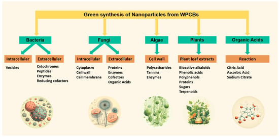
3. Green Organic Acids
The size and well-defined shapes of the nanoparticles depend, to a large extent, on the reducing agent and the action of a stabilizer in the synthesis of the solution phase.
The Turkevich method is a straightforward and reliable approach for synthesizing spherical particles ranging from 10 nm to 30 nm, using WPCBs as a substrate for Ag and Au [
7]. The initial concentration of Ag was reported as 15.7 g·L
−1. In this method, citrates are used as reducing agents [
93,
94].
A stable suspension of gold nanoparticles was achieved by utilizing a combination of sodium citrate and ascorbic acid as reducing agents, along with a stabilizing agent (polyvinylpyrrolidone—PVP), within a temperature range of 25–65 °C and a pH range of 2.5–4.0. This unique combination of reductive agents and a polymeric stabilizer enabled the production of pure gold metallic nanoparticles, featuring well-defined spheroidal and triangular shapes with a size distribution between 5–20 nm [
4].
Ascorbic acid is commonly employed in the synthesis of nanorods. It serves the dual purpose of both reducing and stabilizing during the fabrication of copper nanoparticles (CuNPs) [
95,
96], silver nanoparticles (AgNPs) [
7], and gold nanoparticles (AuNPs) [
97]. Furthermore, it acts as an antioxidant, effectively reducing oxygen free radicals and metal ions. As a result, it is considered as a non-toxic reagent which can influence the growth, aggregation, and interaction of the synthesized particles with the external environment [
95].
Glucose is another agent used to obtain NPs from WPCBs, and its effect is potentiated by ascorbic acid. In a study on the synthesis of CuNPs, this technique was used. The nanoparticles obtained exhibited round shapes, with sizes between 10–50 nm [
98].
4. Plants
The use of plant leaf extract facilitates the biogenic reduction of metal ions into base metals. This process is characterized by its swiftness, as it occurs rapidly, and its ease of execution at room temperature and pressure. Furthermore, it can be conveniently scaled up. Synthesis mediated by plant extracts is environmentally friendly, and most methods used water [
99] and alcohols, such as methanol and ethanol [
100], to extract the main components from the plants. Plant extracts, which comprise bioactive compounds like alkaloids, phenolic acids, polyphenols, proteins, sugars, and terpenoids, are thought to play a crucial role in initially reducing metallic ions and subsequently stabilizing them [
101]. For example,
Cathantharaus roseus plant leaf extract was used to generate copper oxide nanoparticles from WPCB, as it was indicated that the amide groups present in the proteins and enzymes of leaf extract are responsible for the oxidation–reduction process, and that amine groups of the leaf extract act as capping agents. The particle size ranges were from around 5 nm–10 nm, exhibiting a polycrystalline nature [
25].
In a ratio of 1:20 (solid/liquid), an aqueous extract of olive tree leaves was used as a source of polyphenols for the reduction of Cu, Cr, and Sn metals in WPCBS acidic leachates as a greener alternative for the recovery of valuable metals [
99].
An aqueous extract of
Prosopis juliflora leaves obtained by sonication, containing a pool of piperidine alkaloids, was used in equal proportions with PCB-derived copper nitrate at 80 °C to obtain copper oxide nanoparticles (gCON) for the oxidation–reduction process. The outcomes of optical, structural, and morphological analyses verified the existence of monocrystalline, spherical copper oxide nanoparticles, demonstrating an average size of 11 nm [
102].
The leaf extract of
Cassia auriculata was used as a reducing, as well as capping, agent in the synthesis of copper nanoparticles. The findings from this investigation have substantiated the potential of
Cassia auriculata leaf extract for the recovery of copper from printed circuit boards, yielding nanoparticles with a size range of 50–100 nm. The CuNPs exhibited a prominent peak at 300 nm in UV–Vis spectra [
103].
Aqueous extracts derived from aloe vera and geranium (
Pelargonium graveolens) were employed to reduce copper ions present in the leaching solution, a product of copper shale (Kupferschiefier) bioleaching via chemolithotrophic bacteria, including
Acidithiobacillus ferrooxidans. The utilization of
Pelargonium graveolens extract resulted in the formation of copper nanoparticles (CuNPs) with a spherical shape and a size distribution ranging between 100 and 300 nm [
104].
5. Bacteria
The bacterial synthesis of nanoparticles can occur through extracellular and intracellular processes, with various metals being reported for nanoparticle formation using different bacterial components such as biomass, supernatant, cell-free extracts, and their derivatives. Extracellular synthesis is generally preferred due to the ease with which nanoparticles can be recovered [
64,
105].
Cytochromes, peptides, cellular enzymes like nitrate reductase, and reducing cofactors all play crucial roles in nanoparticle synthesis within bacteria. Organic materials released by bacteria serve as natural capping and stabilizing agents for metal nanoparticles, preventing aggregation and ensuring long-term stability [
106]. Consequently, bacteria are recognized as potential biofactories for synthesizing a wide range of nanoparticles, including gold, silver, platinum, copper, nickel, iron, palladium, titanium, titanium dioxide, magnetite, cadmium sulfide, etc. Given the toxicity of many metal ions to bacteria, the bioreduction of ions or the formation of water-insoluble complexes represents a defense mechanism developed by bacteria to mitigate this toxicity [
107].
Intracellular synthesis has been observed, as demonstrated in CuNP synthesis by a bacterium isolated from the marine sponge
Hymeniacidon heliophila. This bacterium exhibited an affinity for crucial metals released from waste printed circuit boards (WPCBs). Notably, at 30 °C, the bacteria secreted substances beneficial for copper bioleaching, while at 40 °C, metallic nanoparticles formed within the cells. This mechanism is believed to neutralize heavy metal toxicity by reducing the ionic force of metals, encapsulating metallic nanoparticles within the vesicles for later release. This transition from metal ions to non-toxic forms is considered to be a survival mechanism in contaminated environments [
27].
Over a three-day period,
Cupriavidus metallidurans and
Delftia acidovorans synthesized gold nanoparticles from e-waste, with diameters ranging from 22 to 33 nm.
C. metallidurans appears to counter gold toxicity through Au-regulated gene expression and the potential methylation of Au complexes, leading to the energy-dependent cellular accumulation of gold nanoparticles. In contrast,
D. acidovorans employs a distinct gold precipitation mechanism involving the secretion of a secondary metabolite called delftibactin to protect itself from the toxic nature of Au
3+. In this case, gold nanoparticles are precipitated in the extracellular medium [
108].
Finally, the management of waste generated in the plant leaf extract is mainly grouped into the following types: thermochemical treatments, such as combustion, gasification, and pyrolysis; biochemical treatments to obtain, for example bioethanol; drying methods; and the condensation of active components [
109].
6. Fungi
Fungi play a crucial role in biogeochemical processes, capable of both dissolving and immobilizing metals. They have gained attention for their potential for synthesizing nanoparticles as part of biotechnological metal recycling from e-waste due to their remarkable resilience to high concentrations of heavy metals [
72]. In contrast, bacterial fermentation processes often entail multiple steps to obtain a clear colloidal, including filtration, solvent extraction, and the use of sophisticated apparatuses that considerably increase the investment costs related to equipment [
4].
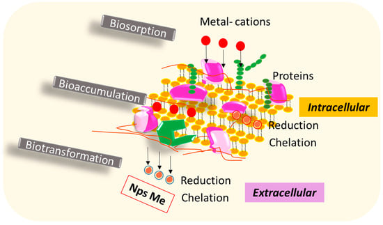
The biosynthesis of metal nanoparticles using fungal cells can employ one of two mechanisms: (i) intracellular or (ii) extracellular synthesis routes. In intracellular synthesis, nanoparticles are formed and localized within the cytoplasm, cell wall, or cell membrane [
110]. Initially, the metal ions, which serve as nanoparticle precursors, interact with oppositely charged cell surface components, where they can be simultaneously reduced to form nanoparticles, while remaining attached to the cell surface. These nanoparticles may then migrate to the cell membrane or cytoplasm. Alternatively, the ions may be internalized through active or passive transport mechanisms and subsequently reduced by intracellular reducing agents [
111]. In the extracellular synthesis routes, fungal proteins, enzymes, cofactors, and metabolites like organic acids (e.g., citric acid, oxalic acid) play vital roles in the organism’s survival and contribute to the reduction of metal ions into nanoparticulate forms.
Figure 2 provides a schematic representation illustrating the interaction between metals and the fungal cell surface.
Figure 2. Biosynthesis of metal nanoparticles using fungal cells from e-waste.
Numerous groups of fungi are employed for the synthesis of nanoparticles from WPCBs, with
Fusarium oxysporum emerging as a prominent organism capable of producing a variety of manufactured nanoparticles. The synthesis of silver nanoparticles (AgNPs) by
F. oxysporum is primarily dependent on a reductase associated with NADH [
4,
112].
A mixture of
Fusarium oxysporum and
Bacillus cereus from e-waste synthesized Cu, Zn, Cd, and Au nanoparticles. The nanoparticles varied in size, but most of them possessed a diameter of less than 10 nm. They exhibited different shapes, including circular, triangular, and complex [
113].
Trichoderma spp.,
Aspergillus spp.,
Penicillium spp., and
Verticillium spp. are additional fungi that offer numerous advantages. These include diverse survival strategies, ease of biomass handling, the capacity to multiply using straightforward culture media, tolerance to high metal concentrations, and enhanced productivity in terms of nanoparticle production from WPCBs [
114].
New research could be carried out, specifically using WPCBs as precursors and employing this fungal route for its multiple advantages in the synthesis of nanoparticles.
7. Algae
Algae represent another species known for its role in the bioreduction of metals, facilitating the production of gold and silver nanoparticles. This capability arises from the presence of specific functional groups, such as carboxyl, on their cell walls [
8], along with the secretion of a polysaccharide called fucoidan, which aids in the intracellular synthesis of gold nanoparticles [
97].
Table 2 summarizes the relevant studies regarding nanoparticle reduction via the use of algae.
Table 2. Studies for nanoparticle reduction employing different algae.
Chlorella vulgaris, a unicellular microalga typically found in carbonate and bicarbonate-rich lakes, is widely recognized for its effectiveness in bioreducing metal ions. This is primarily attributed to the production and secretion of cellular reductases by the microalgal cells into their growth medium. These enzymes demonstrate remarkable efficiency in reducing silver ions in order to form spherical silver nanoparticles (SNPs). Depending on the pH conditions, this process results in monodispersed SNPs at low and neutral pH levels and nanorod structures in alkaline conditions [
117].
Other algal species, including
Rhizoclonium hieroglyphicum,
Lyngbya majuscule, and
Spirulina subsalsa, have also been identified for their capacity to visibly indicate gold reduction, specifically from Au(III) to Au(0), at both the intra- and extracellular levels. This is evidenced by a color change in their biomass to purple [
116].
Green synthesis is presented as a sustainable alternative to traditional methods for the valorization of WPCBs. Within the green techniques mentioned here, some possess important aspects that are presented here as advantages; for example, obtaining nanoparticles using organic acids reduces synthesis times; however, in general, the stability of the nanoparticles obtained, along with their biocompatibility, is reduced. On the other hand, the use of microorganisms, such as bacteria and fungi, provide nanoparticles with defined and stable formats; however, the production cycles are broader, and there are limitations, including their inability to operate at high pulp densities, thus limiting their potential profitability. The use of plant extracts generates stable nanoparticles, but studies report the proportion of the plant extract and the chemical solution as the primary factor affecting the size of the NPs, along with their stability. Impurities of the extracts, limitations in process engineering, and operation stability are considered significant issues. Algae have demonstrated efficiency in the synthesis and stability in regards to the nanoparticles generated, but they present limitations concerning variability in the synthesis processes.
Finally, most of the waste generated in green metal extraction processes, as well as that derived from the synthesis of NPs, can be treated, or valorized, using three major processes, i.e., thermochemical, biological, and mechanical. The thermochemical type includes pyrolysis, gasification, and combustion processes, among others. The biological processes include fermentation, digestion, and the production of enzymes, and finally, the mechanical processes refer to operations such as drying, grinding, and pelletizing, in which added value can be provided to the waste generated in the green metal extraction processes. However, thermochemical processes remain the most required, due to their reduction of waste volumes and their recovery of molten metals [
118].


