Caspase-Dependent Programmed Cell Death: History
Please note this is an old version of this entry, which may differ significantly from the current revision.

Billions of cells die in us every hour, and our tissues do not shrink because there is a natural regulation where Cell Death (CD) is balanced with cell division. The process in which cells eliminate themselves in a controlled manner is called Programmed Cell Death (PCD). The PCD plays an important role during embryonic development, in maintaining homeostasis of the body’s tissues, and in the elimination of damaged cells, under a wide range of physiological and developmental stimuli. Apoptosis is an RCD pathway that occurs inside eukaryotic cells and whose purpose is the death of the cell itself. Apoptosis is a “cellular suicide” in which a protein program of self-destruction triggered by extracellular or intracellular signals is set in motion. RCD means that the steps for cell degeneration are established, but that does not mean that the cell is predetermined to die; that is, there will be no apoptosis if there is no signal to initiate it. The role of apoptosis is important in many physiological and pathological processes of multicellular organisms, such as the morphogenesis of organs and tissues during embryonic development, in the maintenance and regeneration of tissues in the adult animal, in response to pathogens, or as a response to cellular stress and pathologies such as cancer. The number of cells that die by apoptosis is enormous, both during embryonic development and in the adult state, associated with caspases, that not only control apoptosis, but also proliferation, differentiation, cell form and cell migration.

  • Programmed Cell Death
  • caspase-dependent
  • Anoikis
  • Catastrophe Mitotic
  • Pyroptosis
  • Emperitosis
  • Parthanatos

1. Anoikis

Anoikis means “to be without a home” in Greek, and was first coined by Frisch and Francis in 1994 to describe CD induced by the interruption or absence of interactions between epithelial cells and the extracellular matrix (ECM) [1]. Anoikis ultimately leads to intrinsic apoptosis [2]. Anoikis is a mechanism that prevents the formation of cancer cells, preventing cells that have detached from the ECM from colonizing different adjacent organs [3]. In this way, the key episodes that can be observed in Anoikis are downregulation of the β1 integrin and Epidermal Growth Factor Receptor (EGFR), inhibition of extracellular signaling regulated by Extracellular Signal-Regulated Kinase 1 (ERK1), and overexpression of Bcl-2 Interacting Mediator of CD (Bim) protein [4].
In contrast to the aforementioned, “Anoikis resistance” is mediated by two pathways linked to the anchorage-independent growth proteins and the Epithelial–Mesenchymal Transition (EMT) [5]. Oncogenic EMT is necessary for the tumor metastasis [6]. A better understanding of how the EMT occurs oncogenic would enable the creation of pharmacological compounds that suppress EMT, and perhaps Anoikis sensitivity as well, and this would constitute an important therapeutic advance [7]. This research could establish E-cadherin signaling pathways as targets, because they regulate and contribute to tumor progression through [7]: (i) the Wnt way, through β-catenin and T cell-specific transcription factors (TCF); (ii) the signaling pathway that begins in ankyrin; and (iii) the Hippo way.

2. Mitotic Catastrophe

The term “mitotic catastrophe” is a cell death event that results from the premature entry or inappropriate release of the cell into mitosis. In this way, the cell acts as a onco-suppressive mechanism that can occur during or after mitosis; being a mode of cell death that precedes apoptosis, necrosis, or senescence [8][9]. Mitotic catastrophe can be induced by very heterogeneous stimuli; when the cells detect damage to DNA, chromosomes or detect some disturbance in the mitotic apparatus, they arrest the cell cycle and can undergo apoptosis or senescence [10]. However, when cells cannot sustain the cell cycle arrest in the Growth 2 (G2) phase and enter mitosis before the DNA repair process can be terminated, this premature initiation of mitosis leads to mitotic catastrophe and apoptosis [11]. Kimura et al. [10] have investigated the induction of mitotic catastrophe through the disruption of the organization of the spindle [12]; that is, via Small Interfering RNA/Short Interfering RNA/Silencing RNA (siRNA)-mediated depletion of six centrosome proteins (Aurora A, nine in, Aspartate Transaminase (AST/GOT), Transforming Acidic Coiled-Coil Containing Protein 3 (TACC3), and γ-Tubulin, Pericentriolar Material 1 (PCM1)), and observed that this process requires Spindle Assembly Checkpoint (SAC) and Checkpoint Kinase 2 (Chk2) proteins. They also found that while p73 has an important role in mitotic catastrophe, p53 does not stand alone. It is thought that the transcription in M phase is largely inactive because p53 does not regulate transcriptional activation. In contrast, CD after mitosis can be regulated by transcriptional activation of p53 or inducible by p21–p53 protein [10].
Mitotic catastrophe induced by DNA damage presents apoptotic characteristics, such as mitochondrial membrane permeabilization, Annexin V binding, nuclear condensation, and activation of caspases-2, -3, and -9, but not caspase-8; this CD can also cause apoptosis independent of caspases by control spindle activation in Budding Uninhibited by Benzimidazoles 1 (Bub1)-deficient cells [13]. Furthermore, multiple pro-apoptotic B-Cell Lymphoma 2 (BCL-2) family members are involved in the execution of mitotic catastrophe and subsequent apoptosis through the inhibition of Aurora A or Checkpoint Kinase 1 (Chk1) [10][14]. Also, Vakifahmetoglu et al. [15] observed that in the mitotic catastrophe induced by ionizing radiation in HeLa cells instead of nuclear fragmentation, the cells were characterized by a large increase in size due to the accumulation of multiple micronuclei that subsequently present similar characteristics to the necrotic CD. Cell Division Cycle 5-Like Protein (CDC5L) modulates pre-Messenger Ribonucleic Acid (pre-mRNA) splicing expression of a set of genes involved in mitosis and in response to DNA damage. In this way, the suppression or depletion of CDC5L inhibits mitotic progression and would induce mitotic catastrophe [16]. These results would make CDC5L a key regulator of mitotic progression and highlight the potential of CDC5L as a target for cancer therapy [17]. In this sense, CDC5L is highly expressed in cervical tumors, bladder cancer, gliomas, and osteosarcoma [18].

3. Pyroptosis

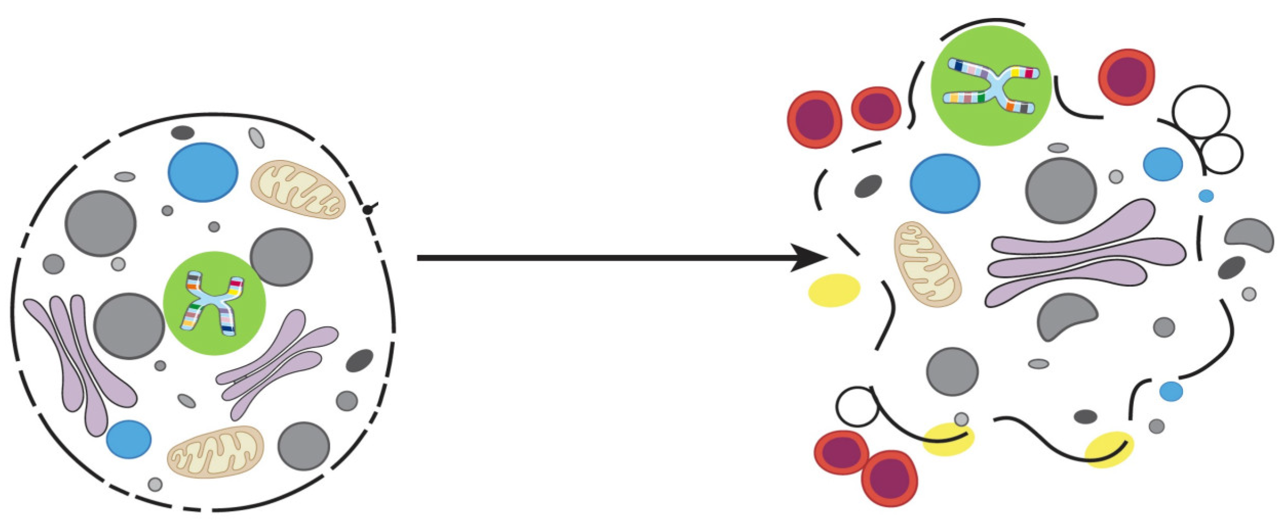
Pyroptosis (Figure 1) was first described in Shigella flexneri-infected macrophages by Zychlinsky et al. [18], and soon after, a similar phenotype was observed in from infection with Salmonella typhimurium [19]. Pyroptosis was subsequently shown to be distinct from apoptosis. Pyro comes from the Greek word for “fire”, and ptosis which means “fall”; the apparent meaning of the combined word is “the fall of fire” which, in this case, refers to the process of pro-inflammatory chemical signals, such as an immune response mechanism, which leads to fever and inflammation and ultimately leads to cell lysis and release of cytosolic contents into extracellular space [20]. Both pyroptosis and apoptosis are PCDs that depend on different caspases. However, caspases-1 and -11 are inflammatory, and are involved only in Pyroptosis and not in apoptosis [21]. Also, Pyroptosis may also require caspase-7, but not caspase-3 [22].
Figure 1. Pyroptosis.
Pyroptosis occurs mainly in inflammatory cells such as macrophages, and can be triggered by bacterial or pathogenic infections [20]. Miao et al. [23] demonstrated in vivo that pyroptosis is an innate immunological mechanism that protects the animal completely from infection with a powerful cleaning which would otherwise be lethal. These investigators [23] have observed that macrophages infected with Salmonella typhimurium rapidly activate caspase-1 and undergo pyroptosis. The lysis of the macrophages infected intracellularly by Salmonella are subsequently phagocytosed and destroyed by neutrophils [19].
The molecular mechanisms involved in pyroptosis depend on two types of pathogen receptors that belong to different families, nucleotide receptors (NLRs) and Toll-like receptors (TLR), though the latter are not enough to trigger pyroptosis by themselves [24]. In this way, inflammasomes, such as NLR family CARD Domain-Containing Protein 3 (NLRP3), NLR Family CARD Domain-Containing Protein 4 (NLRC4), and Interferon-Inducible or Absent in Melanoma 2 Protein (AIM2), are cytosolic sensors that detect pathogens or danger signals and activate caspase-1, which is essential in the process of pyroptosis, since it is responsible for the maturation and the secretion of proinflammatory cytokines such as Interleukin-1β (IL-1β) and Interleukin-18 (IL-18), leading to pyroptosis [21]. Therefore, cells suffering from pyroptosis increase the release of IL-1β and IL-18 [21].

4. Emperitosis

The term emperitosis comes from the contraction of the words “emperiopolesis” and “apoptosis”, derived from the Greek, where em means “within”, peri is “around”, and ptosis is “dropped” [25]. Wang et al. [26] proposed this name to define the process of “cell-in-cell” CD carried out exclusively by immune cell killer cells, with cytotoxic activity that express granzyme B (GzmB), entering the tumor cells to kill them. That makes the GzmB molecule a necessary and essential element in this type of CD [25]. So, emperitosis does not include all cells of the immune system, but only those with cytotoxic activity, such as Cytotoxic T Lymphocytes (CD8+ T cells), natural killer (NK) cells, Cytokine-induced killer (CIK) cells, and lymphokine-activated killer (LAK) cells [26].
The common hallmark during the early stage of all “cell-in-cell” processes leads to the internalized cell enveloping itself in a vacuole within the target cell [26]. Therefore, the released GzmB goes directly into the cytoplasm of the target cell and will bind to caspases that induce fragmentation of the DNA and target cell apoptosis [27][28]. Wang et al. [26] tested to what extent Ala-Ala-Asp-Chloromethylketone (Z-AAD-CMK), an irreversible GzmB-specific inhibitor, interfered with its activity; and observed that it did not affect the formation of “cell-in-cell” structures or the release of GzmB, but inhibited the activation of caspase-3 significantly and, therefore, of apoptotic “cell-in-cell” death of killer cells.

5. Parthanatos

The name parthanatos comes from the poly (ADP-ribose) “PAR”. This molecule occurs mainly in the nucleus, and is a sign of pro-death. In this kind of death cell, PAR is associated with the term “thanatos” which, in Greek mythology, means the personification of death, implying that this CD is caused by said molecule [29]. The term “parthanatos” was first used by Valina Lynn Dawson’s research group [30].
Parthanatos is caspase-independent, and is biochemically and morphologically distinct from features of necrosis and apoptosis [30][31]. The enzyme poly (ADP-ribose) polymerase-1 (PARP-1) is a DNA repair enzyme that is normally activated by the genotoxic stress and DNA damage, along with p53 [31].
The PARP-1 has several functions. It is involved in DNA repair processes by adding multiple polymers of ADP-ribose [30]. It is also involved in the DNA transcription, mitosis, and CD [30], thus regulating a wide variety of physiological processes [32]. But the excessive PARP-1 activation leads to an intrinsic CD program, PARP-1 is translocated from the nucleus to the cytosol and interacts with the mitochondrial outer surface, where the release of mitochondrial Apoptosis-Inducing Factor (AIF) is induced [33][34]. The redox regulation (NADH-dependent oxidoreductase) of AIF [35] induces its dimerization and the formation of degradosome assemblies with histones and cyclophilin A [36] to exert the endonuclease activity and subsequent DNA cleavage. AIF has a high affinity for binding to poly (ADP-ribose), this union is critical and key in the process of parthanatos both in vitro and in vivo, since AIF is released from the mitochondria and translocated to the nucleus, where it triggers pyknosis and DNA fragmentation [30].
It is clear that a greater understanding of parthanatos opens new avenues for therapy in the improvement of diseases related to PARP-1 overactivation such as stroke, diabetes, inflammation, and neurodegeneration [30][31].

6. Cornification

The term cornification or keratinization derives from the Greek Keratos, which means “horn”. The first to describe keratin filaments were Fuchs et al. [37] in 1985. The keratinization process is one in which the epidermal cells undergo the terminal differentiation in which basal keratinocytes transform into highly specialized corneocytes for the formation of the horny layer, which is the outermost skin barrier responsible for keeping the body hydrated and protecting the organism against environmental aggressions excluding pathogens and toxins [38][39]. Cornification is a special form of CD programmed into the skin. Mismatches in this process lead to a variety of diseases, including skin cancers, ichthyosis, and psoriasis [38][40][41].
The epidermis is an organ in continuous self-renewal and differentiation. This process involves the expression of different genes that regulate keratinization [39]; so, the cornification is a well-organized and planned death characterized by: (i) the expression of transglutaminase (TGases), loricrin, involucrin, and keratins [42]; (ii) terminal differentiation. Detachment of keratinocytes from the membrane basal cell is one of the stimuli that initiates terminal differentiation; dead cells are not eliminated, but remain to form the horny barrier [37]; and (iii) the loss of the nucleus and cytoplasmic organelles in the final stages of the cornification [38]. Most importantly, keratinocytes activate the anti-apoptotic and anti-necroptotic pathways to prevent premature CD during terminal differentiation. This view shows cornification as a mode of CD that regulates homeostatic mechanisms in cells of the epidermis [43].

This entry is adapted from the peer-reviewed paper 10.3390/proteomes12010003

References

  1. Frisch, S.M.; Francis, H. Disruption of epithelial cell-matrix interactions induces apoptosis. J. Cell Biol. 1994, 124, 619–626.
  2. Woods, N.T.; Yamaguchi, H.; Lee, F.Y.; Bhalla, K.N.; Wang, H.G. Anoikis, initiated by Mcl-1 degradation and Bim induction, is deregulated during oncogenesis. Cancer Res. 2007, 67, 10744–10752.
  3. Reginato, M.J.; Mills, K.R.; Paulus, J.K.; Lynch, D.K.; Sgroi, D.C.; Debnath, J.; Muthuswamy, S.K.; Brugge, J.S. Integrins and EGFR coordinately regulate the pro-apoptotic protein Bim to prevent anoikis. Nat. Cell Biol. 2003, 5, 733–740.
  4. Frisch, S.M.; Screaton, R.A. Anoikis mechanisms. Curr. Opin. Cell Biol. 2001, 13, 555–562.
  5. Wang, J.; Luo, Z.; Lin, L.; Sui, X.; Yu, L.; Xu, C.; Zhang, R.; Zhao, Z.; Zhu, Q.; An, B.; et al. Anoikis-Associated Lung Cancer Metastasis: Mechanisms and Therapies. Cancers 2022, 14, 4791.
  6. Fernández-Lázaro, D.; Hernández, J.L.G.; García, A.C.; Martínez, A.C.; Mielgo-Ayuso, J.; Cruz-Hernández, J.J. Liquid Biopsy as Novel Tool in Precision Medicine: Origins, Properties, Identification and Clinical Perspective of Cancer’s Biomarkers. Diagnostics 2020, 10, 215.
  7. Frisch, S.M.; Schaller, M.; Cieply, B. Mechanisms that link the oncogenic epithelial-mesenchymal transition to suppression of anoikis. J. Cell Sci. 2013, 126, 21–29.
  8. Portugal, J.; Mansilla, S.; Bataller, M. Mechanisms of drug-induced mitotic catastrophe in cancer cells. Curr. Pharm. Des. 2010, 16, 69–78.
  9. Li, K.; Wu, D.; Chen, X.; Zhang, T.; Zhang, L.; Yi, Y.; Miao, Z.; Jin, N.; Bi, X.; Wang, H.; et al. Current and emerging biomarkers of cell death in human disease. Biomed. Res. Int. 2014, 2014, 690103.
  10. Kimura, M.; Yoshioka, T.; Saio, M.; Banno, Y.; Nagaoka, H.; Okano, Y. Mitotic catastrophe and cell death induced by depletion of centrosomal proteins. Cell Death Dis. 2013, 4, e603.
  11. Bucher, N.; Britten, C.D. G2 checkpoint abrogation and checkpoint kinase-1 targeting in the treatment of cancer. Br. J. Cancer 2008, 98, 523–528.
  12. Jordan, M.A.; Wilson, L. Microtubules as a target for anticancer drugs. Nat. Rev. Cancer 2004, 4, 253–265.
  13. Niikura, Y.; Dixit, A.; Scott, R.; Perkins, G.; Kitagawa, K. BUB1 mediation of caspase-independent mitotic death determines cell fate. J. Cell Biol. 2007, 178, 283–296.
  14. Huang, X.F.; Luo, S.K.; Xu, J.; Li, J.; Xu, D.R.; Wang, L.H.; Yan, M.; Wang, X.R.; Wan, X.B.; Zheng, F.M.; et al. Aurora kinase inhibitory VX-680 increases Bax/Bcl-2 ratio and induces apoptosis in Aurora-A-high acute myeloid leukemia. Blood 2008, 111, 2854–2865.
  15. Vakifahmetoglu, H.; Olsson, M.; Zhivotovsky, B. Death through a tragedy: Mitotic catastrophe. Cell Death Differ. 2008, 15, 1153–1162.
  16. Mu, R.; Wang, Y.B.; Wu, M.; Yang, Y.; Song, W.; Li, T.; Zhang, W.N.; Tan, B.; Li, A.L.; Wang, N.; et al. Depletion of pre-mRNA splicing factor Cdc5L inhibits mitotic progression and triggers mitotic catastrophe. Cell Death Dis. 2014, 5, e1151.
  17. Fernández-Lázaro, D.; Hernández, J.L.G.; García, A.C.; del Castillo, A.C.; Hueso, M.V.; Cruz-Hernández, J.J. Clinical Perspective and Translational Oncology of Liquid Biopsy. Diagnostics 2020, 10, 443.
  18. Zhang, Z.; Mao, W.; Wang, L.; Liu, M.; Zhang, W.; Wu, Y.; Zhang, J.; Mao, S.; Geng, J.; Yao, X. Depletion of CDC5L inhibits bladder cancer tumorigenesis. J. Cancer 2020, 11, 353–363.
  19. Monack, D.M.; Raupach, B.; Hromockyj, A.E.; Falkow, S. Salmonella typhimurium invasion induces apoptosis in infected macrophages. Proc. Natl. Acad. Sci. USA 1996, 93, 9833–9838.
  20. Cookson, B.T.; Brennan, M.A. Pro-inflammatory programmed cell death. Trends Microbiol. 2001, 9, 113–114.
  21. Miao, E.A.; Rajan, J.V.; Aderem, A. Caspase-1-induced pyroptotic cell death. Immunol. Rev. 2011, 243, 206–214.
  22. LaRock, C.N.; Cookson, B.T. Burning down the house: Cellular actions during pyroptosis. PloS Pathog. 2013, 9, e1003793.
  23. Miao, E.A.; Leaf, I.A.; Treuting, P.M.; Mao, D.P.; Dors, M.; Sarkar, A.; Warren, S.E.; Wewers, M.D.; Aderem, A. Caspase-1-induced pyroptosis is an innate immune effector mechanism against intracellular bacteria. Nat. Immunol. 2010, 11, 1136–1142.
  24. Bortoluci, K.R.; Medzhitov, R. Control of infection by pyroptosis and autophagy: Role of TLR and NLR. Cell Mol. Life Sci. 2010, 67, 1643–1651.
  25. Salvesen, G.S. Dying from within: Granzyme B converts entosis to emperitosis. Cell Death Differ. 2014, 21, 3–4.
  26. Wang, S.; He, M.F.; Chen, Y.H.; Wang, M.Y.; Yu, X.M.; Bai, J.; Zhu, H.Y.; Wang, Y.Y.; Zhao, H.; Mei, Q.; et al. Rapid reuptake of granzyme B leads to emperitosis: An apoptotic cell-in-cell death of immune killer cells inside tumor cells. Cell Death Dis. 2013, 4, e856.
  27. Darmon, A.J.; Nicholson, D.W.; Bleackley, R.C. Activation of the apoptotic protease CPP32 by cytotoxic T-cell-derived granzyme B. Nature 1995, 377, 446–448.
  28. Chinnaiyan, A.M.; Hanna, W.L.; Orth, K.; Duan, H.; Poirier, G.G.; Froelich, C.J.; Dixit, V.M. Cytotoxic T-cell-derived granzyme B activates the apoptotic protease ICE-LAP3. Curr. Biol. 1996, 6, 897–899.
  29. Harraz, M.M.; Dawson, T.M.; Dawson, V.L. Advances in neuronal cell death 2007. Stroke 2008, 39, 286–288.
  30. Wang, Y.; Dawson, V.L.; Dawson, T.M. Poly(ADP-ribose) signals to mitochondrial AIF: A key event in parthanatos. Exp. Neurol. 2009, 218, 193–202.
  31. David, K.K.; Andrabi, S.A.; Dawson, T.M.; Dawson, V.L. Parthanatos, a messenger of death. Front. Biosci. 2009, 14, 1116–1128.
  32. Jeggo, P.A. DNA repair: PARP—Another guardian angel? Curr. Biol. 1998, 8, R49–R51.
  33. Yu, S.W.; Andrabi, S.A.; Wang, H.; No, S.K.; Poirier, G.G.; Dawson, T.M.; Dawson, V.L. Apoptosis-inducing factor mediates poly(ADP-ribose) (PAR) polymer-induced cell death. Proc. Natl. Acad. Sci. USA 2006, 103, 18314–18319.
  34. Andrabi, S.A.; No, S.K.; Yu, S.W.; Wang, H.; Koh, D.W.; Sasaki, M.; Klaus, J.A.; Otsuka, T.; Zhang, Z.; Koehler, R.C.; et al. Poly(ADP-ribose) (PAR) polymer is a death signal. Proc. Natl. Acad. Sci. USA 2006, 103, 18308–18313.
  35. Villanueva, R.; Ferreira, P.; Marcuello, C.; Usón, A.; Miramar, M.D.; Peleato, M.L.; Lostao, A.; Susin, S.A.; Medina, M. Key Residues Regulating the Reductase Activity of the Human Mitochondrial Apoptosis Inducing Factor. Biochemistry 2015, 54, 5175–5184.
  36. Novo, N.; Romero-Tamayo, S.; Marcuello, C.; Boneta, S.; Blasco-Machin, I.; Velázquez-Campoy, A.; Villanueva, R.; Moreno-Loshuertos, R.; Lostao, A.; Medina, M.; et al. Beyond a platform protein for the degradosome assembly: The Apoptosis-Inducing Factor as efficient nuclease involved in chromatinolysis. PNAS Nexus 2022, 2, pgac312.
  37. Fuchs, E.; Hanukoglu, I.; Marchuk, D.; Grace, M.P.; Kim, K.H. The nature and significance of differential keratin gene expression. Ann. N. Y. Acad. Sci. 1985, 455, 436–450.
  38. Candi, E.; Schmidt, R.; Melino, G. The cornified envelope: A model of cell death in the skin. Nat. Rev. Mol. Cell Biol. 2005, 6, 328–340.
  39. Elias, P.M. Stratum corneum defensive functions: An integrated view. J. Investig. Dermatol. 2005, 125, 183–200.
  40. Descargues, P.; Sil, A.K.; Karin, M. IKKalpha, a critical regulator of epidermal differentiation and a suppressor of skin cancer. EMBO J. 2008, 27, 2639–2647.
  41. Wu, N.L.; Lee, T.A.; Tsai, T.L.; Lin, W.W. TRAIL-induced keratinocyte differentiation requires caspase activation and p63 expression. J. Investig. Dermatol. 2011, 131, 874–883.
  42. Martens, M.D.; Karch, J.; Gordon, J.W. The molecular mosaic of regulated cell death in the cardiovascular system. Biochim. Biophys. Acta Mol. Basis Dis. 2022, 868, 166297.
  43. Eckhart, L.; Lippens, S.; Tschachler, E.; Declercq, W. Cell death by cornification. Biochim. Biophys. Acta 2013, 1833, 3471–3480.
More
This entry is offline, you can click here to edit this entry!
Video Production Service