CKD Interplay with Comorbidities and Carbohydrate Metabolism: History
Please note this is an old version of this entry, which may differ significantly from the current revision.

Chronic kidney disease (CKD) poses a global health challenge, engendering various physiological and metabolic shifts that significantly impact health and escalate the susceptibility to severe illnesses. It is impacting populations worldwide causing health complications and increasing the risk of serious illnesses, with high mortality rates. CKD is associated with different complex deleterious changes in a patient’s physiology and metabolic activity. They include deteriorating function and/or subsequent kidney failure, uremia, irregularities in metabolism of amino acid, lipids, minerals, and homocysteine (leads to malnutrition, anemia, vitamin deficiency, dementia, stroke and heart diseases), metabolic acidosis, insulin resistance, inflammatory and oxidative stress, dysfunction of skeletal muscle and many more. Further, other diseases or disease-causing factors (diabetes and hypertension) which coexist within CKD are associated with deteriorating the health and mortality.

  • chronic kidney disease
  • cellular growth homeostasis
  • inflammation
  • insulin resistance
  • dietary interventions

1. Carbohydrate Metabolism and Its Impairment in CKD Patients

Carbohydrate metabolism is a fundamental process that regulates the utilization of glucose, a primary energy source, in the human body. This intricate system involves various biochemical pathways and is tightly regulated to maintain energy homeostasis [1]. The process begins with glycolysis, occurring in the cytoplasm, where glucose is broken down into pyruvate, generating a small amount of adenosine triphosphate (ATP) and nicotinamide adenine dinucleotide (NADH). Pyruvate can then enter the mitochondria for further metabolism in the TCA cycle, contributing to the production of more ATP through oxidative phosphorylation [2]. Insulin, a hormone secreted by the pancreas, plays a crucial role in glucose metabolism. Upon elevated blood glucose levels, insulin facilitates glucose uptake by cells, promoting its conversion to glycogen for storage in the liver and muscles. This process is known as glycogenesis, serving as a short-term energy reserve. Conversely, during fasting or periods of low glucose, glycogen undergoes glycogenolysis, releasing glucose back into the bloodstream to maintain blood glucose levels. The liver, in particular, plays a key role in regulating systemic glucose levels by balancing glycogen storage and release. In instances where glucose is scarce, gluconeogenesis occurs, predominantly in the liver and kidneys. This process synthesizes glucose from non-carbohydrate precursors, such as amino acids and glycerol, ensuring a steady supply of glucose even in fasting states [3].
The kidneys play a crucial role in glucose homeostasis by filtering and reabsorbing glucose from the glomerular filtrate. Proximal tubular cells, particularly in the S1 and S2 segments, actively reabsorb glucose through SGLT. This cotransport mechanism couples the uphill movement of sodium ions with the reabsorption of glucose. Subsequently, glucose exits the tubular cells via facilitative glucose transporters (GLUTs), returning to the bloodstream. The SGLT-mediated reabsorption ensures the efficient retrieval of glucose, preventing its excretion and maintaining glucose homeostasis in the body [4]. CKD and metabolic disturbances share a bidirectional relationship. CKD induces insulin resistance, dyslipidemia, and inflammation, promoting diabetes and cardiovascular complications. Simultaneously, metabolic abnormalities exacerbate kidney dysfunction. This intricate interplay emphasizes the importance of holistic management strategies addressing both CKD and metabolic disorders to improve patient outcomes [5]. Insulin resistance, a hallmark of CKD, arises when cells exhibit diminished responsiveness to insulin’s regulatory signals. CKD-induced factors, including inflammation, oxidative stress, and altered hormonal regulation, contribute to insulin resistance. Skeletal muscle, a major insulin target, experiences reduced glucose uptake, while the liver increases gluconeogenesis, elevating blood glucose levels. Insulin resistance in CKD is linked to metabolic complications, such as impaired glucose tolerance and an increased risk of developing diabetes mellitus. Managing insulin resistance becomes crucial in mitigating the progression of both CKD and associated metabolic disorders in affected individuals [6].
In a study, incidence of insulin resistance (IR) and assess impaired fasting glucose (IFG), impaired glucose tolerance (IGT), and diabetes mellitus (DM) in CKD patients was studied. Among 113 CKD patients, 59.3% had IFG, and 69% exhibited a carbohydrate metabolism disorder. Additionally, 23% of CKD patients had IR, with elevated levels of fasting blood glucose, insulin, triglycerides, and HbA1c. Notably, 31.3% of patients with IFG were found to have IR. The findings highlight the prevalence of metabolic disorders and insulin resistance in CKD patients, emphasizing the importance of screening for diabetes in this population [7].

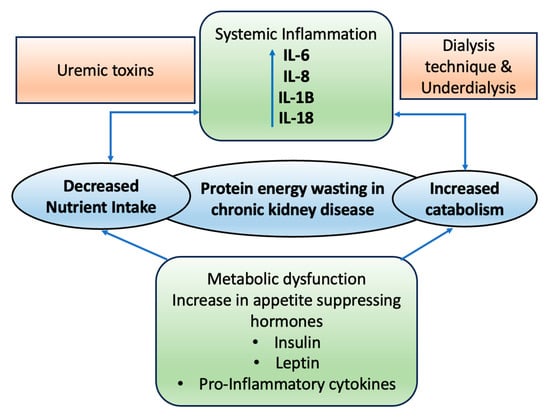
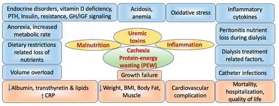
2. Role of CKD on Wasting and Malnutrition

CKD, characterized by the progressive decline in renal function, is an escalating health concern with potential public health implications. Malnutrition and wasting are prevalent issues in this patient population, significantly impacting morbidity, mortality, functional abilities, and overall quality of life [8][9].
Malnutrition typically refers to a poor nutritional state resulting from inadequate nutrient intake. However, in the context of CKD, major malnutrition can also stem from insufficient food intake, coupled with low levels of serum and tissue proteins, irrespective of body weight adherence to standard nutritional guidelines [10]. Various studies in children with CKD have reported a prevalence of malnutrition ranging from 20% to 45%, depending on the clinical criteria used [11][12][13]. Using the subjective global assessment scale (SGA), a recent study found that 31% of adults with CKD, including both dialysis and non-dialysis patients, experienced protein-energy wasting [14].
Wasting syndromes exhibit maladaptive reactions, such as loss of appetite and increased metabolism, which can lead to low serum albumin levels, weight loss, decreased muscle mass, and, intriguingly, the preservation or even an increase in fat mass (Figure 1). This complexity arises from various contributing factors [15][16]. The Society for Cachexia and Wasting Disorders (SCWD) describes cachexia or wasting as a complicated metabolic condition associated with underlying illnesses. It is marked by the loss of muscle, sometimes alongside the loss of fat [17]. In individuals with CKD, wasting can occur due to several reasons, such as not eating enough nutritious food, inflammation throughout the body, disruptions in hormonal balance, and irregular signaling from neuropeptides in the body [15][16][18][19][20][21][22][23].
Figure 1. Illustration depicting the factors leading to protein-energy wasting and the pathophysiological interactions in chronic kidney disease. IL = Interleukins.
The likelihood of muscle loss in CKD patients is linked to the stage of their condition. As CKD advances to stages 4 and 5, a decline in nutritional intake often goes hand in hand with worsening protein and energy levels in the body. This means that as kidney disease becomes more severe, people may struggle more with their nutrition and energy levels, which can contribute to muscle wasting [24]. As a result, muscle loss becomes more common in the later stages of kidney disease. In adults, a significant degree of muscle wasting (in stages 3 and 4) can be found in a range of 18% to 48%, and it can even climb as high as 75% in individuals with end-stage renal disease. This means that as kidney disease progresses, more and more people are affected by muscle loss, and the numbers can be quite substantial in severe cases [24][25][26].
The pathogenesis of malnutrition and wasting in patients with CKD is multifactorial, with several risk factors contributing to these conditions. Figure 2 representing the overview of the various factors involved in the pathophysiology of the CKD.
Figure 2. Schematic representation of protein-energy wasting (PEW) and the pathophysiological effects associated with kidney disease. BMI = Body mass index; CRP = C-reactive protein. Upward arrow = Increased levels; Downward arrow = Reduced levels.
Muscle protein degradation, which is responsible for muscle protein wasting, involves four major proteolytic pathways. These pathways include the ATP-dependent ubiquitin–proteasome system and the cathepsin system, which can facilitate comprehensive breakdown of cellular proteins into amino acids or small peptides. In contrast, the caspase-3 and cytosolic calcium-dependent calpain systems have limited proteolytic capabilities due to their restricted specificity [27].

3. Association of CKD Inflammation, Oxidative Stress and Cardiovascular Diseases

Inflammation and oxidative stress are common in patients with CKD and can exacerbate their clinical symptoms. Oxidative stress occurs when there is a disruption in the balance between harmful substances like reactive oxygen species (ROS) or reactive nitrogen species (RNS) and protective antioxidants in the body. These harmful substances, because of their molecular instability (such as having unpaired electrons), tend to trigger oxidation reactions with different molecules like proteins, lipids, and DNA in an attempt to stabilize themselves [28].
Key ROS include free radicals like hydroperoxide, the highly reactive hydroxyl radical and superoxide anion radical. Additionally, there are redox signaling agents like hydrogen peroxide (H2O2) and singlet oxygen (1O3), which, although lacking unpaired electrons, possess significant oxidizing properties. Oxidative stress reactions can also include oxygen compounds that team up with other elements, not just free radicals like the alkoxy radical, but also non-radical compounds such as peroxynitrite (ONOO) and hypochlorous acid (HOCl). These reactions demonstrate how various molecules can become involved in oxidative stress, not just those with unpaired electrons [29].
Oxidative stress can manifest through four distinct pathways: chlorinated stress, carbonyl stress classical oxidative stress, and nitrosative stress [30]. People with CKD/ESRD often experience higher levels of oxidative stress because their natural defense mechanisms against it are not working as well as they should. This heightened oxidative stress can lead to nucleic acid damage, increasing the risk of developing subsequent tumors. Oxidative stress in kidney and vascular tissues can also contribute to hypertension, and vice versa, creating a vicious circle of oxidative stress and hypertension [31].
Several factors contribute to heightened oxidative stress in individuals with CKD, including gut dysbiosis, hyperglycemia, dialysis, and inflammation [32][33][34][35].
CKD is known to be a condition marked by widespread inflammation, which is closely linked to increased oxidative stress. When the body experiences inflammation, cells that fight off infections release harmful reactive substances at these inflamed sites. On the flip side, the byproducts of oxidation and ROS encourage immune cells like macrophages and neutrophils to release inflammatory molecules and even more ROS. This sets up a continuous cycle where inflammation and oxidative stress fuel each other, creating a feedback loop. When immune cells, known as phagocytic cells, release ROS, they can prompt nearby non-immune cells to release inflammatory substances [36]. This means that when something causes a rise in oxidative stress, it can also trigger an inflammatory reaction, which in turn worsens oxidative stress, creating a back-and-forth cycle. Inflammation is characterized by increased levels of markers of inflammation, such as cytokines, acute phase proteins, and adhesion molecules. In this process, cells responsible for the body’s initial immune response play a central role. Many factors play a part in the ongoing state of inflammation in CKD, including things like the body making more inflammatory substances, oxidative stress, acidity in the body, frequent and long-lasting infections, disruptions in gut bacteria, and changes in how fat tissue works [37].
Research involving patients has shown connections between markers of inflammation and various complications associated with CKD. These complications include problems like malnutrition, calcium buildup in the coronary arteries, hardening of the arteries, irregular heart rhythms (atrial fibrillation), an enlarged left ventricle in the heart (left ventricular hypertrophy), heart failure, and a higher risk of death in CKD patients [38][39][40]. Furthermore, inflammation plays a role in worsening CKD, making the body less responsive to insulin, impairing the function of blood vessel linings, affecting the balance of minerals and bones, causing anemia, and making the body less receptive to erythropoietin (a hormone involved in red blood cell production) [41][42][43][44].
Chronic inflammation triggers oxidative stress, which is linked to an intensified oxygen reaction when immune cells like monocytes/macrophages and neutrophils are activated. The respiratory burst is a vital weapon in the body’s defense against harmful invaders like bacteria, fungi, and parasites. However, when the body is stuck in a state of ongoing inflammation, immune cells are overstimulated and become overzealous, producing an excessive amount of ROS. In CKD, the proinflammatory factors present cause oxidative stress levels to rise, and the body’s disrupted balance of redox compounds amplifies inflammation even more. These two processes effectively feed off each other, making the situation worse. For example, when the body releases pro-inflammatory cytokines like interleukin 6 (IL-6), it triggers an increase in the expression of Nox4. Conversely, elevated Nox4 expression stimulates the synthesis of IL-6 [45].
The connection between oxidative stress and inflammation is intricate and can work through different pathways, extending beyond well-known proinflammatory substances like tumor necrosis factor-alpha (TNF-α) or IL-6. For example, in the group undergoing hemodialysis (HD), researchers like Uchimura et al. (2001) and Santhanam et al. (2014) discovered a notable connection between advanced glycation end products (AGEs), specifically substances like pentosidine and N(6)-carboxymethyl lysine (which result from both the breakdown of fats and glycation reactions), and the levels of two proinflammatory cytokines: interleukin 18 (IL-18) and macrophage colony-stimulating factor (M-CSF) [46][47].
Several mechanisms are known to underlie the connection between elevated oxidative stress and the progression of both CKD and cardiovascular disease (CVD) in CKD patients. When a patient has kidney failure, it becomes an especially risky situation, particularly if they already had cardiovascular disease. Even in individuals with high blood pressure without kidney problems, there is a connection between their blood creatinine levels and the risk of heart and blood vessel issues, even when those creatinine levels are considered normal [28].
In a study with 1829 patients who had high blood pressure and healthy kidney function, the researchers found that over an 11-year period of monitoring, a rise in blood creatinine levels by 0.23 mg/dL was linked to a 30% increased likelihood of experiencing cardiovascular problems like heart attacks and strokes [48].

4. Effect of CKD on Functioning

4.1. Cognitive Dysfunction

Individuals dealing with CKD and ESRD are much more likely to experience cognitive difficulties compared to the general population, even when considering factors like age, diabetes, cardiovascular health, and other underlying health conditions as noted by Hailpern et al. (2007) and Yaffe et al. (2010) [49][50]. Additionally, multiple studies have established a link between dementia, mild cognitive impairment, and higher mortality rates among ESRD patients. For instance, in a study conducted by Kurella and colleagues in 2006 [51], they found that among patients undergoing HD, those who had dementia had a higher risk of death. Specifically, they reported that the risk of mortality was 1.48 times higher (with a confidence interval of 95%, ranging from 1.32 to 1.66) for HD patients with dementia compared to those without dementia. Similarly, in a study led by Griva and her team in 2010 [52], they followed 145 patients receiving dialysis who had not previously experienced dementia or strokes. Their findings showed that when comparing patients with mild-to-moderate cognitive issues to those without any cognitive deficits, there was an adjusted hazard ratio for all-cause mortality of 2.53. This means that the risk of death over 7 years was 2.53 times higher (with a 95% confidence interval ranging from 1.03 to 6.22) for those with cognitive impairment compared to those without such deficits.
It is important to note that routine cognitive testing is not typically conducted among CKD and ESRD patients. As a result, the prevalence of cognitive impairment in this population is estimated to range from 30% to 70% and is closely linked to the severity of kidney disease [53][54]. In a study led by Murray and colleagues in 2006 [55], they examined 338 hemodialysis patients aged 55 years or older. Surprisingly, only 2.9% of them had a history of prior cognitive impairment. However, when they assessed these patients for memory, executive function, and language skills, they found that 13.1% had mild cognitive issues, 36.1% had moderate impairment, and 37.3% had severe impairment. Shockingly, only 12.7% of the participants showed normal cognitive function. The exact pathophysiology of dementia and cognitive impairment in CKD remains incompletely understood but is likely multifactorial.
Apart from factors like toxins and metabolic imbalances that can lead to cognitive problems, it is important to consider the role of cerebrovascular disease, which can be a significant contributor. Cerebrovascular disease is more common in people with all stages of CKD compared to those without kidney problems, especially in individuals who are on long-term dialysis treatments. Although some of the higher risk of stroke in CKD and ESRD patients can be linked to common risk factors like diabetes, high blood pressure, cardiovascular disease, and prior strokes, it is important to note that these typical risk factors do not entirely explain the significantly increased risk observed in these individuals [56][57].
Apart from strokes, when researchers conducted brain magnetic resonance imaging (MRI) on dialysis patients, they found signs of cerebral atrophy and other issues in the white matter of the brain. These white matter problems have been linked to cognitive difficulties, even in patients who have not had a stroke before [58]. Individuals with ESRD appear to undergo changes in the structure of their blood vessels and experience increased stiffness in their arteries. These changes may be connected to factors such as having too much fluid in their bodies and disruptions in how calcium and phosphate are managed. Furthermore, the fluctuations in blood flow that occur during HD sessions can potentially make matters worse, possibly causing problems with blood supply to the brain and increasing the risk of stroke or other brain injuries [59][60].
In a cross-sectional study comparing 45 maintenance dialysis patients to 67 controls without known kidney disease, patients undergoing hemodialysis showed higher rates of white matter problems, brain shrinkage, and a significant presence of unnoticed areas of damage in the brain when compared to individuals in the control group [58]. Additionally, in research by Chou et al. (2013) [61], using advanced magnetic resonance imaging (MRI) methods that can detect subtle changes, researchers found that individuals with ESRD had more damage to the protective covering of nerve fibers (axonal demyelination) compared to people of a similar age who were healthy.

4.2. On Emotional Functioning

People dealing with CKD can experience an increased risk of developing or worsening psychological issues like depression and anxiety. This often happens because they face various stressors that take a toll on their emotional well-being. These stressors can involve adapting to strict dietary and fluid limitations, worrying about starting dialysis, and concerns about feeling like a burden to their caregivers [62][63]. Depression is a particularly common mental health challenge among people with CKD. A comprehensive review conducted by Palmer and colleagues in 2013 [64] evaluated depressive symptoms by using questionnaires administered by healthcare professionals for individuals at all stages of CKD. They discovered that the rate of depressive symptoms was the highest among those undergoing dialysis, reaching 39.3%. Among individuals with CKD, including those at stages 1–5, and among kidney transplant recipients, about 26.5% and 26.6%, respectively, reported experiencing symptoms of depression. Beyond the emotional distress that comes with these symptoms, it is worth noting that they are also linked to negative health consequences. When individuals with CKD also grapple with depression, it often leads to a lower overall quality of life. They tend to see a faster decline in their kidney function, measured as estimated glomerular filtration rate (eGFR), which accelerates the progression to ESRD. Additionally, they face a greater likelihood of hospitalization and a higher risk of mortality [65][66][67][68]. Information gathered from the CRIC study showed that the presence of depressive symptoms varied depending on the level of kidney function. Specifically, as kidney function, as measured by eGFR, decreased, the chances of experiencing depression became more likely [69].

4.3. On Taste Perception

Researchers had indicated the disturbance of taste (5 basic taste qualities) in CKD patients [70][71][72]. There are several factors believed to affect the sense of taste in CKD. These include the impact of kidney-related issues, the use of medications, changes in saliva composition, and variations in dietary habits and nutrition status. Additionally, factors like zinc deficiency can play a role in this as well [73]. Changes in taste are quite common in CKD, and they can influence how much people enjoy their food and what they choose to eat, which, in turn, can impact their nutritional well-being. Research has shown that these taste alterations can happen in CKD regardless of a person’s age or gender. Specifically, there tends to be difficulty in detecting sour, umami, and salty tastes. Manley et al. (2012) [72] and McMahon et al. (2014) [74] had reported that consuming more salt in your diet might raise the amount of sodium in your saliva for CKD patients. This, in turn, could make it harder for them to taste salt, as well as affect their ability to perceive other tastes due to increased solutes in their saliva, including sodium. These factors might have an impact on how someone perceives the overall flavor and how much they enjoy certain foods. For instance, difficulties in detecting umami could affect their interest in protein-rich foods, while trouble with salty taste perception might influence their salt intake. The information about how common taste problems are and what they are like for CKD patients can help guide future efforts to improve their sense of taste. It is important to note that the existing data on taste issues in CKD is somewhat limited. Most of the studies involved small groups of participants, and they mainly focused on individuals in the later stages of CKD.

This entry is adapted from the peer-reviewed paper 10.3390/life14010013

References

  1. Maughan, R.; McCommis, K.S.; Finck, B.N. Carbohydrate metabolism. Surgery 2009, 27, 6–10.
  2. McCommis, K.S.; Finck, B.N. Mitochondrial pyruvate transport: A historical perspective and future research directions. Biochem. J. 2015, 466, 443–454.
  3. Exton, J.H.; Park, C.R. Control of gluconeogenesis in liver: I. General features of gluconeogenesis in the perfused livers of rats. J. Biol. Chem. 1967, 242, 2622–2636.
  4. Vallon, V. Glucose transporters in the kidney in health and disease. Pflügers Archiv Eur. J. Physiol. 2020, 472, 1345–1370.
  5. Thomas, R.; Kanso, A.; Sedor, J.R. Chronic kidney disease and its complications. Prim. Care 2008, 35, 329–344.
  6. Yuen, D.A.; Stead, B.E.; Zhang, Y.; White, K.E.; Kabir, M.G.; Thai, K.; Advani, S.L.; Connelly, K.A.; Takano, T.; Zhu, L.; et al. eNOS deficiency predisposes podocytes to injury in diabetes. J. Am. Soc. Nephrol. 2012, 23, 1810–1823.
  7. Basturk, T.; Unsal, A. What is the frequency of carbohydrate metabolism disorder in CKD? J. Ren. Care 2012, 38, 15–21.
  8. Snyder, S.; Pendergraph, B. Detection and evaluation of chronic kidney disease. Am. Fam. Physician 2005, 72, 1723–1732.
  9. Stevens, P.E.; O’Donoghue, D.J.; De Lusignan, S.; Van Vlymen, J.; Klebe, B.; Middleton, R.; Hague, N.; New, J.; Farmer, C.K. Chronic kidney disease management in the United Kingdom: NE-OERICA project results. Kidney Int. 2007, 72, 92–99.
  10. Pupim, L.B.; Cuppari, L.; Ikizler, T.A. Nutrition and metabolism in kidney disease. Semin. Nephrol. 2006, 26, 134–157.
  11. Brem, A.S.; Lambert, C.; Hill, C.; Kitsen, J.; Shemin, D.G. Prevalence of protein malnutrition in children maintained on peritoneal dialysis. Pediatr. Nephrol. 2002, 17, 527–530.
  12. Sozeri, B.; Mir, S.; Kara, O.D.; Dincel, N. Growth impairment and nutritional status in children with chronic kidney disease. Iran J. Pediatr. 2011, 21, 271–277.
  13. Apostolou, A.; Printza, N.; Karagiozoglou-Lampoudi, T.; Dotis, J.; Papachristou, F. Nutrition assessment of children with advanced stages of chronic kidney disease-A single center study. Hippokratia 2014, 18, 212–216.
  14. Dai, L.; Mukai, H.; Lindholm, B.; Heimbürger, O.; Barany, P.; Stenvinkel, P.; Qureshi, A.R. Clinical global assessment of nutritional status as predictor of mortality in chronic kidney disease patients. PLoS ONE 2017, 12, e0186659.
  15. Mak, R.H.; Ikizler, A.T.; Kovesdy, C.P.; Raj, D.S.; Stenvinkel, P.; Kalantar-Zadeh, K. Wasting in chronic kidney disease. J. Cachexia Sarcopenia Muscle 2011, 2, 9–25.
  16. Mak, R.H.; Cheung, W.W.; Zhan, J.Y.; Shen, Q.; Foster, B.J. Cachexia and protein-energy wasting in children with chronic kidney disease. Pediatr. Nephrol. 2012, 27, 173–181.
  17. Evans, W.J.; Morley, J.E.; Argiles, J.; Bales, C.; Baracos, V.; Guttridge, D.; Jatoi, A.; Kalantar-Zadeh, K.; Lochs, H.; Mantovani, G.; et al. Cachexia: A new definition. Clin. Nutr. 2008, 27, 793–799.
  18. Mitch, W.E. Malnutrition: A frequent misdiagnosis for hemodialysis patients. J. Clin. Investig. 2002, 110, 437–439.
  19. Mitch, W.E. Insights into the abnormalities of chronic renal disease attributed to malnutrition. J. Am. Soc. Nephrol. 2002, 13 (Suppl. S1), S22–S27.
  20. Kalantar-Zadeh, K.; Ikizler, T.A.; Block, G.; Avram, M.M.; Kopple, J.D. Malnutrition-inflammation complex syndrome in dialysis patients: Causes and consequences. Am. J. Kidney Dis. 2003, 42, 864–881.
  21. Mak, R.H.; Cheung, W.; Cone, R.D.; Marks, D.L. Leptin and inflammation-associated cachexia in chronic kidney disease. Kidney Int. 2006, 69, 794–797.
  22. Ingulli, E.G.; Mak, R.H. Growth in children with chronic kidney disease: Role of nutrition, growth hormone, dialysis, and steroids. Curr. Opin. Pediatr. 2014, 26, 187–192.
  23. Mak, R.H. Cachexia in children with chronic kidney disease: Challenges in diagnosis and treatment. Curr. Opin. Support Palliat. Care 2016, 10, 293–297.
  24. Kovesdy, C.P.; Kopple, J.D.; Kalantar-Zadeh, K. Management of protein-energy wasting in non-dialysis-dependent chronic kidney disease: Reconciling low protein intake with nutritional therapy. Am. J. Clin. Nutr. 2013, 97, 1163–1177.
  25. Kopple, J.D.; Berg, R.; Houser, H.; Steinman, T.I.; Teschan, P. Nutritional status of patients with different levels of chronic renal insufficiency. Modification of diet in renal disease (MDRD) study group. Kidney Int. Suppl. 1989, 27, S184–S194.
  26. Hollinger, D.; Maroni, B.J.; Merrill, D.; Scherch, L.K.; Schulman, G.; Wang, S.R.; Zimmer, G.S. Relationship between nutritional status and the glomerular filtration rate: Results from the MDRD study. Kidney Int. 2000, 57, 1688–1703.
  27. Costelli, P.; Reffo, P.; Penna, F.; Autelli, R.; Bonelli, G.; Baccino, F.M. Ca-dependent proteolysis in muscle wasting. Int. J. Biochem. Cell Biol. 2005, 37, 2134–2146.
  28. Podkowińska, A.; Formanowicz, D. Chronic kidney disease as oxidative stress- and inflammatory-mediated cardiovascular disease. Antioxidants 2020, 9, 752.
  29. Poulianiti, K.P.; Kaltsatou, A.; Mitrou, G.I.; Jamurtas, A.Z.; Koutedakis, Y.; Maridaki, M.; Stefanidis, I.; Sakkas, G.K.; Karatzaferi, C. Systemic redox imbalance in chronic kidney disease. Oxid. Med. Cell. Longev. 2016, 2016, 8598253.
  30. Ling, X.C.; Kuo, K. Oxidative stress in chronic kidney disease. Ren. Replace. Ther. 2018, 4, 53.
  31. Descamps-Latscha, B.; Drüeke, T.; Witko-Sarsat, V. Dialysis-induced oxidative stress: Biological aspects, clinical consequences, and therapy. Semin. Dial. 2001, 14, 193–199.
  32. Wu, C.C.; Chen, J.S.; Wu, W.M.; Liao, T.N.; Chu, P.; Lin, S.H.; Chuang, C.H.; Lin, Y.F. Myeloperoxidase serves as a marker of oxidative stress during single haemodialysis session using two different biocompatible dialysis membranes. Nephrol. Dial. Transplant. 2005, 20, 1134–1139.
  33. Miranda-Díaz, A.G.; Pazarín-Villaseñor, L.; Yanowsky-Escatell, F.G.; Andrade-Sierra, J. Oxidative stress in diabetic nephropathy with early chronic kidney disease. J. Diabetes Res. 2016, 2016, 7047238.
  34. Mahmoodpoor, F.; Saadat, Y.R.; Barzegari, A.; Ardalan, M.; Vahed, S.Z. The impact of gut microbiota on kidney function and pathogenesis. Biomed. Pharmacother. 2017, 93, 412–419.
  35. San, A.; Fahim, M.; Campbell, K.; Hawley, C.M.; Johnson, D.W. The role of oxidative stress and systemic inflammation in kidney disease and its associated cardiovascular risk. Nov. Prospect. Oxidative Nitrosative Stress 2018, 8, 3.
  36. Canton, M.; Sánchez-Rodríguez, R.; Spera, I.; Venegas, F.C.; Favia, M.; Viola, A.; Castegna, A. Reactive oxygen species in macrophages: Sources and targets. Front. Immunol. 2021, 12, 734229.
  37. Mihai, S.; Codrici, E.; Popescu, I.D.; Enciu, A.M.; Albulescu, L.; Necula, L.G.; Mambet, C.; Anton, G.; Tanase, C. Inflammation-Related Mechanisms in Chronic Kidney Disease Prediction, Progression and Outcome. J. Immunol. Res. 2018, 2018, 2180373.
  38. Stenvinkel, P.; Heimburger, O.; Paultre, F.; Diczfalusy, U.; Wang, T.; Berglund, L.; Jogestrand, T. Strong association between malnutrition, inflammation and atherosclerosis in chronic renal failure. Kidney Int. 1999, 55, 1899–1911.
  39. Shlipak, M.G.; Fried, L.F.; Crump, C.; Bleyer, A.J.; Manolio, T.A.; Tracy, R.P.; Furberg, C.D.; Psaty, B.M. Elevations of inflammatory and procoagulant biomarkers in elderly persons with renal insufficiency. Circulation 2002, 107, 87–92.
  40. Gupta, J.; Mitra, N.; Kanetsky, P.A.; Devaney, J.; Wing, M.R.; Reilly, M.; Shah, V.O.; Balakrishnan, V.S.; Guzman, N.J.; Girndt, M.; et al. Association between albuminuria, kidney function and inflammatory biomarker profile in CKD in CRIC. Clin. J. Am. Soc. Nephrol. 2012, 7, 1938–1946.
  41. Clapp, B.R.; Hingorani, A.D.; Kharbanda, R.K.; Mohamed-Ali, V.; Stephens, J.W.; Vallance, P.; MacAllister, R.J. Inflammation-induced endothelial dysfunction involves reduced nitric oxide bioavailability and increased oxidant stress. Cardiovasc. Res. 2004, 64, 172–178.
  42. Shoelson, S.E.; Lee, J.; Goldfine, A.B. Inflammation and insulin resistance. J. Clin. Investig. 2006, 116, 1793–1801.
  43. Navarro-Gonzalez, J.F.; Mora-Fernandez, C.; Muros de Fuentes, M.; García-Pérez, J. Inflammatory molecules and pathways in the pathogenesis of diabetic nephropathy. Nat. Rev. Nephrol. 2011, 7, 327–340.
  44. Akchurin, O.M.; Kaskel, F. Update on inflammation in chronic kidney disease. Blood Purif. 2015, 39, 84–92.
  45. Biswas, S.K. Does the interdependence between oxidative stress and inflammation explain the antioxidant paradox? Oxid. Med. Cell. Longev. 2016, 2016, 5698931.
  46. Uchimura, T.; Nakano, K.; Hashiguchi, T.; Iwamoto, H.; Miura, K.; Yoshimura, Y.; Hanyu, N.; Hirata, K.; Imakuma, M.; Motomiya, Y.; et al. Elevation of N-(carboxymethyl) valine residue in hemoglobin of diabetic patients: Its role in the development of diabetic nephropathy. Diabetes Care 2001, 24, 891–896.
  47. Santhanam, A.V.; d’Uscio, L.V.; Katusic, Z.S. Erythropoietin increases bioavailability of tetrahydrobiopterin and protects cerebral microvasculature against oxidative stress induced by eNOS uncoupling. J. Neurochem. 2014, 131, 521–529.
  48. Schillaci, G.; Reboldi, G.; Verdecchia, P. High-normal serum creatinine concentration is a predictor of cardiovascular risk in essential hypertension. Arch. Intern. Med. 2001, 161, 886–891.
  49. Hailpern, S.M.; Melamed, M.L.; Cohen, H.W.; Hostetter, T.H. Moderate chronic kidney disease and cognitive function in adults 20 to 59 years of age: Third National Health and Nutrition Examination Survey (NHANES III). J. Am. Soc. Nephrol. 2007, 18, 2205–2213.
  50. Yaffe, K.; Ackerson, L.; Tamura, M.K.; Le Blanc, P.; Kusek, J.W.; Sehgal, A.R.; Cohen, D.; Anderson, C.; Appel, L.; DeSalvo, K.; et al. Chronic Renal Insufficiency Cohort Investigators. Chronic kidney disease and cognitive function in older adults: Findings from the Chronic Renal Insufficiency Cohort Cognitive Study. J. Am. Geriatr. Soc. 2010, 58, 338–345.
  51. Kurella, M.; Mapes, D.L.; Port, F.K.; Chertow, G.M. Correlates and outcomes of dementia among dialysis patients: The Dialysis Outcomes and Practice Patterns Study. Nephrol. Dial. Transplant. 2006, 21, 2543–2548.
  52. Griva, K.; Stygall, J.; Hankins, M.; Davenport, A.; Harrison, M.; Newman, S.P. Cognitive impairment and 7-year mortality in dialysis patients. Am. J. Kidney Dis. 2010, 56, 693–703.
  53. Kurella, M.C.; Glenn, M.; Luan, J.; Yaffe, K. Cognitive impairment in chronic kidney disease. J. Am. Geriatr. Soc. 2004, 52, 1863–1869.
  54. Murray, A.M.; Knopman, D.S. Cognitive impairment in CKD: No longer an occult burden. Am. J. Kidney Dis. 2010, 56, 615–618.
  55. Murray, A.M.; Tupper, D.E.; Knopman, D.S.; Gilbertson, D.T.; Pederson, S.L.; Li, S.; Smith, G.E.; Hochhalter, A.K.; Collins, A.J.; Kane, R.L. Cognitive impairment in hemodialysis patients is common. Neurology 2006, 67, 216–223.
  56. Weiner, D.E.; Tabatabai, S.; Tighiouart, H.; Elsayed, E.; Bansal, N.; Griffith, J.; Salem, D.N.; Levey, A.S.; Sarnak, M.J. Cardiovascular outcomes and all-cause mortality: Exploring the interaction between CKD and cardiovascular disease. Am. J. Kidney Dis. 2006, 48, 392–401.
  57. Matta, S.M.; Moreira, J.M.; Kummer, A.M.; Barbosa, I.G.; Teixeira, A.L.; Silva, A.C.S. Cognitive alterations in chronic kidney disease: An update. J. Bras. Nefrol. 2014, 36, 241–245.
  58. Drew, D.A.; Bhadelia, R.; Tighiouart, H.; Bovak, V.; Scott, T.M.; Lou, K.V.; Shaffi, K.; Weiner, D.E.; Sarnak, M.J. Anatomic brain disease in hemodialysis patients: A cross-sectional study. Am. J. Kidney Dis. 2013, 61, 271–278.
  59. Pringle, E.; Phillips, C.; Thijs, L.; Davidson, C.; Staessen, J.A.; de Leeuw, P.W.; Jaaskivi, M.; Nachev, C.; Parati, G.; T O’Brien, E.; et al. Systolic blood pressure variability as a risk factor for stroke and cardiovascular mortality in the elderly hypertensive population. J. Hypertens. 2003, 21, 2251–2257.
  60. Flythe, J.E.; Inrig, J.K.; Shafi, T.; Chang, T.I.; Cape, K.; Dinesh, K.; Kunaparaju, S.; Brunelli, S.M. Association of intradialytic blood pressure variability with increased all-cause and cardiovascular mortality in patients treated with long-term hemodialysis. Am. J. Kidney Dis. 2013, 61, 966–974.
  61. Chou, M.C.; Hsieh, T.J.; Lin, Y.L.; Hsieh, Y.T.; Li, W.Z.; Chang, J.M.; Ko, C.H.; Kao, E.F.; Jaw, T.S.; Liu, G.C. Widespread white matter alterations in patients with end-stage renal disease: A voxelwise diffusion tensor imaging study. Am. J. Neuroradiol. 2013, 34, 1945–1951.
  62. Kimmel, P.L. Depression in patients with chronic renal disease: What we know and what we need to know. J. Psychosom. Res. 2002, 53, 951–956.
  63. Davison, S.N.; Jhangri, G.S. Impact of pain and symptom burden on the health-related quality of life of hemodialysis patients. J. Pain Symptom Manag. 2010, 39, 477–485.
  64. Palmer, S.; Vecchio, M.; Craig, J.C.; Tonelli, M.; Johnson, D.W.; Nicolucci, A.; Pellegrini, F.; Saglimbene, V.; Logroscino, G.; Fishbane, S.; et al. Prevalence of depression in chronic kidney disease: Systematic review and meta-analysis of observational studies. Kidney Int. 2013, 84, 179–191.
  65. Hedayati, S.S.; Minhajuddin, A.T.; Afshar, M.; Toto, R.D.; Trivedi, M.H.; Rush, A.J. Association between major depressive episodes in patients with chronic kidney disease and initiation of dialysis, hospitalization, or death. JAMA 2010, 303, 1946–1953.
  66. Cukor, D.; Fruchter, Y.; Ver Halen, N.; Naidoo, S.; Patel, A.; Saggi, S.J. A preliminary investigation of depression and kidney functioning in patients with chronic kidney disease. Nephron Clin. Pract. 2012, 122, 139–145.
  67. Chiang, H.H.; Guo, H.R.; Livneh, H.; Lu, M.C.; Yen, M.L.; Tsai, T.Y. Increased risk of progression to dialysis or death in CKD patients with depressive symptoms: A prospective 3-year follow-up cohort study. J. Psychosom. Res. 2015, 79, 228–232.
  68. Kittiskulnam, P.; Sheshadri, A.; Johansen, K.L. Consequences of CKD on functioning. Semin. Nephrol. 2016, 36, 305–318.
  69. Fischer, M.J.; Xie, D.; Jordan, N.; Kop, W.J.; Krousel-Wood, M.; Tamura, M.K.; Kusek, J.W.; Ford, V.; Rosen, L.K.; Strauss, L.; et al. CRIC Study Group Investigators CRIC Study Group Investigators. Factors associated with depressive symptoms and use of antidepressant medications among participants in the Chronic Renal Insufficiency Cohort (CRIC) and Hispanic-CRIC Studies. Am. J. Kidney Dis. 2012, 60, 27–38.
  70. Middleton, R.A.; Allman-Farinelli, M.A. Taste sensitivity is altered in patients with chronic renal failure receiving continuous ambulatory peritoneal dialysis. J. Nutr. 1999, 129, 122–125.
  71. Kusaba, T.; Mori, Y.; Masami, O.; Hiroko, N.; Adachi, T.; Sugishita, C.; Sonomura, K.; Kimura, T.; Kishimoto, N.; Nakagawa, H.; et al. Sodium restriction improves the gustatory threshold for salty taste in patients with chronic kidney disease. Kidney Int. 2009, 76, 638–643.
  72. Manley, K.J.; Haryono, R.Y.; Keast, R.S. Taste changes and saliva composition in chronic kidney disease. Ren. Soc. Australas J. 2012, 8, 56–60.
  73. van der Eijk, I.; Allman Farinelli, M.A. Taste testing in renal patients. J. Ren. Nutr. 1997, 7, 3–9.
  74. McMahon, E.J.; Campbell, K.L.; Bauer, J.D. Taste perception in kidney disease and relationship to dietary sodium intake. Appetite 2014, 83, 236–241.
More
This entry is offline, you can click here to edit this entry!
Video Production Service