1. Scanning Electron Microscopy and Mineral Characterization
SEM makes use of secondary electron imaging to analyze the surface topology and morphology of micron/nanometer-scale minerals [
64]. For a comprehensive understanding of the microstructure and mineral components, SEM is usually combined with X-ray techniques to complement the acquired information [
65,
66,
67,
68]. The infrared spectroscopy method is helpful in identifying chemical species and determining the molecular structure of minerals. This technique has been widely used in mineral characterization [
69,
70,
71,
72].
One of the major quantitative analysis methods in mineral analysis is X-ray diffraction (XRD). It correlates the content of minerals with diffraction density, which helps in identifying and quantifying the minerals present in the sample [
73,
74]. For example, XRD can be used to analyze calcite and nahcolite in saline brine [
75], evaluate deposits by identifying minerals in phyllite [
76], examine the order degree of dolomite [
77], and study the content of calcite and dolomite in carbonate rocks [
78]. XRD is a rapid and accurate method for quantitative mineral analysis; however, some mineral compositional structures could lead to errors in analytical results [
73].
Combining qualitative analysis with quantification assessment methods can provide a better understanding of the investigated minerals. Such methods include SEM energy-dispersive spectroscopy (SEM–EDS) [
78,
79,
80,
81,
82], automated SEM mineral liberation analysis (SEM-MLA) [
83,
84,
85], and quantitative evaluation of minerals by scanning electron microscopy (QEMSCAN) [
86,
87,
88]. These methods incorporate a mineral quantitative analysis system by using an energy spectrometer and SEM. For accurate identification of minerals, backscattered electron (BSE) images are used, which can reflect the difference between the X-ray energy spectrum and mineral phase composition [
89,
90,
91]. The quantitative analysis of rare earth minerals is a challenging task with conventional identification methods, and the abovementioned techniques have attained rare earth mineral identification. The problems associated with the usage of these methods pertain to difficulties in application, promotion, and high measurement costs.
1.1. SEM Energy-Dispersive X-ray Spectroscopy (SEM–EDS)
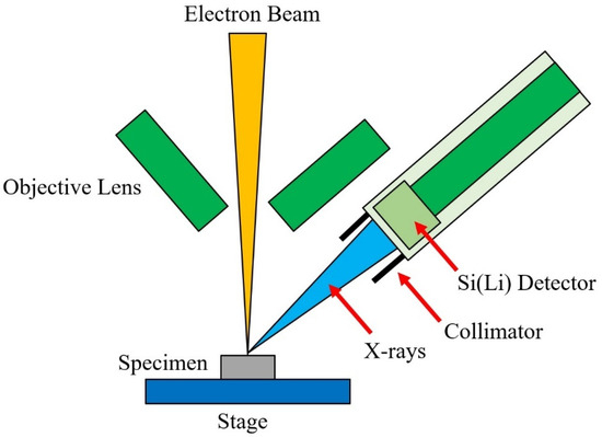
When the electron beam emitted from the gun penetrates and interacts with the volume beneath the sample surface, X-rays are generated. This is a well-established principle in physics: the deceleration of electrons due to their entrance into the Coulomb field of the specimen results in a loss of electron energy and emits photons. In SEM analysis, similar X-ray photons are emitted, which are characteristic of the sample under investigation [
89], as shown in
Figure 2.
The quantification scheme is achieved by measuring the X-ray intensity. This was illustrated by Heinrich and Yakowitz in 1968 in their publication,
Quantitative Electron Probe Microanalysis [
92], which later became the standard for developing X-ray fields. At that time, X-ray absorption, determination of correction factors at the instant of electron penetration and scattering, and conversion of X-ray intensity to the relative concentration were missing. Many problems pertaining to the electron probe field were solved with the development of energy-dispersive spectrometry (EDS). At present, various studies have incorporated SEM–EDS for qualitative and semiquantitative analysis in a variety of subject areas [
93,
94,
95,
96,
97,
98,
99].
A schematic diagram of an energy-dispersive spectrometer is shown in
Figure 5. The X-ray detection system (which is a solid-state detector) separates the characteristic X-rays of various elements present in the sample. Then, the EDS system software analyzes the energy spectrum to determine the amplitudes of particular elements, and electrical signals are generated from the respective photon energies. This results in qualitative and quantitative determination of a chemical composition map of the elements present in the sample [
89]. SEM–EDS has been used in a variety of fields for mineral characterization [
100,
101,
102,
103,
104,
105,
106].
Figure 5. Schematic diagram of an energy-dispersive spectrometer.
1.2. SEM-Based Automated Mineralogy (SEM-AM)
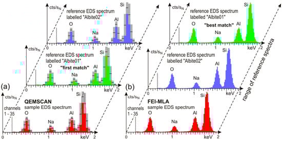
SEM-AM is a tool that was initially designed to characterize mineral processing products and ores. The measurement process starts with collecting backscattered electron (BSE) images, which are analyzed using image analysis software procedures. Based on BSE image adjustments, the energy-dispersive X-ray spectra (EDS) are obtained at selected points. The EDS spectra of the sample are then classified based on the list of approved reference EDS spectra. Relevant software providers offer services such as particle analysis, EDS spectral mapping, sparse phase search, and point counting modal analysis using four principal SEM-AM measurement routines and different classification algorithms, which can be used based on the analysis requirements. The main challenges associated with the process are materials with very different hardnesses, polishing relief surfaces of particles, electron beam stability, and appropriate nonevaporating epoxy resin mixtures [
84].
SEM-based automated mineralogy (SEM-AM) is still underutilized, although SEM instruments are widely distributed in industry, geosciences, and materials research. SEM-AM can produce valuable results for a variety of major applications by characterizing the primary ores and optimizing mineral concentration, flotation, comminution, and metallurgical processes in the mining industry through the generation of quantified reliable data [
107,
108,
109,
110,
111]. Beyond the classical fields, the potential of SEM-AM has gained further interest on scientific and economic grounds. Some closely related topics are ore fingerprinting, metallurgy, and applications in petrology [
112,
113,
114].
SEM-AM systems are a combination of hardware platforms, processing software, and specific image analysis. Any SEM with minor adjustments can be used as a hardware tool for SEM-AM. These adjustments include a high vacuum operation mode and additional internal mainboards. A vacuum pressure of 10
−5 to 10
−7 Pa is needed for its operation. Electron sources of tungsten cathodes and field emission guns can be employed. Tungsten cathodes can be used for economical operation; however, field emission guns are recommended for the long-term stability of electron beams for automated measurements. The speed of analysis and X-ray count rate are increased in SEM-AM by employing two or more EDS spectrometers in the SEM hardware. Multiple samples can be accommodated in a large sample chamber for simultaneous analysis in a single measurement session. A very accurate stage movement of SEM allows precise positioning using small intervals. For valuable analysis results, a fine-quality backscattered electron (BSE) detector is needed. In SEM-AM analysis, BSE image quality and stability are important factors, as the resultant image (in combination with the EDS spectrum) is used for phase or mineral discrimination. Prior to measurement, fixed working distances must be set to keep the BSE image gray levels constant [
84].
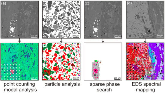
Keeping the image calibration constant ensures that a specific phase or mineral always possesses the same BSE image gray level. The calibration process can be conducted with various BSE image gray levels of reference materials such as quartz (dark gray), copper (intermediate), and gold (very bright) [
84]. The choice of calibration reference material should be made based on the sample material to be investigated. For example, many slags, industrial ash, or particulate materials are investigated using SEM-AM, and quartz or copper are used for calibration with dark gray to intermediate BSE image gray levels. This results in SEM images with better resolution and quality. For SEM-AM technology, four principal measurement routines can be outlined, which starts with collecting BSE gray-level images with respect to the calibrated gray level, as shown in
Figure 6. The upper row represents the BSE images, while the bottom row indicates the EDS images of SEM-AM of one measurement frame. White or black crosses denote the points of X-ray analyses (only some points are shown).
Figure 6 presents the EDS point counting technique used for the quantification of modal composition.
Figure 6 shows particle analysis by EDS, which has been developed for fast automated characterization of grain mounts with up to 10
6 particles, such as milled products from mineral processing and mining.
Figure 6 illustrates the sparse phase search method, which combines single spot EDS spectral analysis of grains with a BSE gray tone value trigger. It is valuable in massive rock applications, such as drill cores and thin sections.
Figure 6 demonstrates EDS spectral mapping, which combines BSE image levels with mapping of the EDS spectrum. This method is helpful, especially in cases where fine details of mineral intergrowth are considered. In summary, SEM-AM is a powerful tool for mineral characterization and has actively been used in recent literature [
115,
116,
117,
118].
Figure 6. SEM-AM methods of one measurement frame showing BSE (
upper row,
a–
d) and EDS (
lower row) images. Numerous single EDS analysis points map each grain with a distinguishable BSE gray level and are visualized as color-coded pixels, such as the garnet grain, which is indicated by red-colored pixels [
84]. CC-BY.
1.3. Automated SEM Mineral Liberation Analysis (SEM-MLA)
Recent software developments in SEM have incited dominant growth in its application in solid matter investigations. One of the economic solutions is the use of mineral liberation analysis (MLA) for optimizing the mineral processing methodology of metallic ores. SEM-MLA has been an important driver in transforming numerous software versions for SEM applications [
84]. SEM-MLA was designed to quantify the mineralogy of ores. After the mining process, the ore is processed to increase the concentration of minerals of interest (and value). The processing of ores is also important for removing minerals of no value or those with detrimental effects on the needed mineral products. This processing of grinding the ores and liberating the minerals of interest provided rapid automated analysis of target minerals and extensively improved the process.
A mineral liberation analyzer (MLA) based on SEM was developed in the late 1990s by the JKRMC (Julius Kruttschnitt Mineral Research Centre, Australia), and it is currently commercially available [
118]. In MLA, minerals are differentiated by attaining and combining the information gathered from EDS and BSE. Depending on the size range of the particles in the sample, size fractions from the sample are produced. Then, liberation is measured in each size fraction, followed by liberation reconstruction of the whole sample. The measurement of mineral liberation is usually carried out through one of two methods, i.e., either the area method or the linear intercept method. Liberation by area measurement has shown lower stereological error compared to linear measurement.
It is important to note that liberation measurements by the linear intercept method are known as one-dimensional, while area method measurements are called two-dimensional liberations. Both of the measured liberations are lower dimensional projections of the true volumetric liberation, which is three-dimensional. Stereological correction is based on stereological transformation and prediction of liberation measurements. This stereological correction can be based on entropy regularization [
119]. Correction of the apparent liberation and production of three-dimensional liberations have also been described in several other investigations [
120,
121,
122]. Various operating modes for the MLA system are available, i.e., X-ray modal analysis (XMOD), particle X-ray mapping (PXMAP), selected particle X-ray mapping (SXMAP), sparse phase liberation analysis (SPL), standard BSE liberation analysis (BSE), extended BSE liberation analysis (XBSE), and rare phase search (RPS) [
123]. The use of SEM-MLA is shown in
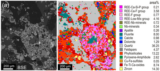
Figure 7 for quantifying the mineralogy of a hydrothermally overprinted alkali plutonite [
85].
Figure 7. (
a) SEM-MLA measurement of a hydrothermally overprinted alkali plutonite showing the backscattered electron (BSE) image and (
b) color-coded, grouped, and classified presentation of the frame presented in (
a) [
85]. CC-BY.
1.4. Quantitative Evaluation of Minerals by Scanning Electron Microscopy (QEMSCAN)
Traditional mineral analysis based on microscopy cannot provide the needed data because of the absence of quantitative information and the very small size of the particles of interest. QEMSCAN technology, initially termed QEM*SEM, demonstrated the potential to revolutionize automated mineralogy [
124]. In a mold, the particulate mineral sample is mixed with epoxy resin, and the sample surface is prepared using cutting, polishing, and carbon coating. The sample is scanned using SEM in backscatter mode, enabling the differentiation of particles from the background. After the identification of particles on the resin block, their composition is systematically mapped using EDS. In contrast to the most modern SEMs coupled with single EDX, QEMSCAN possesses the attribute of having multiple EDXs at the same time, enabling rapid quantitative mineralogy. The acquired EDX signals are then compared with reference known materials in the database and assigned a mineral name or to a chemical compositional grouping. With this process, the mineralogy of the sample can be determined by particle-by-particle analysis [
124]. QEMSCAN locates the particles using a BSE signal, while identifying the mineral by an EDS signal. It can be compared to SEM-MLA, which makes more use of the BSE signal than EDS for identifying the mineral. SEM-MLA works very well for bright phases (such as platinum group element minerals).
Among SEM–EDS techniques, QEMSCAN is one of the most widely used and offers quantitative characterization of minerals, ores, and other mineralogical compounds [
125,
126,
127,
128,
129]. QEMSCAN is usually used in conjunction with other analytical techniques, such as electron probe microanalysis (EPMA) and X-ray diffraction (XRD), as shown in
Table 1 [
130].
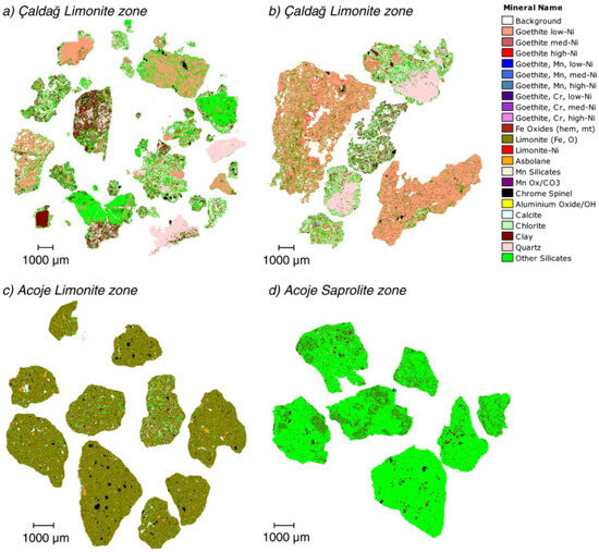
Figure 8 indicates the use of QEMSCAN for identifying the mineral distribution of four samples [
130]. It shows the presence of geothite, quartz, clay, limonite, and other silicate minerals.
Figure 8. QEMSCAN analysis indicating mineral distribution in four different zones [
130]. CC-BY.
Table 1. Techniques used for investigating mineralogy and their comparison. ✔ indicates good, ● represents poor, while ▲ suggests it is possible but not recommended [
130].
| Investigation |
Electron Microprobe |
XRD |
QEMSCAN |
| Mineral texture |
▲ |
● |
✔✔✔ |
| Mineral distribution and associations |
▲ |
● |
✔✔ |
| Mineral-specific particle size information |
▲ |
● |
✔✔ |
| Mineral abundance |
● |
✔✔✔ |
✔✔✔ |
| Amorphous minerals (geothite, silica) |
✔✔✔ |
● |
✔✔✔ |
| Distribution of minor metals within minerals |
✔✔✔ |
● |
✔ |
| Crystallinity (clay, silica, geothite, and limonite) |
● |
✔✔✔ |
● |
This entry is adapted from the peer-reviewed paper 10.3390/app132312600