Role of Akt/Protein Kinase B in Cancer Metastasis: History
Please note this is an old version of this entry, which may differ significantly from the current revision.
Contributor: , ,

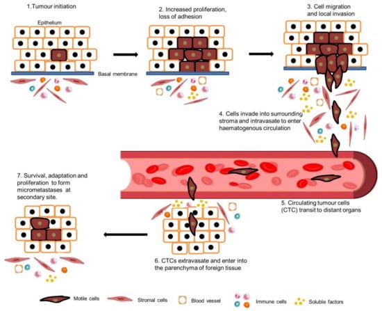
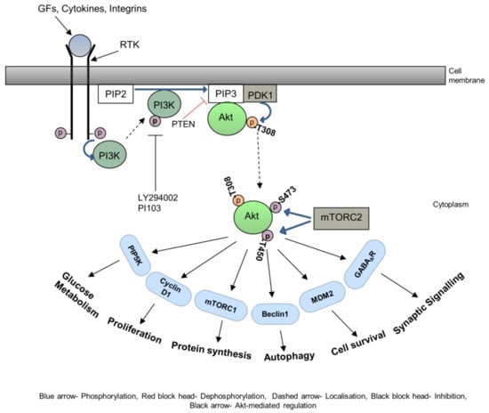
Metastasis is a critical step in the process of carcinogenesis and a vast majority of cancer-related mortalities result from metastatic disease that is resistant to current therapies. Cell migration and invasion are the first steps of the metastasis process, which mainly occurs by two important biological mechanisms, i.e., cytoskeletal remodelling and epithelial to mesenchymal transition (EMT). Akt (also known as protein kinase B) is a central signalling molecule of the PI3K-Akt signalling pathway. Aberrant activation of this pathway has been identified in a wide range of cancers. Several studies have revealed that Akt actively engages with the migratory process in motile cells, including metastatic cancer cells. The downstream signalling mechanism of Akt in cell migration depends upon the tumour type, sites, and intracellular localisation of activated Akt.

  • :Akt
  • cancer
  • metastasis
  • HNSCC
  • EMT

1. Introduction

The primary reason for cancer-related deaths is metastatic disease [1]. The spreading of tumour cells from the primary lesion is the main cause for the mortality and morbidity of cancer patients, whether it exists at the time of diagnosis, progresses during treatment, or happens at the time of disease relapse [2]. The metastasis process involve a series of sequential, interconnected steps including: separation of tumour cells from the primary lesion and invasion of neighbouring, healthy connective tissue, intravasation into the blood and lymphatic vessels, circulation through the blood vessels (circulating tumour cells) to other tissues in the body, extravasation from the blood vessel into the new tissue, growth in specific distant organs, and building a secondary tumour [3][4][5] (Figure 1).
Figure 1. Metastasis cascade. Tumour cells proliferate uncontrollably and eventually lose their adhesive phenotype. Tumour cells then migrate and invade into surrounding tissues induced by the tumour microenvironment and intravasate to lymphatic and blood vessels. Circulating tumour cells then extravasate, enter into another tissue, and form micro-metastases at the secondary site.
Recently, a novel ecological dispersal model of multidirectional cancer progression is proposed by Luo [6]. Taking nasopharyngeal cancer metastasis as an example, Luo hypothesized that the “nature of NPC is not a genetic disease but an ecological disease: A multidimensional spatiotemporal unity of ecological and evolution pathological ecosystem”. To adapt to the selective pressure from the remodelling microenvironment, NPC cells with cancer stem cells (CSCs) characteristics undergo EMT to dissociate from budding cells (tumour–host interface) and interplay with the local primary ecosystem (various stroma components); intravasate and survive into the circulation, and extravasate to circulating ecosystem (lymph node or a distant metastatic site); developing a distant metastatic ecosystem by entering slow-cycling states of dormancy, evading immune response, constructing organ-specific niches to colonise micro/macro metastases and later spread; self-feeding by CTCs or metastatic cells seeding at a distant site or secreting exosomes, cytokines, and chemokines and creating a multidirectional ecosystem by host cells including CAFs and immune cells to return to the primary tumour [6].
Cell migration through tissues results from highly integrated multistep cellular events [7][8][9]. First, the moving cell polarises, elongates, and extends protrusions in the way of migration reacting to migration-promoting agents. There are two types of protrusions, which can be spike-like filopodia, or large and broad lamellipodia. Protrusions are typically guided by actin polymerisation and are stabilised by adhering to the extracellular matrix or adjacent cells via related transmembrane receptors [10].
Variable experimental behaviour and histological patterns of tumour cells suggest that tumour cells can utilise different cellular and molecular modes of migration based on cell-type-specific autonomous mechanisms and reactive mechanisms stimulated by the local microenvironments [11][12]. Tumour cells are detected as both single cells and organized collective sheets in malignant cancer patients, indicating that cancer cells exhibit the plasticity to switch between single and collective cell migration. Studies on single cell migration have founded the cellular and molecular basis, providing a significant understanding into the spreading of tumours whose cells migrate constitutively as single cells such as leukaemia or lymphomas, after separation from cohesive lesions through the epithelial to mesenchymal transition (EMT) [9][13]. Collective cell migration occurs when the junctions between cells are retained over extended periods of time, so cells are adherent to their neighbours. The efficiency of the metastatic process is increased by the transition to single cell migration. However, circulating grouped tumour cells detected in the patient peripheral blood samples suggests that the intravasation process can also be enacted by a cell cluster [14][15]. Cell migration is the first step to invasion. The extracellular matrix is degraded by invasive cells via proteolysis before entering neighbouring tissues [16][17].
Highly integrated multistep cellular events lead to cell migration and invasion through tissues that are regulated by various cell signalling pathways, including the PI3K-Akt signalling pathway. The serine/threonine kinase Akt is also known as protein kinase B (PKB). It was originally discovered as a proto-oncogene. Akt plays a significant regulatory role in various cellular activities including cell survival, cell migration and invasion progression, insulin metabolism, and protein synthesis and has thus become a focus of major attention. The Akt signalling pathway is activated by receptor tyrosine kinases (RTK), cytokine receptors, G-protein coupled receptors, integrins, B and T cells receptors, and other stimuli that stimulate the production of phosphatidylinositol 3,4,5, triphosphates (PIP3) through phosphoinositide 3-kinase (PI3K) [18]. The PI3 kinases are a set of lipid kinases that phosphorylate the membrane phospholipid, phosphatidylinositol 4,5 biphosphate (PIP2), generating phosphatidylinositol 3,4,5, triphosphates (PIP3). PIP3 controls a range of effector molecules including the Akt group of oncogenic kinases termed Akt1, Akt2, and Akt3. The activation of Akt1, a 60 kDa kinase, depends on PI3K [19]. An increase in cellular PIP3 by PI3K eventually allows the activation of Akt1 by phosphorylation at Thr308 and Ser473 residues [20]. This activation is completed by structural modification stimulated by PI3K-dependent kinase-1 (PDK-1)-dependent phosphorylation at Thr308 and stabilisation by mTORC2 or DNA-PK (DNA-activated protein kinase) dependent phosphorylation at Ser473 [21][22][23]. A third phosphorylation site on Akt1 has been identified at Thr450 [24]. This site is referred to as the turn phosphorylation site and is controlled by mTORC2 activity [25][26]. The activation of the three Akt isoforms plays a pivotal role in fundamental cellular functions such as protein synthesis, cell survival, proliferation, and autophagy by regulating a variety of downstream substrates such as mTORC1, MDM2, Cyclin D1, and Beclin1, respectively [18][27][28] (Figure 2).
Figure 2. PI3K-Akt signalling pathway. Upon ligand binding, conformational changes occur in the receptor tyrosine kinase (RTK), the PI3 kinases are then activated by RTK and translocate to the plasma membrane. Activated PI3K then converts PIP2 to PIP3. Pleckstrin homology (PH) domain containing protein, Akt then translocate to the membrane, bind to PIP3, and phosphorylate at the Threonine 308 residue by PDK1. Akt translocates back to the cytoplasm and is phosphorylated further at Serine 473 and Threonine 450 residues by mTORC2. Activated Akt is responsible for initiating various cellular activities such as proliferation, protein synthesis, autophagy, cell survival, etc.
There are frequent alterations of the PI3K-Akt pathway in various types of human cancers. Amplification of the PIK3C gene encoding PI3K or the Akt gene lead to the constitutive activation of the PI3K-Akt pathway. PTEN (phosphatase and tensin homologue deleted on chromosome 10) can inhibit the Akt activation, and mutation in the PTEN gene also causes the constitutive activation of Akt [29][30][31]. Recent evidence has also suggested that Akt plays an important role in cancer cell migration and invasion [32][33]

2. Akt in Cytoskeletal Rearrangements

The cytoskeleton is the supporting structure of cells which is composed of a filamentous network of micro filaments such as actin and myosin, intermediate filaments such as vimentin and keratin, and microtubules such as tubulin [34]. The main purpose of the cytoskeleton is to maintain cellular structure, intracellular transport, and supporting cell division. Cytoskeletal rearrangements occur in various physiological and pathological events such as cell movement, wound healing, and cancer metastasis [35]. Cellular motility either in physiological events or in pathological conditions is driven by cytoskeletal remodelling, initiated by various signalling pathways. The synergistic effect of all the three basic elements—filamentous actin, microtubules, and the intermediate filament vimentin—is the potential basis for a cell to migrate [32]. Wide-ranging studies have focused on how the stabilisation of intracellular filaments and dynamic polymerisation control cell migration [36][37].
Growth of the vascular network is essential for the spread of cancer cells. Angiogenesis is the process whereby new vessels are formed and involved in the supply of nutrients, oxygen, and immune cells and also the removal of waste products [38]. Angiogenic factors play a huge role in neoplastic vascularisation, thus increasingly gaining attention. Vascular endothelial cell migration is a vital step for angiogenesis. Vascular endothelial growth factor (VEGF) activates Akt and stimulates the migration of endothelial cells by increasing actin polymerisation. Abrogated Akt activity by expression of a kinase-dead mutant inhibits actin bundle formation and blocks cell migration. This effect is enhanced when myristylated Akt is expressed [39], demonstrating that Akt is a critical mediator of VEGF-induced endothelial cell migration through actin reorganisation.
In chicken embryonic fibroblasts (CEF), PI3K-transduced migratory signal was blocked by inhibiting Akt activity. PI3K also activated p70S6K1 via Rac and induced actin filament remodelling and cell migration in CEF cells. This study confirms that the activation of PI3K activity alone is adequate to remodel actin filaments to increase cell migration through the activation of Akt and p70S6K1 in CEF cells [40].
The actin-rich structure of highly motile cells like invadopodia, filopodia, and pseudopodia needs to be stabilised to function properly. Actin-associated proteins are responsible for stabilising this actin structure by blocking the degradation of newly formed actin filaments [41]. ALE (the Akt phosphorylation enhancer), also termed the ‘girder’ of actin filaments (Girdin), is one of the best examples of this type of protein. APE/Girdin provides the integrity of the actin meshwork (actin filament) at the leading edge of migrating cells. Reduction in APE/Girdin destabilises the actin bundles, triggering the ablation of stress fibres and actin structure. This results in the loss of directional migratory ability and establishes the vital activity of APE/Girdin in the regulation of cell migration. Enomoto et al. proved that APE/Girdin is phosphorylated by Akt on Serine 1416 (S1416) [42].
An actin-associated structural (cross-linker) protein, filamin A, is phosphorylated by Akt on residue S2152 [43][44][45]. In turn, phosphorylated filamin A mediates caveolin-1-induced cancer cell migration through the IGF signalling pathway [46][47]. Akt has been shown to phosphorylate NHE1 (sodium-hydrogen exchanger isoform 1), a key mediator of stress fibre disassembly on S648 and suggested to be critical for the growth factor-induced cytoskeletal rearrangements that favour cell migration and invasion [48]
Extensive studies have been carried out to investigate the role of intermediate filaments in cell motility [49][50]. The most abundant intermediate protein that maintains normal cell and tissue integrity is called vimentin, a type three filamentous protein. It is phosphorylated by Akt1 on residue S39, stabilised, and thereby regulates cancer cell invasion in aggressive sarcoma [51]. It has also been shown that vimentin is highly expressed in breast cancer lung metastases [52][53]; however, the specific mechanisms to control cell migration by some Akt substrates are still undefined. For example, S-phase kinase-associated protein 2 (skp2), a component of E3 ligase, is phosphorylated by Akt on the S72 residue, stimulates Skp-2 dependent ligase activity, and induces cell migration [54][55].
Akt interacts with promigratory proteins, in addition to targeting cytoskeletal proteins, thus facilitating crosstalk between associated signalling axes. The VEGFR/eNOS signalling pathway-controlled cell migration is dependent on Akt-mediated phosphorylation on S1177 [56]. Accumulating evidence has indicated the importance of nitric oxide (NO) in pathological conditions, especially in malignant tumours [57][58]. Furthermore, VEGFR signalling often cooperates with the G-protein coupled receptor, sphingosine-1-phosphate receptor 1 (SIPR1, also known as endothelial differentiation gene 1, EDG-1). SIP/SIPR1 activation leads to the phosphor-activation of VEGFR which phosphorylates Src kinase, consequently activating the PI3K/Akt/eNOS axis [59]. Akt-mediated phosphorylation of SIPR1 on T236 further enhances their activity and stimulates cortical actin assembly, angiogenesis, and chemotaxis [60][61]. Thus, Akt plays a vital role in regulating VEGFR and the SIP/SIPR1 signalling pathway and actively regulates cell migration. EphA2 (Ephrin receptor tyrosine kinase A2), a member of the largest tyrosine kinase family, is also phosphorylated by Akt on S897 residue.
It is now well established that membrane redistribution of integrin by various signalling pathways is a critical mediator of cellular movement. The ANK repeat and pleckstrin homology domain-containing protein 1 (ACAP 1) is a GTPase activating protein (GAP) for ADP ribosylation factor 6 (ARF6) known to participate in integrin beta recycling. ACAP1 is phosphorylated by Akt on S554, stimulates integrin recycling, and therefore promotes cell migration [62]. Another GTPase activation protein, RhoGAP22, is shown to be phosphorylated by Akt on S16, upon stimulation by insulin or possibly PDGF, and this plays a vital role in regulating cell migration, leading to modulation of Rac1 activity [63]. Various studies have established the role of the mammalian targets of rapamycin complex 1 (mTORC1) in the cell migration and relationship with Akt [64][65].

3. Akt in EMT

Epithelial cells are tightly connected to their adjacent cells via E-cadherin and with actin filaments via α- or β-catenin. Epithelial tumour cells must break these intercellular junctions before migrating as single cells and invading stromal tissues. Epithelial tumour cells undergo a process named epithelial to mesenchymal transition (EMT) to facilitate the invasion as a single cell. The EMT process can be induced either by extracellular growth factors, for example EGF, TGF-α and β, FGF, or by intracellular cues, such as oncogenic Ras [66][67]. Epithelial cells gain a mesenchymal phenotype by losing their polarity and cell–cell contacts during EMT. Functional loss of E-Cadherin and downregulation of epithelial cell markers such as cytokeratins and ZO-1, and the overexpression of mesenchymal or fibroblast cell markers such as N-cadherin, vimentin, and fibronectin are the main characteristics of EMT [68][69]. EMT is a complex biological process that plays a critical role in cancer metastasis. In head and neck cancer, EMT can be involved in the dissemination of cancer cells to distant sites. However, it is important to understand that EMT is not an all-or-nothing phenomenon; there are partial or hybrid states of EMT that can have unique implications for cancer metastasis.
Partial EMT (p-EMT) is a term used to describe a state in which cancer cells exhibit some, but not all, of the characteristics associated with a full EMT [70]. p-EMT can enhance the invasive capacity of cancer cells. These cells may have an increased ability to break away from the primary tumour as a group of cells and infiltrate surrounding tissues, which is a crucial step in metastasis. Partially EMT-activated cancer cells might be less susceptible to apoptosis. This allows them to survive in the bloodstream and at distant site, where they might otherwise be eliminated by the body’s natural defences [71][72]. Cells which undergo p-EMT may also evade the immune system to some extent, making it more challenging for the body to recognize and destroy these cells. Partial EMT can also contribute to the formation of a pre-metastatic niche at distant sites.
EMT is reversible and, sometimes, cells undergo the reciprocal mesenchymal to epithelial transition (MET). During the development process, EMT plays an essential role in the development of various tissues and organs such as the heart, neural crest, and peripheral nervous and musculoskeletal systems. Only a small number of cells in adult organisms have the ability to go through the EMT process in specific physiological or pathological events such as wound healing. Nevertheless, tumour cells often gain the ability to reactivate the EMT process during metastasis, which enhances the migration and invasion capacity of cancer cells [69][73]. A number of studies have reported that Akt is frequently activated in human carcinomas [74][75][76][77][78]. Akt2 has been shown to be activated in ovarian carcinoma, affecting epithelial cell morphology, tumorigenicity, cell motility, and invasiveness, which is characterised by the loss of histological features of epithelial differentiation [79]. Evidence that Akt was shown to regulateEMT was first published in 2003, where squamous cell carcinoma cells, overexpressing an activated mutant of Akt, were shown to undergo EMT and downregulate E-cadherin [80]. Loss of E-cadherin and relocalisation of β-catenin from the membrane to the nucleus is frequently detected in tumour cells undergoing EMT [81][82]. Several transcription factors have been recognized that can induce and maintain the EMT process, including Snail, Twist, and Zeb. The definitive molecular signalling mechanisms of normal regulation of these transcription factors are still uncertain; however, they are apparently deregulated in many invasive cancers [68][83]. Evidence suggests a strong relationship between Akt and EMT-inducing transcription factors. Snail is phosphorylated by GSK3β (glycogen synthase kinase 3 beta) in normal epithelial cells but is very unstable and hardly detectable. Expression of Snail in epithelial cells strongly induces morphological changes associated with enhanced migratory capacity [84][85].
Y-box binding protein-1 (YB-1), a transcription/translation regulatory protein, is reported to be activated by Akt and translocated to the nucleus. Nuclear YB-1 thus phosphorylates Snail and decreases E-cadherin expression, which in turn induces EMT in invasive breast carcinoma [86]. Furthermore, upregulated Snail could, in turn, increase Akt activity. Snail increases the binding of Akt2 to the E-cadherin (CDH1) promoter and Akt2 interference unexpectedly inhibits Snail repression of the CDH1 gene [87]. Akt2 could also be activated by another EMT-inducer, Twist, in invasive breast cancer cells [88]. Inhibition of Akt also downregulates Twist in cancer cells [89]. Furthermore, Akt phosphorylates and activates Twist1, which in turn enhances the phosphorylation of Akt because of increased TGFβ signalling in human breast cancer [90][91][92]. Data also suggest that the polycomb group protein named B lymphoma Mo-MLV insertion region 1 homolog (Bmi1) is a downstream target of Twist1 and is crucial for EMT and cancer metastasis [93]. Akt can phosphorylate Bmi1 directly in high-grade prostate tumours [94]. Promotion of Akt activity by Bmi1 was also found to promote EMT by blocking the GSK3β-mediated degradation of Snail in HNSCC and breast cancer [95][96].

4. Akt in HNSCC Metastasis

Head and neck squamous cell carcinoma (HNSCC) denotes epithelial tumours that develop in the oral cavity, pharynx, larynx, and nasal cavity. The main risk factors of HNSCC are alcohol and tobacco use and HPV infection [97][98]. It is the seventh most common cancer worldwide, with more than 887,000 cases and 450,000 deaths every year (accumulation of different head and neck cancer sites) [99]. It has recently been shown that Akt is persistently activated in the vast majority of HNSCC cases. Active forms of Akt (phosphorylated) can readily be detected in both experimental and human HNSCCs and HNSCC-derived cell lines [100][101][102]. Akt can be phosphorylated, hence activated by different growth factors, chemokines, integrins, etc., and their respective receptors, ras mutations, PI3Ka gene amplification, overexpression, or activating mutations. Akt can also be activated by aberrant PTEN activity due to genetic alterations or reduced expression in HNSCC [103][104]. Akt activation is an early event in HNSCC progression which can be identified in as many as 50% of tongue preneoplastic lesions [105]. Akt activation also represents an independent prognostic marker of poor clinical outcome in both tongue and oropharyngeal HNSCCs [105][106] and is linked with the conversion of a potentially malignant oral lesion to OSCC (oral squamous cell carcinoma) [107].
Akt is known to induce morphological changes associated with EMT, loss of cell–cell adhesion, and increased motility and invasion in HNSCC [68]. Oral carcinoma cells, of epithelial origin, ectopically express a mesenchyme-specific transcription factor (HMGA2) at the invasive front, which has a significant impact on tumour progression and patient survival [108]. However, the definitive evidence that EMT was induced by Akt was provided by a study in which oral squamous cell carcinoma cell lines overexpressing activated mutant Akt were shown to undergo EMT and downregulate E-cadherin [80]. Snail and SIP1 exhibit an inverse correlation with E-cadherin expression levels in oral carcinoma cells [109][110]. An OSCC clone with stable Snail overexpression displayed spindle morphology, amplified expression of vimentin, and reduced expression of E-cadherin [111].

5. Conclusions

Extensive studies have demonstrated that the activation of Akt by phosphorylation of different amino acid residues determines substrate selectivity and thus exerts different biological activity in different cell types. Three highly homologous Akt isoforms have non-overlapping and opposing functions in different cancer types. As Akt is the central signalling node that incorporates cell membrane, cytoplasmic and nuclear signals regulating cell fate, analysing Akt isoforms and cell-type-specific signalling pathways and targeting them will contribute to personalised targeted HNSCC therapy. Thus, carefully designing a clinical study using a combination of a PI3K-Akt pathway inhibitor and another signalling molecule inhibitor or receptor inhibitor during the early stages of HNSCC might result in an expected positive outcome.

This entry is adapted from the peer-reviewed paper 10.3390/biomedicines11113001

References

  1. Dillekås, H.; Rogers, M.S.; Straume, O. Are 90% of deaths from cancer caused by metastases? Cancer Med. 2019, 8, 5574–5576.
  2. Palmer, T.D.; Ashby, W.J.; Lewis, J.D.; Zijlstra, A. Targeting tumor cell motility to prevent metastasis. Adv. Drug Deliv. Rev. 2011, 63, 568–581.
  3. Leber, M.F.; Efferth, T. Molecular principles of cancer invasion and metastasis (review). Int. J. Oncol. 2009, 34, 881–895.
  4. Robert, J. Biology of cancer metastasis. Bull. Cancer 2013, 100, 333–342.
  5. Woodhouse, E.C.; Chuaqui, R.F.; Liotta, L.A. General mechanisms of metastasis. Cancer 1997, 80, 1529–1537.
  6. Luo, W. Nasopharyngeal carcinoma ecology theory: Cancer as multidimensional spatiotemporal “unity of ecology and evolution” pathological ecosystem. Theranostics 2023, 13, 1607–1631.
  7. Friedl, P.; Brocker, E.B. The biology of cell locomotion within three-dimensional extracellular matrix. Cell. Mol. Life Sci. 2000, 57, 41–64.
  8. Lauffenburger, D.A.; Horwitz, A.F. Cell migration: A physically integrated molecular process. Cell 1996, 84, 359–369.
  9. Ridley, A.J.; Schwartz, M.A.; Burridge, K.; Firtel, R.A.; Ginsberg, M.H.; Borisy, G.; Parsons, J.T.; Horwitz, A.R. Cell Migration: Integrating Signals from Front to Back. Science 2003, 302, 1704–1709.
  10. Mitchison, T.J.; Cramer, L.P. Actin-based cell motility and cell locomotion. Cell 1996, 84, 371–379.
  11. Ahmed, H.; Ghoshal, A.; Jones, S.; Ellis, I.; Islam, M. Head and Neck Cancer Metastasis and the Effect of the Local Soluble Factors, from the Microenvironment, on Signalling Pathways: Is It All about the Akt? Cancers 2020, 12, 2093.
  12. Ellis, I.R.; Islam, M.R.; Aljorani, L.; Jones, S.J. Fibronectin: The N-terminal region and its role in cell migration- implications for disease and healing. In Fibronectin: Current Concepts in Structure, Function and Pathology; Beattie, J., Ed.; Protein Biochemistry, Synthesis, Structure and Cellular Functions; Nova Science Publishers: New York, NY, USA, 2012; pp. 35–69.
  13. Friedl, P. Prespecification and plasticity: Shifting mechanisms of cell migration. Curr. Opin. Cell Biol. 2004, 16, 14–23.
  14. Inaki, M.; Vishnu, S.; Cliffe, A.; Rorth, P. Effective guidance of collective migration based on differences in cell states. Proc. Natl. Acad. Sci. USA 2012, 109, 2027–2032.
  15. Rorth, P. Whence directionality: Guidance mechanisms in solitary and collective cell migration. Dev. Cell 2011, 20, 9–18.
  16. Friedl, P.; Wolf, K. Tumour-cell invasion and migration: Diversity and escape mechanisms. Nat. Rev. Cancer 2003, 3, 362–374.
  17. Friedl, P.; Locker, J.; Sahai, E.; Segall, J.E. Classifying collective cancer cell invasion. Nat. Cell Biol. 2012, 14, 777–783.
  18. Manning, B.D.; Cantley, L.C. AKT/PKB signaling: Navigating downstream. Cell 2007, 129, 1261–1274.
  19. Datta, S.R.; Brunet, A.; Greenberg, M.E. Cellular survival: A play in three Akts. Genes Dev. 1999, 13, 2905–2927.
  20. Alessi, D.R.; Cohen, P. Mechanism of activation and function of protein kinase B. Curr. Opin. Genet. Dev. 1998, 8, 55–62.
  21. Bozulic, L.; Hemmings, B.A. PIKKing on PKB: Regulation of PKB activity by phosphorylation. Curr. Opin. Cell Biol. 2009, 21, 256–261.
  22. Feng, J.; Park, J.; Cron, P.; Hess, D.; Hemmings, B.A. Identification of a PKB/Akt Hydrophobic Motif Ser-473 Kinase as DNA-dependent Protein Kinase. J. Biol. Chem. 2004, 279, 41189–41196.
  23. Sarbassov, D.D.; Guertin, D.A.; Ali, S.M.; Sabatini, D.M. Phosphorylation and regulation of Akt/PKB by the rictor-mTOR complex. Science 2005, 307, 1098–1101.
  24. Bellacosa, A.; Chan, T.O.; Ahmed, N.N.; Datta, K.; Malstrom, S.; Stokoe, D.; McCormick, F.; Feng, J.; Tsichlis, P. Akt activation by growth factors is a multiple-step process: The role of the PH domain. Oncogene 1998, 17, 313–325.
  25. Hart, J.R.; Vogt, P.K. Phosphorylation of AKT: A mutational analysis. Oncotarget 2011, 2, 467–476.
  26. Ikenoue, T.; Inoki, K.; Yang, Q.; Zhou, X.; Guan, K.-L. Essential function of TORC2 in PKC and Akt turn motif phosphorylation, maturation and signalling. EMBO J. 2008, 27, 1919–1931. Available online: http://www.nature.com/emboj/journal/v27/n14/suppinfo/emboj2008119a_S1.html (accessed on 5 September 2023).
  27. Liu, P.; Cheng, H.; Roberts, T.M.; Zhao, J.J. Targeting the phosphoinositide 3-kinase pathway in cancer. Nat. Rev. Drug Discov. 2009, 8, 627–644.
  28. Wang, R.C.; Wei, Y.; An, Z.; Zou, Z.; Xiao, G.; Bhagat, G.; White, M.; Reichelt, J.; Levine, B. Akt-mediated regulation of autophagy and tumorigenesis through Beclin 1 phosphorylation. Science 2012, 338, 956–959.
  29. De Marco, C.; Rinaldo, N.; Bruni, P.; Malzoni, C.; Zullo, F.; Fabiani, F.; Losito, S.; Scrima, M.; Marino, F.Z.; Franco, R.; et al. Multiple genetic alterations within the PI3K pathway are responsible for AKT activation in patients with ovarian carcinoma. PLoS ONE 2013, 8, e55362.
  30. Gonzalez-Angulo, A.M.; Ferrer-Lozano, J.; Stemke-Hale, K.; Sahin, A.; Liu, S.; Barrera, J.A.; Burgues, O.; Lluch, A.M.; Chen, H.; Hortobagyi, G.N. PI3K pathway mutations and PTEN levels in primary and metastatic breast cancer. Mol. Cancer Ther. 2011, 10, 1093–1101.
  31. Wu, R.; Baker, S.J.; Hu, T.C.; Norman, K.M.; Fearon, E.R.; Cho, K.R. Type I to Type II Ovarian Carcinoma Progression: Mutant Trp53 or Pik3ca Confers a More Aggressive Tumor Phenotype in a Mouse Model of Ovarian Cancer. Am. J. Pathol. 2013, 182, 1391–1399.
  32. Xue, G.; Hemmings, B.A. PKB/Akt-Dependent Regulation of Cell Motility. J. Natl. Cancer Inst. 2013, 105, 393–404.
  33. Yoeli-Lerner, M.; Toker, A. Akt/PKB Signaling in Cancer: A Function in Cell Motility and Invasion. Cell Cycle 2006, 5, 603–605.
  34. Frixione, E. Recurring views on the structure and function of the cytoskeleton: A 300-year epic. Cell Motil. Cytoskelet. 2000, 46, 73–94.
  35. Bonello, T.; Coombes, J.; Schevzov, G.; Gunning, P.; Stehn, J. Therapeutic Targeting of the Actin Cytoskeleton in Cancer. In Cytoskeleton and Human Disease; Kavallaris, M., Ed.; Humana Press: New York, NY, USA, 2012; pp. 181–200.
  36. Pollard, T.D.; Borisy, G.G. Cellular motility driven by assembly and disassembly of actin filaments. Cell 2003, 112, 453–465.
  37. Bugyi, B.; Carlier, M.F. Control of actin filament treadmilling in cell motility. Annu. Rev. Biophys. 2010, 39, 449–470.
  38. Folkman, J. Angiogenesis in cancer, vascular, rheumatoid and other disease. Nat. Med. 1995, 1, 27–31.
  39. Morales-Ruiz, M.; Fulton, D.; Sowa, G.; Languino, L.R.; Fujio, Y.; Walsh, K.; Sessa, W.C. Vascular endothelial growth factor-stimulated actin reorganization and migration of endothelial cells is regulated via the serine/threonine kinase Akt. Circ. Res. 2000, 86, 892–896.
  40. Qian, Y.; Corum, L.; Meng, Q.; Blenis, J.; Zheng, J.Z.; Shi, X.; Flynn, D.C.; Jiang, B.H. PI3K induced actin filament remodeling through Akt and p70S6K1: Implication of essential role in cell migration. Am. J. Physiol. Cell Physiol. 2004, 286, C153–C163.
  41. Chhabra, E.S.; Higgs, H.N. The many faces of actin: Matching assembly factors with cellular structures. Nat. Cell Biol. 2007, 9, 1110–1121.
  42. Enomoto, A.; Murakami, H.; Asai, N.; Morone, N.; Watanabe, T.; Kawai, K.; Murakumo, Y.; Usukura, J.; Kaibuchi, K.; Takahashi, M. Akt/PKB regulates actin organization and cell motility via Girdin/APE. Dev. Cell 2005, 9, 389–402.
  43. Feng, Y.; Walsh, C.A. The many faces of filamin: A versatile molecular scaffold for cell motility and signalling. Nat. Cell Biol. 2004, 6, 1034–1038.
  44. Ravid, D.; Chuderland, D.; Landsman, L.; Lavie, Y.; Reich, R.; Liscovitch, M. Filamin A is a novel caveolin-1-dependent target in IGF-I-stimulated cancer cell migration. Exp. Cell Res. 2008, 314, 2762–2773.
  45. Stossel, T.P.; Condeelis, J.; Cooley, L.; Hartwig, J.H.; Noegel, A.; Schleicher, M.; Shapiro, S.S. Filamins as integrators of cell mechanics and signalling. Nat. Rev. Mol. Cell Biol. 2001, 2, 138–145.
  46. Ravid, D.; Maor, S.; Werner, H.; Liscovitch, M. Caveolin-1 inhibits cell detachment-induced p53 activation and anoikis by upregulation of insulin-like growth factor-I receptors and signaling. Oncogene 2005, 24, 1338–1347.
  47. Nallapalli, R.K.; Ibrahim, M.X.; Zhou, A.X.; Bandaru, S.; Sunkara, S.N.; Redfors, B.; Pazooki, D.; Zhang, Y.; Boren, J.; Cao, Y.; et al. Targeting filamin A reduces K-RAS-induced lung adenocarcinomas and endothelial response to tumor growth in mice. Mol. Cancer 2012, 11, 50.
  48. Meima, M.E.; Webb, B.A.; Witkowska, H.E.; Barber, D.L. The sodium-hydrogen exchanger NHE1 is an Akt substrate necessary for actin filament reorganization by growth factors. J. Biol. Chem. 2009, 284, 26666–26675.
  49. Chang, L.; Goldman, R.D. Intermediate filaments mediate cytoskeletal crosstalk. Nat. Rev. Mol. Cell Biol. 2004, 5, 601–613.
  50. Helfand, B.T.; Chang, L.; Goldman, R.D. Intermediate filaments are dynamic and motile elements of cellular architecture. J. Cell Sci. 2004, 117, 133–141.
  51. Zhu, Q.S.; Rosenblatt, K.; Huang, K.L.; Lahat, G.; Brobey, R.; Bolshakov, S.; Nguyen, T.; Ding, Z.; Belousov, R.; Bill, K.; et al. Vimentin is a novel AKT1 target mediating motility and invasion. Oncogene 2011, 30, 457–470.
  52. Lahat, G.; Zhu, Q.S.; Huang, K.L.; Wang, S.; Bolshakov, S.; Liu, J.; Torres, K.; Langley, R.R.; Lazar, A.J.; Hung, M.C.; et al. Vimentin is a novel anti-cancer therapeutic target; insights from in vitro and in vivo mice xenograft studies. PLoS ONE 2010, 5, e10105.
  53. Satelli, A.; Li, S. Vimentin in cancer and its potential as a molecular target for cancer therapy. Cell Mol. Life Sci. 2011, 68, 3033–3046.
  54. Gao, D.; Inuzuka, H.; Tseng, A.; Chin, R.Y.; Toker, A.; Wei, W. Phosphorylation by Akt1 promotes cytoplasmic localization of Skp2 and impairs APCCdh1-mediated Skp2 destruction. Nat. Cell Biol. 2009, 11, 397–408.
  55. Lin, H.K.; Wang, G.; Chen, Z.; Teruya-Feldstein, J.; Liu, Y.; Chan, C.H.; Yang, W.L.; Erdjument-Bromage, H.; Nakayama, K.I.; Nimer, S.; et al. Phosphorylation-dependent regulation of cytosolic localization and oncogenic function of Skp2 by Akt/PKB. Nat. Cell Biol. 2009, 11, 420–432.
  56. Dimmeler, S.; Dernbach, E.; Zeiher, A.M. Phosphorylation of the endothelial nitric oxide synthase at ser-1177 is required for VEGF-induced endothelial cell migration. FEBS Lett. 2000, 477, 258–262.
  57. Ridnour, L.A.; Barasch, K.M.; Windhausen, A.N.; Dorsey, T.H.; Lizardo, M.M.; Yfantis, H.G.; Lee, D.H.; Switzer, C.H.; Cheng, R.Y.; Heinecke, J.L.; et al. Nitric oxide synthase and breast cancer: Role of TIMP-1 in NO-mediated Akt activation. PLoS ONE 2012, 7, e44081.
  58. Xu, W.; Liu, L.Z.; Loizidou, M.; Ahmed, M.; Charles, I.G. The role of nitric oxide in cancer. Cell Res. 2002, 12, 311–320.
  59. Spiegel, S.; Milstien, S. Sphingosine-1-phosphate: An enigmatic signalling lipid. Nat. Rev. Mol. Cell Biol. 2003, 4, 397–407.
  60. Lee, M.J.; Thangada, S.; Paik, J.H.; Sapkota, G.P.; Ancellin, N.; Chae, S.S.; Wu, M.; Morales-Ruiz, M.; Sessa, W.C.; Alessi, D.R.; et al. Akt-mediated phosphorylation of the G protein-coupled receptor EDG-1 is required for endothelial cell chemotaxis. Mol. Cell 2001, 8, 693–704.
  61. Ozaki, H.; Hla, T.; Lee, M.J. Sphingosine-1-phosphate signaling in endothelial activation. J. Atheroscler. Thromb. 2003, 10, 125–131.
  62. Li, J.; Ballif, B.A.; Powelka, A.M.; Dai, J.; Gygi, S.P.; Hsu, V.W. Phosphorylation of ACAP1 by Akt regulates the stimulation-dependent recycling of integrin beta1 to control cell migration. Dev. Cell 2005, 9, 663–673.
  63. Rowland, A.F.; Larance, M.; Hughes, W.E.; James, D.E. Identification of RhoGAP22 as an Akt-dependent regulator of cell motility in response to insulin. Mol. Cell Biol. 2011, 31, 4789–4800.
  64. Berven, L.A.; Willard, F.S.; Crouch, M.F. Role of the p70(S6K) pathway in regulating the actin cytoskeleton and cell migration. Exp. Cell Res. 2004, 296, 183–195.
  65. Sakakibara, K.; Liu, B.; Hollenbeck, S.; Kent, K.C. Rapamycin inhibits fibronectin-induced migration of the human arterial smooth muscle line (E47) through the mammalian target of rapamycin. Am. J. Physiol. Heart Circ. Physiol. 2005, 288, H2861–H2868.
  66. Nieto, M.A. The ins and outs of the epithelial to mesenchymal transition in health and disease. Annu. Rev. Cell Dev. Biol. 2011, 27, 347–376.
  67. Yang, J.; Weinberg, R.A. Epithelial-mesenchymal transition: At the crossroads of development and tumor metastasis. Dev. Cell 2008, 14, 818–829.
  68. Larue, L.; Bellacosa, A. Epithelial-mesenchymal transition in development and cancer: Role of phosphatidylinositol 3’ kinase/AKT pathways. Oncogene 2005, 24, 7443–7454.
  69. Zheng, H.; Kang, Y. Multilayer control of the EMT master regulators. Oncogene 2013, 33, 1755–1763.
  70. Pal, A.; Barrett, T.F.; Paolini, R.; Parikh, A.; Puram, S.V. Partial EMT in head and neck cancer biology: A spectrum instead of a switch. Oncogene 2021, 40, 5049–5065.
  71. Saitoh, M. Involvement of partial EMT in cancer progression. J. Biochem. 2018, 164, 257–264.
  72. Pastushenko, I.; Blanpain, C. EMT Transition States during Tumor Progression and Metastasis. Trends Cell Biol. 2019, 29, 212–226.
  73. Thompson, E.W.; Williams, E.D. EMT and MET in carcinoma--clinical observations, regulatory pathways and new models. Clin. Exp. Metastasis 2008, 25, 591–592.
  74. Bellacosa, A.; Kumar, C.C.; Di Cristofano, A.; Testa, J.R. Activation of AKT kinases in cancer: Implications for therapeutic targeting. Adv. Cancer Res. 2005, 94, 29–86.
  75. Fresno Vara, J.A.; Casado, E.; de Castro, J.; Cejas, P.; Belda-Iniesta, C.; Gonzalez-Baron, M. PI3K/Akt signalling pathway and cancer. Cancer Treat. Rev. 2004, 30, 193–204.
  76. Ringel, M.D.; Hayre, N.; Saito, J.; Saunier, B.; Schuppert, F.; Burch, H.; Bernet, V.; Burman, K.D.; Kohn, L.D.; Saji, M. Overexpression and overactivation of Akt in thyroid carcinoma. Cancer Res. 2001, 61, 6105–6111.
  77. Testa, J.R.; Bellacosa, A. AKT plays a central role in tumorigenesis. Proc. Natl. Acad. Sci. USA 2001, 98, 10983–10985.
  78. Wu, H.T.; Ko, S.Y.; Fong, J.H.; Chang, K.W.; Liu, T.Y.; Kao, S.Y. Expression of phosphorylated Akt in oral carcinogenesis and its induction by nicotine and alkaline stimulation. J. Oral Pathol. Med. 2009, 38, 206–213.
  79. Bellacosa, A.; de Feo, D.; Godwin, A.K.; Bell, D.W.; Cheng, J.Q.; Altomare, D.A.; Wan, M.; Dubeau, L.; Scambia, G.; Masciullo, V.; et al. Molecular alterations of the AKT2 oncogene in ovarian and breast carcinomas. Int. J. Cancer 1995, 64, 280–285.
  80. Grille, S.J.; Bellacosa, A.; Upson, J.; Klein-Szanto, A.J.; Van Roy, F.; Lee-Kwon, W.; Donowitz, M.; Larue, L. The protein kinase Akt induces epithelial mesenchymal transition and promotes enhanced motility and invasiveness of squamous cell carcinoma lines. Cancer Res. 2003, 63, 2172–2178.
  81. Kalluri, R.; Weinberg, R.A. The basics of epithelial-mesenchymal transition. J. Clin. Investig. 2009, 119, 1420–1428.
  82. Thiery, J.P.; Acloque, H.; Huang, R.Y.; Nieto, M.A. Epithelial-mesenchymal transitions in development and disease. Cell 2009, 139, 871–890.
  83. Bellacosa, A.; Larue, L. PI3K/AKT Pathway and the Epithelial–Mesenchymal Transition. In Cancer Genome and Tumor Microenvironment; Thomas-Tikhonenko, A., Ed.; Springer Science+Business Media: New York, NY, USA, 2010; pp. 11–31.
  84. Katoh, M.; Katoh, M. Cross-talk of WNT and FGF signaling pathways at GSK3beta to regulate beta-catenin and SNAIL signaling cascades. Cancer Biol. Ther. 2006, 5, 1059–1064.
  85. Zhou, B.P.; Deng, J.; Xia, W.; Xu, J.; Li, Y.M.; Gunduz, M.; Hung, M.C. Dual regulation of Snail by GSK-3beta-mediated phosphorylation in control of epithelial-mesenchymal transition. Nat. Cell Biol. 2004, 6, 931–940.
  86. Evdokimova, V.; Tognon, C.; Ng, T.; Ruzanov, P.; Melnyk, N.; Fink, D.; Sorokin, A.; Ovchinnikov, L.P.; Davicioni, E.; Triche, T.J.; et al. Translational activation of snail1 and other developmentally regulated transcription factors by YB-1 promotes an epithelial-mesenchymal transition. Cancer Cell 2009, 15, 402–415.
  87. Villagrasa, P.; Diaz, V.M.; Vinas-Castells, R.; Peiro, S.; Del Valle-Perez, B.; Dave, N.; Rodriguez-Asiain, A.; Casal, J.I.; Lizcano, J.M.; Dunach, M.; et al. Akt2 interacts with Snail1 in the E-cadherin promoter. Oncogene 2012, 31, 4022–4033.
  88. Cheng, G.Z.; Chan, J.; Wang, Q.; Zhang, W.; Sun, C.D.; Wang, L.H. Twist transcriptionally up-regulates AKT2 in breast cancer cells leading to increased migration, invasion, and resistance to paclitaxel. Cancer Res. 2007, 67, 1979–1987.
  89. Hong, K.O.; Kim, J.H.; Hong, J.S.; Yoon, H.J.; Lee, J.I.; Hong, S.P.; Hong, S.D. Inhibition of Akt activity induces the mesenchymal-to-epithelial reverting transition with restoring E-cadherin expression in KB and KOSCC-25B oral squamous cell carcinoma cells. J. Exp. Clin. Cancer Res. CR 2009, 28, 28.
  90. Xue, G.; Restuccia, D.F.; Lan, Q.; Hynx, D.; Dirnhofer, S.; Hess, D.; Ruegg, C.; Hemmings, B.A. Akt/PKB-mediated phosphorylation of Twist1 promotes tumor metastasis via mediating cross-talk between PI3K/Akt and TGF-beta signaling axes. Cancer Discov. 2012, 2, 248–259.
  91. Yao, K.; Ye, P.P.; Tan, J.; Tang, X.J.; Shen Tu, X.C. Involvement of PI3K/Akt pathway in TGF-beta2-mediated epithelial mesenchymal transition in human lens epithelial cells. Ophthalmic Res. 2008, 40, 69–76.
  92. Yokoyama, K.; Kimoto, K.; Itoh, Y.; Nakatsuka, K.; Matsuo, N.; Yoshioka, H.; Kubota, T. The PI3K/Akt pathway mediates the expression of type I collagen induced by TGF-beta2 in human retinal pigment epithelial cells. Graefe’s Arch. Clin. Exp. Ophthalmol. 2012, 250, 15–23.
  93. Yang, M.H.; Hsu, D.S.; Wang, H.W.; Wang, H.J.; Lan, H.Y.; Yang, W.H.; Huang, C.H.; Kao, S.Y.; Tzeng, C.H.; Tai, S.K.; et al. Bmi1 is essential in Twist1-induced epithelial-mesenchymal transition. Nat. Cell Biol. 2010, 12, 982–992.
  94. Nacerddine, K.; Beaudry, J.B.; Ginjala, V.; Westerman, B.; Mattiroli, F.; Song, J.Y.; van der Poel, H.; Ponz, O.B.; Pritchard, C.; Cornelissen-Steijger, P.; et al. Akt-mediated phosphorylation of Bmi1 modulates its oncogenic potential, E3 ligase activity, and DNA damage repair activity in mouse prostate cancer. J. Clin. Investig. 2012, 122, 1920–1932.
  95. Guo, B.H.; Feng, Y.; Zhang, R.; Xu, L.H.; Li, M.Z.; Kung, H.F.; Song, L.B.; Zeng, M.S. Bmi-1 promotes invasion and metastasis, and its elevated expression is correlated with an advanced stage of breast cancer. Mol. Cancer 2011, 10, 10.
  96. Song, L.B.; Li, J.; Liao, W.T.; Feng, Y.; Yu, C.P.; Hu, L.J.; Kong, Q.L.; Xu, L.H.; Zhang, X.; Liu, W.L.; et al. The polycomb group protein Bmi-1 represses the tumor suppressor PTEN and induces epithelial-mesenchymal transition in human nasopharyngeal epithelial cells. J. Clin. Investig. 2009, 119, 3626–3636.
  97. Gillison, M.L.; Koch, W.M.; Capone, R.B.; Spafford, M.; Westra, W.H.; Wu, L.; Zahurak, M.L.; Daniel, R.W.; Viglione, M.; Symer, D.E.; et al. Evidence for a causal association between human papillomavirus and a subset of head and neck cancers. J. Natl. Cancer Inst. 2000, 92, 709–720.
  98. Neville, B.W.; Day, T.A. Oral cancer and precancerous lesions. CA Cancer J. Clin. 2002, 52, 195–215.
  99. Bray, F.; Ferlay, J.; Soerjomataram, I.; Siegel, R.L.; Torre, L.A.; Jemal, A. Global cancer statistics 2018: GLOBOCAN estimates of incidence and mortality worldwide for 36 cancers in 185 countries. CA Cancer J. Clin. 2018, 68, 394–424.
  100. Islam, M.R.; Ellis, I.R.; Macluskey, M.; Cochrane, L.; Jones, S.J. Activation of Akt at T308 and S473 in alcohol, tobacco and HPV-induced HNSCC: Is there evidence to support a prognostic or diagnostic role? Exp. Hematol. Oncol. 2014, 3, 25.
  101. Islam, M.R.; Jones, S.J.; Macluskey, M.; Ellis, I.R. Is there a pAkt between VEGF and oral cancer cell migration? Cell. Signal. 2014, 26, 1294–1302.
  102. Amornphimoltham, P.; Sriuranpong, V.; Patel, V.; Benavides, F.; Conti, C.J.; Sauk, J.; Sausville, E.A.; Molinolo, A.A.; Gutkind, J.S. Persistent activation of the Akt pathway in head and neck squamous cell carcinoma: A potential target for UCN-01. Clin. Cancer Res. 2004, 10, 4029–4037.
  103. Amornphimoltham, P.; Patel, V.; Molinolo, A.; Gutkind, J.S. Head and Neck Cancer and PI3K/Akt/mTOR Signaling Network: Novel Molecular Targeted Therapy. In Signaling Pathways in Squamous Cancer; Glick, A.B., Van Waes, C., Eds.; Springer Science+Business Media, LLC: New York, NY, USA, 2011; pp. 407–430.
  104. Marquard, F.E.; Jücker, M. PI3K/AKT/mTOR signaling as a molecular target in head and neck cancer. Biochem. Pharmacol. 2020, 172, 113729.
  105. Massarelli, E.; Liu, D.D.; Lee, J.J.; El-Naggar, A.K.; Lo Muzio, L.; Staibano, S.; De Placido, S.; Myers, J.N.; Papadimitrakopoulou, V.A. Akt activation correlates with adverse outcome in tongue cancer. Cancer 2005, 104, 2430–2436.
  106. Yu, Z.; Weinberger, P.M.; Sasaki, C.; Egleston, B.L.; Speier, W.F.t.; Haffty, B.; Kowalski, D.; Camp, R.; Rimm, D.; Vairaktaris, E.; et al. Phosphorylation of Akt (Ser473) predicts poor clinical outcome in oropharyngeal squamous cell cancer. Cancer Epidemiol. Biomark. Prev. 2007, 16, 553–558.
  107. Pontes, H.A.; de Aquino Xavier, F.C.; da Silva, T.S.; Fonseca, F.P.; Paiva, H.B.; Pontes, F.S.; dos Santos Pinto, D., Jr. Metallothionein and p-Akt proteins in oral dysplasia and in oral squamous cell carcinoma: An immunohistochemical study. J. Oral Pathol. Med. 2009, 38, 644–650.
  108. Miyazawa, J.; Mitoro, A.; Kawashiri, S.; Chada, K.K.; Imai, K. Expression of Mesenchyme-Specific Gene HMGA2 in Squamous Cell Carcinomas of the Oral Cavity. Cancer Res. 2004, 64, 2024–2029.
  109. Maeda, G.; Chiba, T.; Okazaki, M.; Satoh, T.; Taya, Y.; Aoba, T.; Kato, K.; Kawashiri, S.; Imai, K. Expression of SIP1 in oral squamous cell carcinomas: Implications for E-cadherin expression and tumor progression. Int. J. Oncol. 2005, 27, 1535–1541.
  110. Yokoyama, K.; Kamata, N.; Hayashi, E.; Hoteiya, T.; Ueda, N.; Fujimoto, R.; Nagayama, M. Reverse correlation of E-cadherin and snail expression in oral squamous cell carcinoma cells in vitro. Oral Oncol. 2001, 37, 65–71.
  111. Taki, M.; Kamata, N.; Yokoyama, K.; Fujimoto, R.; Tsutsumi, S.; Nagayama, M. Down-regulation of Wnt-4 and up-regulation of Wnt-5a expression by epithelial-mesenchymal transition in human squamous carcinoma cells. Cancer Sci. 2003, 94, 593–597.
More
This entry is offline, you can click here to edit this entry!
Video Production Service