Regulation of NFR2 in Chronic Liver Diseases: History
Please note this is an old version of this entry, which may differ significantly from the current revision.
Contributor: , ,

Chronic liver disease (CLD) affects a significant portion of the global population, leading to a substantial number of deaths each year. Distinct forms like non-alcoholic fatty liver disease (NAFLD) and alcoholic fatty liver disease (ALD), though they have different etiologies, highlight shared pathologies rooted in oxidative stress. Central to liver metabolism, mitochondria are essential for ATP production, gluconeogenesis, fatty acid oxidation, and heme synthesis. However, in diseases like NAFLD, ALD, and liver fibrosis, mitochondrial function is compromised by inflammatory cytokines, hepatotoxins, and metabolic irregularities. This dysfunction, especially electron leakage, exacerbates the production of reactive oxygen species (ROS), augmenting liver damage. Amidst this, nuclear factor erythroid 2-related factor 2 (NRF2) emerges as a cellular protector. It not only counters oxidative stress by regulating antioxidant genes but also maintains mitochondrial health by overseeing autophagy and biogenesis.

  • oxidative stress
  • CLD
  • ROS
  • mitochondria
  • NRF2

1. Introduction

Chronic liver diseases (CLDs), which encompass a range of conditions, are a major health burden affecting millions of people worldwide. These include NAFLD, nonalcoholic steatohepatitis (NASH), ALD, cirrhosis, and hepatocellular carcinoma (HCC) [1]. These diseases share the common features of progressive ROS production, oxidative stress, liver deterioration, inflammation, and fat accumulation within the liver, ultimately leading to compromised liver function and potential long-term complications [2][3]. The impact of CLDs on public health is substantial, with an estimated 1.5 billion people affected globally [4]. Tragically, these diseases contribute to approximately 2 million deaths each year [4]. Within the realm of CLD, NAFLD stands out because of its alarming prevalence and strong correlation with conditions such as metabolic syndrome, oxidative stress, mitochondrial dysfunction, and obesity [5][6]. Approximately 25% of the global population is affected by NAFLD [7].
These diseases are characterized by their intricate pathogeneses, highlighting the need for a comprehensive understanding of the mechanisms underlying their pathophysiologies. Interestingly, their progression provides crucial insights into the development of targeted interventions. The development of NAFLD is initiated via the accumulation of excess fat within hepatocytes, a state known as simple steatosis [8]; this accumulation results from an imbalance between the influx and clearance of triglycerides [9]. Insulin resistance and increased lipogenesis contribute to enhanced lipid accumulation [10]. Dysfunctional adipose tissue leads to an increased release of free fatty acids, contributing to hepatic fat accumulation [11]. As simple steatosis progresses to NASH, cellular stress and inflammation intensify [12]. Concurrently, adipokines and pro-inflammatory cytokines drive inflammation and oxidative stress, further disrupting liver homeostasis.
In contrast, ALD is primarily caused by chronic excessive alcohol consumption, resulting in a continuum of liver conditions that may evolve into cirrhosis and HCC [13]. The initial stage of ALD is liver steatosis driven by alcohol-induced metabolic changes. Hepatocytes metabolize ethanol through various enzymatic pathways, and alcohol dehydrogenase (ADH) and cytochrome P450 2E1 (CYP2E1) convert ethanol into acetaldehyde, a highly toxic substance that damages liver cells and impairs DNA [14]. Moreover, ethanol metabolism generates ROS, triggering oxidative stress that leads to cellular damage [15], protein and DNA modifications [16], and lipid peroxidation [17].
Although NAFLD and ALD have distinct triggers, they share pivotal mechanisms that accelerate liver disease [18]. Both these conditions involve hepatocellular injury and inflammation. Inflammatory cytokines contribute to the progression of both NAFLD and ALD by fostering a pro-inflammatory microenvironment that fuels disease progression. Cellular stress is amplified as NAFLD and ALD progress, setting the stage for subsequent stages. Moreover, the progression of NAFLD and ALD is associated with an increased risk of cirrhosis [19]. Cirrhosis is the end-stage manifestation of both conditions. Continued inflammation, oxidative stress, and fibrosis drive the transformation of healthy liver tissue into fibrous scar tissue, which compromises liver function and culminates in cirrhosis [20][21]. Cirrhosis, often a result of long-standing NAFLD or ALD, creates an environment conducive to the development of HCC. Chronic inflammation and cellular stress drive genetic and epigenetic changes, promoting the transition of hepatocytes toward malignant growth [22].

2. Oxidative Stress and NRF2 Signaling Pathways in Chronic Liver Disease

2.1. The NRF2-KEAP1 Pathway as a Key Regulator of Oxidative Stress in Liver Health

NRF2 is a critical transcription factor that plays a pivotal role in cellular defense against oxidative stress resulting from a high level of ROS [23]. NRF2 is involved in various cellular functions, including detoxification and the regulation of cell metabolism [24]. It has particular relevance considering that oxidative stress is a leading cause of liver disease [25].
Under normal physiological conditions, NRF2 is tightly regulated by its interaction with Kelch-like ECH-associated protein 1 (KEAP1), a primary inhibitor of NRF2. KEAP1 acts as a sensor for redox reactions, and together with CULLIN3, forms an E3 ubiquitin ligase complex responsible for targeting NRF2 for ubiquitination and subsequent degradation. This tight regulation ensures that NRF2 activity is precisely controlled, preventing the uncontrolled activation of cytoprotective genes [26] (Figure 1).
Figure 1. NRF2 regulation and NRF2-mitochondrial interplay in chronic liver disease. In chronic liver disease, elevated levels of ROS lead to the oxidation of membranes, particularly polyunsaturated fatty acids (PUFAs), culminating in lipid peroxidation. Subsequently, lipid peroxides are converted into aldehydes, such as malondialdehyde (MDA) and 4-hydroxynonenal (4-HNE), which further inflict damage on mitochondria and DNA. This cycle results in an accumulation of dysfunctional mitochondria and DNA mutation. Normally, NRF2 is complexed with Keap1-Cul3, targeting it for proteasomal degradation. NRF2 activators (oxidative stress/NRF2-activator drugs) modify KEAP1, stabilizing NRF2 and facilitating its translocation to the nucleus. In the nucleus, NRF2 binds to the antioxidant response element (ARE), initiating the transcription of proteins that reduce the levels of ROS, inhibit lipid peroxidation, neutralize lipid peroxides and aldehydes, and bolster mitochondrial biogenesis, activity, and turnover. This cascade also enhances the expression of proteins like CD36, CPT1A, ACAD, and CACT, promoting beta-oxidation and mitophagy. Mitochondria produce the protein NAMPT, which further activates NRF2, establishing a feedback loop between NRF2 and mitochondrial function. All graphical figures are created by using BioRender (https://biorender.com; accessed on 27 September 2023).
Notably, critical cysteine residues on KEAP1 are modified in response to oxidative stress or increased ROS, leading to the disruption of the KEAP1–NRF2 complex; additionally, certain chemicals, known as NRF2 activators, can also modify this complex, illustrating the diverse regulatory mechanisms in the KEAP1–NRF2 pathway [27][28]. Consequently, NRF2 is stabilized and accumulates within cells. The accumulated NRF2 translocates to the nucleus, where it forms a heterodimer with a small musculoaponeurotic fibrosarcoma oncogene homolog (sMAF). The NRF2–sMAF complex binds to antioxidant response elements (AREs) within the promoter regions of target genes, initiating their transcription. The transcriptional program orchestrated by NRF2 activation encompasses a broad range of cytoprotective genes, including those encoding phase II detoxification enzymes, such as NAD(P)H quinone oxidoreductase 1 (NQO1), glutathione S-transferases (GSTs), and heme oxygenase-1 (HO-1), which play crucial roles in neutralizing toxic compounds and reactive metabolites [29]. This regulatory network encompasses antioxidant metabolism, lipid metabolism, protein degradation, and inflammation regulation and reduces the activation of hepatic stellate cells (HSCs), thereby ensuring the maintenance of cellular homeostasis and resilience against diverse stressors and thus contributing to liver health and protection against oxidative damage [30].

2.2. The NRF2-Mediated Regulation of Lipid Metabolism in CLDs

In addition to NRF2’s primary function in regulating antioxidant pathways, it has also been shown to impact lipid metabolism, making it a topic of growing interest, especially in the context of CLDs [31][32].
The dysregulation of lipid metabolism plays a significant role in CLDs such as NAFLD and NASH. The activation of NRF2 because of oxidative stress or corresponding activators has emerged as a promising therapeutic approach to addressing lipid metabolism abnormalities and mitigating disease progression in chronic liver conditions [33]. It exerts its effects on lipid metabolism via several interconnected mechanisms. A notable study emphasized Nrf2’s regulatory influence on hepatic lipid accumulation induced via a high-fat diet. It was observed that an Nrf2 deficiency augments lipogenesis primarily through enhancing the activity of sterol regulatory element-binding protein-1c (SREBP-1c), a fundamental molecule in lipid synthesis. Moreover, the absence of Nrf2 diminishes autophagic flux and hinders the fusion of autophagosomes with lysosomes, causing a reduction in lipolysis in the liver and leading to lipid accumulation [32]. Conversely, when examining the effects of a methionine- and choline-deficient diet, which typically induces fatty liver, it was found that amplifying Nrf2 expression in mice counteracted the fatty liver condition, suggesting Nrf2’s protective or remedial role against fatty liver disease under particular dietary circumstances. Additionally, in high-fat-diet-resultant liver diseases, NRF2 activation prevented the adverse effects of the diet, including increased body weight, adipose mass, and hepatic lipid accumulation in wild-type mice. In cells undergoing adipogenesis, the activation of NRF2 inhibited lipid accumulation, and its influence led to a marked downregulation of the genes responsible for fatty acid synthesis in the liver [34].
Another significant effect of NRF2 activation is the promotion of β-oxidation, the process through which fatty acids are broken down in the mitochondria to produce energy [35]. NRF2 induces the expression of genes associated with fatty acid transport, such as carnitine palmitoyl transferase 1A (CPT1A) and acyl-CoA dehydrogenases (ACADs), thereby facilitating the transport of fatty acids into the mitochondria for β-oxidation [36]. Furthermore, NRF2 enhances the expression of key enzymes in the β-oxidation pathway, such as carnitine acylcarnitine translocase (CACT), which results in an increased breakdown of triglycerides into fatty acids and glycerol. This heightened β-oxidation helps metabolize accumulated lipids and reduces the hepatic lipid content, thus ameliorating hepatic steatosis [37]. Moreover, NRF2 activation plays a crucial role in reducing oxidative stress and is strongly associated with the pathogeneses of CLDs. Oxidative stress leads to lipid peroxidation, which causes cellular damage and impairs lipid metabolism [38]. By mitigating oxidative stress, NRF2 protects hepatocytes from lipid-induced injury and maintains lipid homeostasis.

2.3. NRF2-Mediated Protection against Lipid Peroxidation

NRF2 plays a crucial role in regulating lipid peroxidation and protecting cells from its detrimental effects. Lipid peroxidation, a result of ROS action and oxidative stress, plays a pivotal role in driving pathological processes in the liver and contributes to inflammation, fibrosis, and cellular damage [39]. The excessive accumulation of ROS within cells initiates a series of chemical reactions that lead to the oxidation of the polyunsaturated fatty acids (PUFAs) present in lipid molecules. PUFAs with multiple double bonds are particularly susceptible to oxidation [40]. During lipid peroxidation, ROS, such as the hydroxyl radical (OH), abstract a hydrogen atom from neighboring PUFAs in lipid molecules, leading to the formation of highly reactive lipid radicals (L) and water (H2O). The lipid radicals (L) then react with molecular oxygen (O2) to generate lipid peroxyl radicals (LOO). This initiates a chain reaction that propagates lipid peroxidation throughout the cellular membranes [41]. As lipid peroxidation progresses, reactive aldehydes, such as MDA and 4-hydroxynonenal (4-HNE), are produced [42]. These highly reactive aldehydes interact with cellular macromolecules, including nucleic acids [43], and other lipids [44]. The accumulation of lipid peroxidation products intensifies cellular damage and perpetuates oxidative stress, further contributing to the onset of CLDs such as NAFLD, NASH, and ALD [44][45][46]. However, amidst this oxidative onslaught, NRF2 strengthens cellular defenses, particularly by upregulating enzymes such as glutathione S-transferases (GSTs). These crucial enzymes attach GSH to the products of lipid peroxidation, mitigating their damaging effects and emphasizing NRF2’s pivotal role in cellular protection [47] (Figure 1).
In the absence of adequate NRF2 response, lipid peroxidation can induce inflammation via the NF-κB pathway [48], a key regulator of inflammation. Reactive aldehydes from lipid peroxidation can activate NF-κB, a central regulator of inflammation [49]. Under such circumstances, NRF2 serves as a counteractive force. NRF2 not only upregulates the expression of antioxidant enzymes like HO-1 and NQO1 but also plays a role in maintaining redox homeostasis through the regulation of glutathione synthesis and recycling, further helping to detoxify reactive aldehydes and other byproducts of oxidative stress [50]. This robust antioxidant response orchestrated by NRF2 effectively reduces the pro-inflammatory response instigated by NF-κB [31][51].
Continued inflammation from lipid-peroxidation-derived aldehydes significantly contributes to the activation of HSCs, a defining event in fibrosis [52]. Inflammatory signals within the liver microenvironment stimulate the transition of quiescent HSCs into an activated myofibroblast-like phenotype. Activated HSCs become the main source of excess ECM proteins like collagen and fibronectin, leading to ECM accumulation and remodeling. This disrupts the liver’s structure and impairs its function. These HSCs also intensify inflammation by producing proinflammatory substances [53]. The ongoing inflammation and fibrogenesis form a cycle that exacerbates liver damage and fibrosis. Persistent liver injury can advance fibrosis to cirrhosis, further affecting liver function and raising the risk of complications like HCC [54]. NRF2’s protective role in this scenario is pivotal. Apart from promoting the elimination of reactive lipid derivatives, NRF2 activation also downregulates profibrogenic genes in HSCs, thereby reducing their activation potential [55]. Together, these NRF2-mediated responses not only curb the immediate threats posed by lipid peroxidation but also play a more long-term role in safeguarding liver health and preventing fibrotic changes [56].

2.4. The Interplay of NRF2 and NF-κB in the Modulation of Inflammation via Kupffer Cells

In chronic liver disease, Kupffer cells, the resident macrophages of the liver, play a pivotal role in inflammation. The activation and subsequent inflammation of Kupffer cells can be initiated via various stimuli, such as hepatotoxic agents, damaged liver cells, or pathogens [57]. Within a Kupffer cell, NRF2 acts as a key modulator of the inflammatory response and contributes to cellular homeostasis and tissue integrity in mitigating liver inflammation [58][59].
NRF2 regulates inflammation through multiple mechanisms. It promotes the expression of genes encoding antioxidant enzymes, including HO-1, SOD, and GPx. These enzymes play crucial roles in neutralizing ROS and reducing oxidative stress, which are known to trigger inflammation [60][61]. Additionally, NRF2 promotes the production of anti-inflammatory mediators that help suppress inflammatory responses. One such mediator is interleukin-10 (IL-10), an immunomodulatory cytokine that inhibits the production of pro-inflammatory cytokines. NRF2 stimulates the expression of IL-10 by binding to AREs in the IL-10 promoter. IL-10 acts as a negative feedback regulator that attenuates the inflammatory cascade and promotes tissue resolution [62][63].
In Kupffer cells, NRF2 and NF-κB represent two pivotal transcription factors that play contrasting roles in the regulation of inflammation. NRF2 curtails the inflammatory effects of NF-κB by impeding its signaling pathway [64][65]. NRF2 interferes with NF-κB activation through multiple mechanisms. It also competes with NF-κB for coactivators required for gene transcription that enhance the activity of transcription factors by facilitating their interactions with the transcriptional machinery. By effectively competing with NF-κB for these coactivators, NRF2 limits their availability, resulting in reduced NF-κB transcriptional activity and dampening the NF-κB-dependent expression of pro-inflammatory genes [66][67][68]. Thus, NRF2 attenuates the pro-inflammatory response by reducing NF-κB-mediated gene expression.
Another mechanism through which NRF2 inhibits NF-κB signaling involves the regulation of IκB (inhibitor of NF-κB) proteins. IκB proteins play a crucial role in controlling NF-κB activation. They sequester NF-κB in the cytoplasm, thereby preventing its translocation to the nucleus and inhibiting its transcription [64][69]. NRF2 activation induces IκB proteins, including IκBα, leading to the enhanced retention of NF-κB in the cytoplasm and restricting its nuclear translocation, thereby reducing pro-inflammatory signaling [69] (Figure 2).
Figure 2. The interplay of NRF2 in Kupffer cells and HSCs during the progression of chronic liver disease. In the context of CLD, elevated levels of ROS play a pivotal role in amplifying inflammation. An increase in ROS accelerates the degradation of IκB, a natural inhibitor of NF-κB. Therefore, more NF-κB translocates to the nucleus, fostering the production of pro-inflammatory and growth factors such as IL-1B, IL-6, and TNF-a. Kupffer cells then secrete these cytokines, among which growth factors are recognized by hepatic stellate cells (HSC) via receptors. This recognition process subsequently triggers the formation of SMAD complexes that migrate to the nucleus, promoting fibrotic responses. NRF2 has a dual role in both inhibiting the pro-inflammatory NF-κB signaling pathway and modulating HSC activation. NRF2 prevents the degradation of IκB and curtails the translocation of NF-κB. This transcription factor further dampens the inflammatory landscape by inhibiting the release of pro-inflammatory cytokines and bolstering the formation of IL-10, an anti-inflammatory cytokine. Specifically, within HSCs, NRF2 thwarts the formation of fibrosis-promoting SMAD complexes and diminishes the expression of fibrogenic mediators like PDGF, CTGF, and ET-1. All graphical figures are created by using BioRender (https://biorender.com; accessed on 27 September 2023).
Furthermore, the degradation of IκB proteins is tightly regulated and critical for NF-κB activation [70]. Cellular exposure to pro-inflammatory stimuli causes IκB phosphorylation, followed by ubiquitination and proteasomal degradation. This frees NF-κB, allowing for its nuclear translocation, where it activates the transcription of pro-inflammatory mediators. However, NRF2 inhibits the phosphorylation and subsequent degradation of IκB proteins. By preventing IκB degradation, NRF2 aids the cytoplasmic sequestration of NF-κB, restricting its activity and reducing its pro-inflammatory signaling [71].
Through these mechanisms, NRF2 effectively modulates NF-κB signaling and the subsequent expression of pro-inflammatory genes. This crosstalk between NRF2 and NF-κB contributes to the fine-tuned regulation of inflammation by NRF2.

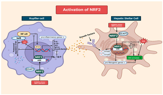
2.5. The Role of NRF2 in Mitigating Oxidative-Stress-Induced HSC Activation

NRF2 activation plays a crucial role in ameliorating liver fibrosis by interfering with the activation of HSCs, which are the key drivers of excessive ECM production. NRF2 exerts its inhibitory effects through multiple interconnected mechanisms [72]. One of the major mechanisms though which NRF2 inhibits HSC activation is the modulation of the transforming growth factor-beta (TGF-β) signaling pathway [73]. TGF-β is a potent profibrogenic cytokine that promotes HSC activation and ECM synthesis. NRF2 activation disrupts the Smad signaling pathway downstream of TGF-β, preventing the formation of Smad complexes and their translocation to the nucleus [74]. Consequently, the expression of fibrotic genes is suppressed, leading to reduced HSC activation and ECM production. In addition to interfering with TGF-β signaling, NRF2 activation modulates the expression of various fibrogenic mediators involved in HSC activation. For example, NRF2 downregulates the production of platelet-derived growth factor, connective tissue growth factor, and endothelin-1, which are potent stimulators of HSC activation and ECM synthesis. Thus, NRF2 limits HSC activation and attenuates fibrosis progression by inhibiting the expression of these fibrogenic mediators [75] (Figure 2).
Moreover, NRF2 activation exerts anti-inflammatory effects on HSCs, which are crucial for the development of fibrosis. It inhibits the release of pro-inflammatory cytokines and chemokines such as interleukin-6 (IL-6) and tumor necrosis factor-alpha (TNF-α), thereby reducing the inflammatory milieu that promotes HSC activation [76]. The anti-inflammatory effect of NRF2 attenuates fibrogenesis and limits ECM production in HSCs [77]. Additionally, NRF2 activation reduces oxidative stress in HSCs, which is another key driver of the activation and progression of fibrosis. By enhancing the cellular antioxidant defense system, NRF2 upregulates the expression of antioxidant enzymes that scavenge ROS, thereby reducing oxidative damage in HSCs [78][79]. Reduced oxidative stress inhibits HSC activation and subsequent ECM production. Furthermore, NRF2 activation modulates epigenetic mechanisms in HSCs. It influences DNA methylation patterns and histone modifications, which can lead to the suppression of fibrotic gene expression in HSCs [80].
In summary, NRF2 activation ameliorates liver fibrosis by interfering with HSC activation through modulating TGF-β signaling, downregulating fibrogenic mediators, suppressing inflammation, reducing oxidative stress, and epigenetic regulation. These interconnected mechanisms work together to limit HSC activation, ECM production, and fibrosis progression in CLDs.

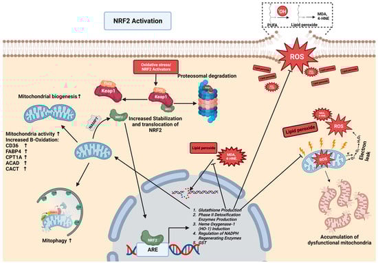
3. Crosstalk between NRF2 and Mitochondria Quality Control in Chronic Liver Disease

3.1. The Role of Mitochondria in the Formation of ROS

Mitochondria, often referred to as the powerhouses of cells, play a pivotal role in producing ROS and driving oxidative stress, especially in the context of chronic liver disease [81]. One primary source of ROS in mitochondria is electron leakage from the electron transport chain (ETC), which is fundamental in generating cellular energy through oxidative phosphorylation. While this chain mostly transfers electrons efficiently to molecular oxygen, a small fraction can escape prematurely, resulting in the creation of superoxide anion (O2•−) as a primary ROS. Components such as complex I and complex III of the ETC are recognized as significant sites for the production of ROS. Electron leakage, along with inefficient electron transfer and compromised mitochondrial membrane potential, can escalate the output of ROS [82]. Several factors can induce these dysfunctions, including mitochondrial DNA mutations, calcium accumulation, the opening of the mitochondrial permeability transition pore (mPTP), oxidative damage to ETC components diminishing the availability of electron carriers, and issues with protein assembly [83][84]. Mitochondrial DNA (mtDNA), which is situated close to ROS production locations, is exceptionally vulnerable to oxidative harm. Damaged mtDNA can further debilitate mitochondrial functionality, promoting the generation of ROS and establishing a cycle of increasing mitochondrial malfunction and oxidative stress [85].
In the context of chronic liver disease, mitochondria-derived ROS cause hepatocellular injury, inflammation, and fibrosis, thereby intensifying the progression of liver pathology. Understanding the intricate mechanisms through which mitochondria contribute to the production of ROS and oxidative stress is crucial for the development of strategies for countering mitochondrial dysfunction, restoring redox balance, and alleviating oxidative-stress-related damage in disease conditions [2][86].

3.2. The Importance of Mitochondrial Metabolism in the Progression of Chronic Diseases

In the liver, mitochondria play a pivotal role both in metabolic functions and in disease pathology. Chronic conditions such as NAFLD, ALD, and liver fibrosis are characterized by mitochondrial dysfunction, oxidative stress, and weakened antioxidant defense mechanisms [87].
Mitochondrial dysfunction arises from various adversities like inflammation and hepatotoxic substances. The liver, a key metabolic organ, requires substantial energy to maintain its crucial functions, especially while experiencing the strain of a chronic disease. Mitochondria meet this demand by producing ATP through oxidative phosphorylation, a highly efficient process. This ATP is fundamental for various liver functions. For instance, the syntheses of proteins and lipids, which are energy-intensive processes, rely heavily on ATP. Additionally, the liver’s role in detoxification, a process that neutralizes and removes toxins and waste, is also ATP-dependent. Any disruption in mitochondrial ATP production can impede the liver’s detoxification capabilities, disrupt the synthesis of essential molecules, and affect ionic balance. Thus, ensuring mitochondrial health and adequate ATP production is vital for managing and potentially mitigating the impacts of liver diseases [88].
Beyond their role in energy production, the mitochondria in liver cells carry out a crucial function in gluconeogenesis. This process involves the conversion of non-carbohydrate substrates, such as lactate derived from anaerobic glycolysis, amino acids from protein catabolism, and glycerol from fat breakdown, into glucose [89]. Mitochondrial enzymes play a pivotal role in this pathway. Enzymes like PEPCK (phosphoenolpyruvate carboxykinase) and G6Pase (glucose-6-phosphatase) are instrumental in the final stages of this pathway, converting oxaloacetate to glucose which can then be released into the bloodstream to maintain blood glucose levels [90]. In the context of chronic liver diseases, this pathway becomes even more significant, particularly during prolonged fasting or under conditions in which the glucose supply is limited [91].
Moreover, the role of mitochondria in fatty acid oxidation holds immense significance in liver metabolism. During this process, fatty acids are broken down, leading to the production of acetyl-CoA. This molecule subsequently enters the citric acid cycle to produce ATP. In the context of chronic liver diseases like NAFLD or alcoholic liver disease, there might be an accumulation of lipids in the liver [92]. Here, efficient mitochondrial fatty acid oxidation becomes imperative to prevent excessive lipid buildup. A malfunction in this process can contribute to the progression of fatty liver diseases as accumulated lipids can cause liver inflammation, fibrosis, and even cirrhosis in prolonged cases. For patients with chronic liver disease, the optimal function of mitochondria in the liver is paramount. They support energy production, regulate glucose and lipid metabolism, assist in the synthesis of ketone bodies and heme, and participate in detoxification pathways [93]. Disruptions in these mitochondrial functions could worsen conditions like NAFLD and metabolic syndrome. Recognizing the central role of mitochondria in liver metabolism is crucial for the effective management and treatment of chronic liver diseases [94].

3.3. NRF2 Mediates Mitophagy and Mitochondrial Turnover

NRF2 is a crucial factor in maintaining cellular homeostasis. Its activation can lead to increases in mitophagy and mitochondrial turnover, which are important processes for maintaining mitochondrial quality control [95][96].
Mitophagy is a selective form of autophagy that involves the removal and degradation of damaged or dysfunctional mitochondria. It is a critical process for maintaining a healthy mitochondrial population within cells. Dysfunctional mitochondria can escalate the production of ROS, intensifying oxidative stress, which is a documented antagonist in the progression of CLD [97]. By mediating the removal of dysfunctional mitochondria, mitophagy acts as a defense mechanism, thereby potentially alleviating liver damage. In the context of CLD, this is of paramount importance given the role oxidative stress plays in the disease’s progression [98].
The activation of NRF2 is empirically linked to the enhancement of mitophagy in liver cells. It achieves this by promoting the expression of genes fundamental to both autophagy and mitophagy. Genes like p62/SQSTM1 and LC3, which are central to the process of recognizing and isolating damaged mitochondria during mitophagy, are directly influenced by NRF2. By bolstering these mechanisms, NRF2 reinforces liver health and resilience against CLD [99].
In addition, NRF2 plays a broader role in augmenting mitochondrial turnover, which encompasses the creation and degradation of mitochondria [100]. Key genes involved in mitochondrial biogenesis, such as peroxisome proliferator-activated receptor gamma coactivator 1-alpha (PGC-1α) and mitochondrial transcription factor A (TFAM), witness an uptick in their expression due to NRF2 activation [101]. This, in turn, boosts the replication and synthesis of mitochondrial DNA (mtDNA), fostering the growth of new, healthy mitochondria in liver cells [102].
Healthy mitochondria, in addition playing a role in combating ROS, have another significant function: they contribute to reducing free fatty acids (FFA) and bolster insulin sensitivity. A build-up of FFAs can lead to lipid toxicity, further exacerbating liver damage, while improved insulin sensitivity is critical for glucose metabolism. Both these functions are vital in the context of CLD, with lipid accumulation and insulin resistance among the hallmarks of conditions like NAFLD [103]. Thus, NRF2’s role in promoting healthy mitochondrial populations indirectly supports these two pivotal functions, further underscoring its therapeutic potential in preventing and treating CLD [104][105].

3.4. The Interplay of Mitochondria and NRF2 in CLD

In protecting the liver from oxidative stress, mitochondria, in turn, influence the regulatory dynamics of NRF2.
One way mitochondria achieve this is through an enzyme called NAMPT (nicotinamide phosphoribosyl transferase), which is an enzyme that is important to mitochondrial function and instrumental in the biosynthesis of NAD+ (nicotinamide adenine dinucleotide) [106]. NAD+ is vital in cellular redox reactions and bioenergetics, processes which are quintessential for liver cells, especially when dealing with problems like NAFLD or alcoholic liver disease [107]. With reduced mitochondrial health, which is often seen in liver problems due to factors like inflammation, harmful substances, and viral infections, there is a clear change in NAMPT activity [108]. This change results in a drop in NAD+ levels. Later effects impact the SIRT1 pathway, a known controller of NRF2 in liver cells. SIRT1, depending on NAD+, works with NRF2, affecting its defense roles [109].
This relationship shows a two-way connection where in which NRF2 helps keep mitochondria healthy. At the same time, mitochondrial health, guided by the NAMPT-NAD+-SIRT1 path, influences NRF2 activity [106][110].

This entry is adapted from the peer-reviewed paper 10.3390/antiox12111928

References

  1. Asrani, S.K.; Devarbhavi, H.; Eaton, J.; Kamath, P.S. Burden of liver diseases in the world. J. Hepatol. 2019, 70, 151–171.
  2. Mansouri, A.; Gattolliat, C.H.; Asselah, T. Mitochondrial Dysfunction and Signaling in Chronic Liver Diseases. Gastroenterology 2018, 155, 629–647.
  3. Seen, S. Chronic liver disease and oxidative stress—A narrative review. Expert. Rev. Gastroenterol. Hepatol. 2021, 15, 1021–1035.
  4. Moon, A.M.; Singal, A.G.; Tapper, E.B. Contemporary Epidemiology of Chronic Liver Disease and Cirrhosis. Clin. Gastroenterol. Hepatol. 2020, 18, 2650–2666.
  5. Juanola, O.; Martinez-Lopez, S.; Frances, R.; Gomez-Hurtado, I. Non-Alcoholic Fatty Liver Disease: Metabolic, Genetic, Epigenetic and Environmental Risk Factors. Int. J. Environ. Res. Public Health 2021, 18, 5227.
  6. Powell, E.E.; Wong, V.W.; Rinella, M. Non-alcoholic fatty liver disease. Lancet 2021, 397, 2212–2224.
  7. Paik, J.M.; Kabbara, K.; Eberly, K.E.; Younossi, Y.; Henry, L.; Younossi, Z.M. Global burden of NAFLD and chronic liver disease among adolescents and young adults. Hepatology 2022, 75, 1204–1217.
  8. Pierantonelli, I.; Svegliati-Baroni, G. Nonalcoholic Fatty Liver Disease: Basic Pathogenetic Mechanisms in the Progression from NAFLD to NASH. Transplantation 2019, 103, e1–e13.
  9. Kawano, Y.; Cohen, D.E. Mechanisms of hepatic triglyceride accumulation in non-alcoholic fatty liver disease. J. Gastroenterol. 2013, 48, 434–441.
  10. Sakurai, Y.; Kubota, N.; Yamauchi, T.; Kadowaki, T. Role of Insulin Resistance in MAFLD. Int. J. Mol. Sci. 2021, 22, 4156.
  11. Wang, X.; Rao, H.; Liu, F.; Wei, L.; Li, H.; Wu, C. Recent Advances in Adipose Tissue Dysfunction and Its Role in the Pathogenesis of Non-Alcoholic Fatty Liver Disease. Cells 2021, 10, 3300.
  12. Wree, A.; Broderick, L.; Canbay, A.; Hoffman, H.M.; Feldstein, A.E. From NAFLD to NASH to cirrhosis-new insights into disease mechanisms. Nat. Rev. Gastroenterol. Hepatol. 2013, 10, 627–636.
  13. Buchanan, R.; Sinclair, J.M.A. Alcohol use disorder and the liver. Addiction 2021, 116, 1270–1278.
  14. Hyun, J.; Han, J.; Lee, C.; Yoon, M.; Jung, Y. Pathophysiological Aspects of Alcohol Metabolism in the Liver. Int. J. Mol. Sci. 2021, 22, 5717.
  15. Tan, H.K.; Yates, E.; Lilly, K.; Dhanda, A.D. Oxidative stress in alcohol-related liver disease. World J. Hepatol. 2020, 12, 332–349.
  16. Kruman, I.I.; Henderson, G.I.; Bergeson, S.E. DNA damage and neurotoxicity of chronic alcohol abuse. Exp. Biol. Med. 2012, 237, 740–747.
  17. Smathers, R.L.; Galligan, J.J.; Stewart, B.J.; Petersen, D.R. Overview of lipid peroxidation products and hepatic protein modification in alcoholic liver disease. Chem. Biol. Interact. 2011, 192, 107–112.
  18. Malnick, S.D.H.; Alin, P.; Somin, M.; Neuman, M.G. Fatty Liver Disease-Alcoholic and Non-Alcoholic: Similar but Different. Int. J. Mol. Sci. 2022, 23, 16226.
  19. Mallet, M.; Silaghi, C.A.; Sultanik, P.; Conti, F.; Rudler, M.; Ratziu, V.; Thabut, D.; Pais, R. Current challenges and future perspectives in treating patients with NAFLD-related cirrhosis. Hepatology 2023.
  20. Gines, P.; Krag, A.; Abraldes, J.G.; Sola, E.; Fabrellas, N.; Kamath, P.S. Liver cirrhosis. Lancet 2021, 398, 1359–1376.
  21. Louvet, A.; Mathurin, P. Alcoholic liver disease: Mechanisms of injury and targeted treatment. Nat. Rev. Gastroenterol. Hepatol. 2015, 12, 231–242.
  22. Ramakrishna, G.; Rastogi, A.; Trehanpati, N.; Sen, B.; Khosla, R.; Sarin, S.K. From cirrhosis to hepatocellular carcinoma: New molecular insights on inflammation and cellular senescence. Liver Cancer 2013, 2, 367–383.
  23. Ngo, V.; Duennwald, M.L. Nrf2 and Oxidative Stress: A General Overview of Mechanisms and Implications in Human Disease. Antioxidants 2022, 11, 2345.
  24. He, F.; Ru, X.; Wen, T. NRF2, a Transcription Factor for Stress Response and Beyond. Int. J. Mol. Sci. 2020, 21, 4777.
  25. Galicia-Moreno, M.; Lucano-Landeros, S.; Monroy-Ramirez, H.C.; Silva-Gomez, J.; Gutierrez-Cuevas, J.; Santos, A.; Armendariz-Borunda, J. Roles of Nrf2 in Liver Diseases: Molecular, Pharmacological, and Epigenetic Aspects. Antioxidants 2020, 9, 980.
  26. Baird, L.; Yamamoto, M. The Molecular Mechanisms Regulating the KEAP1-NRF2 Pathway. Mol. Cell. Biol. 2020, 40, e00099-20.
  27. Bellezza, I.; Giambanco, I.; Minelli, A.; Donato, R. Nrf2-Keap1 signaling in oxidative and reductive stress. Biochim. Biophys. Acta Mol. Cell. Res. 2018, 1865, 721–733.
  28. Robledinos-Anton, N.; Fernandez-Gines, R.; Manda, G.; Cuadrado, A. Activators and Inhibitors of NRF2: A Review of Their Potential for Clinical Development. Oxid. Med. Cell. Longev. 2019, 2019, 9372182.
  29. Shaw, P.; Chattopadhyay, A. Nrf2-ARE signaling in cellular protection: Mechanism of action and the regulatory mechanisms. J. Cell. Physiol. 2020, 235, 3119–3130.
  30. Zhou, J.; Zheng, Q.; Chen, Z. The Nrf2 Pathway in Liver Diseases. Front. Cell. Dev. Biol. 2022, 10, 826204.
  31. Shin, S.; Wakabayashi, J.; Yates, M.S.; Wakabayashi, N.; Dolan, P.M.; Aja, S.; Liby, K.T.; Sporn, M.B.; Yamamoto, M.; Kensler, T.W. Role of Nrf2 in prevention of high-fat diet-induced obesity by synthetic triterpenoid CDDO-imidazolide. Eur. J. Pharmacol. 2009, 620, 138–144.
  32. Qiu, S.; Liang, Z.; Wu, Q.; Wang, M.; Yang, M.; Chen, C.; Zheng, H.; Zhu, Z.; Li, L.; Yang, G. Hepatic lipid accumulation induced by a high-fat diet is regulated by Nrf2 through multiple pathways. FASEB J. 2022, 36, e22280.
  33. Akl, M.G.; Li, L.; Baccetto, R.; Phanse, S.; Zhang, Q.; Trites, M.J.; McDonald, S.; Aoki, H.; Babu, M.; Widenmaier, S.B. Complementary gene regulation by NRF1 and NRF2 protects against hepatic cholesterol overload. Cell Rep. 2023, 42, 112399.
  34. Zhang, Y.K.; Yeager, R.L.; Tanaka, Y.; Klaassen, C.D. Enhanced expression of Nrf2 in mice attenuates the fatty liver produced by a methionine- and choline-deficient diet. Toxicol. Appl. Pharmacol. 2010, 245, 326–334.
  35. Tanaka, Y.; Ikeda, T.; Yamamoto, K.; Ogawa, H.; Kamisako, T. Dysregulated expression of fatty acid oxidation enzymes and iron-regulatory genes in livers of Nrf2-null mice. J. Gastroenterol. Hepatol. 2012, 27, 1711–1717.
  36. Dinkova-Kostova, A.T.; Abramov, A.Y. The emerging role of Nrf2 in mitochondrial function. Free Radic. Biol. Med. 2015, 88, 179–188.
  37. Chambel, S.S.; Santos-Goncalves, A.; Duarte, T.L. The Dual Role of Nrf2 in Nonalcoholic Fatty Liver Disease: Regulation of Antioxidant Defenses and Hepatic Lipid Metabolism. Biomed. Res. Int. 2015, 2015, 597134.
  38. Morita, M.; Ishida, N.; Uchiyama, K.; Yamaguchi, K.; Itoh, Y.; Shichiri, M.; Yoshida, Y.; Hagihara, Y.; Naito, Y.; Yoshikawa, T.; et al. Fatty liver induced by free radicals and lipid peroxidation. Free Radic. Res. 2012, 46, 758–765.
  39. Martin-Fernandez, M.; Arroyo, V.; Carnicero, C.; Siguenza, R.; Busta, R.; Mora, N.; Antolin, B.; Tamayo, E.; Aspichueta, P.; Carnicero-Frutos, I.; et al. Role of Oxidative Stress and Lipid Peroxidation in the Pathophysiology of NAFLD. Antioxidants 2022, 11, 2217.
  40. Su, L.J.; Zhang, J.H.; Gomez, H.; Murugan, R.; Hong, X.; Xu, D.; Jiang, F.; Peng, Z.Y. Reactive Oxygen Species-Induced Lipid Peroxidation in Apoptosis, Autophagy, and Ferroptosis. Oxid. Med. Cell. Longev. 2019, 2019, 5080843.
  41. Yin, H.; Xu, L.; Porter, N.A. Free radical lipid peroxidation: Mechanisms and analysis. Chem. Rev. 2011, 111, 5944–5972.
  42. Ayala, A.; Munoz, M.F.; Arguelles, S. Lipid peroxidation: Production, metabolism, and signaling mechanisms of malondialdehyde and 4-hydroxy-2-nonenal. Oxid. Med. Cell. Longev. 2014, 2014, 360438.
  43. Winczura, A.; Zdzalik, D.; Tudek, B. Damage of DNA and proteins by major lipid peroxidation products in genome stability. Free Radic. Res. 2012, 46, 442–459.
  44. Yesilova, Z.; Yaman, H.; Oktenli, C.; Ozcan, A.; Uygun, A.; Cakir, E.; Sanisoglu, S.Y.; Erdil, A.; Ates, Y.; Aslan, M.; et al. Systemic markers of lipid peroxidation and antioxidants in patients with nonalcoholic Fatty liver disease. Am. J. Gastroenterol. 2005, 100, 850–855.
  45. Spiteller, G. Is lipid peroxidation of polyunsaturated acids the only source of free radicals that induce aging and age-related diseases? Rejuvenation Res. 2010, 13, 91–103.
  46. Negre-Salvayre, A.; Auge, N.; Ayala, V.; Basaga, H.; Boada, J.; Brenke, R.; Chapple, S.; Cohen, G.; Feher, J.; Grune, T.; et al. Pathological aspects of lipid peroxidation. Free Radic. Res. 2010, 44, 1125–1171.
  47. Dodson, M.; Castro-Portuguez, R.; Zhang, D.D. NRF2 plays a critical role in mitigating lipid peroxidation and ferroptosis. Redox Biol. 2019, 23, 101107.
  48. Busch, C.J.; Hendrikx, T.; Weismann, D.; Jackel, S.; Walenbergh, S.M.; Rendeiro, A.F.; Weisser, J.; Puhm, F.; Hladik, A.; Goderle, L.; et al. Malondialdehyde epitopes are sterile mediators of hepatic inflammation in hypercholesterolemic mice. Hepatology 2017, 65, 1181–1195.
  49. Yadav, U.C.; Ramana, K.V. Regulation of NF-κB-induced inflammatory signaling by lipid peroxidation-derived aldehydes. Oxid. Med. Cell. Longev. 2013, 2013, 690545.
  50. Ma, F.; Luo, S.; Lu, C.; Jiang, X.; Chen, K.; Deng, J.; Ma, S.; Li, Z. The role of Nrf2 in periodontal disease by regulating lipid peroxidation, inflammation and apoptosis. Front. Endocrinol. 2022, 13, 963451.
  51. Gruber, F.; Ornelas, C.M.; Karner, S.; Narzt, M.S.; Nagelreiter, I.M.; Gschwandtner, M.; Bochkov, V.; Tschachler, E. Nrf2 deficiency causes lipid oxidation, inflammation, and matrix-protease expression in DHA-supplemented and UVA-irradiated skin fibroblasts. Free Radic. Biol. Med. 2015, 88, 439–451.
  52. Koyama, Y.; Brenner, D.A. Liver inflammation and fibrosis. J. Clin. Investig. 2017, 127, 55–64.
  53. Parola, M.; Pinzani, M. Liver fibrosis: Pathophysiology, pathogenetic targets and clinical issues. Mol. Aspects Med. 2019, 65, 37–55.
  54. Bataller, R.; Brenner, D.A. Liver fibrosis. J. Clin. Investig. 2005, 115, 209–218.
  55. Hao, W.; Li, M.; Cai, Q.; Wu, S.; Li, X.; He, Q.; Hu, Y. Roles of NRF2 in Fibrotic Diseases: From Mechanisms to Therapeutic Approaches. Front. Physiol. 2022, 13, 889792.
  56. Gong, Y.; Yang, Y. Activation of Nrf2/AREs-mediated antioxidant signalling, and suppression of profibrotic TGF-beta1/Smad3 pathway: A promising therapeutic strategy for hepatic fibrosis—A review. Life Sci. 2020, 256, 117909.
  57. Yang, H.; Luo, F.; Wei, Y.; Jiao, Y.; Qian, J.; Chen, S.; Gong, Y.; Tang, L. TGR5 protects against cholestatic liver disease via suppressing the NF-κB pathway and activating the Nrf2/HO-1 pathway. Ann. Transl. Med. 2021, 9, 1158.
  58. Thimmulappa, R.K.; Lee, H.; Rangasamy, T.; Reddy, S.P.; Yamamoto, M.; Kensler, T.W.; Biswal, S. Nrf2 is a critical regulator of the innate immune response and survival during experimental sepsis. J. Clin. Investig. 2006, 116, 984–995.
  59. Xue, R.; Qiu, J.; Wei, S.; Liu, M.; Wang, Q.; Wang, P.; Sha, B.; Wang, H.; Shi, Y.; Zhou, J.; et al. Lycopene alleviates hepatic ischemia reperfusion injury via the Nrf2/HO-1 pathway mediated NLRP3 inflammasome inhibition in Kupffer cells. Ann. Transl. Med. 2021, 9, 631.
  60. Liu, X.; Wang, T.; Liu, X.; Cai, L.; Qi, J.; Zhang, P.; Li, Y. Biochanin A protects lipopolysaccharide/D-galactosamine-induced acute liver injury in mice by activating the Nrf2 pathway and inhibiting NLRP3 inflammasome activation. Int. Immunopharmacol. 2016, 38, 324–331.
  61. Rushworth, S.A.; Chen, X.L.; Mackman, N.; Ogborne, R.M.; O’Connell, M.A. Lipopolysaccharide-induced heme oxygenase-1 expression in human monocytic cells is mediated via Nrf2 and protein kinase C. J. Immunol. 2005, 175, 4408–4415.
  62. Liang, W.; Greven, J.; Qin, K.; Fragoulis, A.; Horst, K.; Blasius, F.; Wruck, C.; Pufe, T.; Kobbe, P.; Hildebrand, F.; et al. Sulforaphane Exerts Beneficial Immunomodulatory Effects on Liver Tissue via a Nrf2 Pathway-Related Mechanism in a Murine Model of Hemorrhagic Shock and Resuscitation. Front. Immunol. 2022, 13, 822895.
  63. Sierra-Filardi, E.; Vega, M.A.; Sanchez-Mateos, P.; Corbi, A.L.; Puig-Kroger, A. Heme Oxygenase-1 expression in M-CSF-polarized M2 macrophages contributes to LPS-induced IL-10 release. Immunobiology 2010, 215, 788–795.
  64. Wang, L.; He, C. Nrf2-mediated anti-inflammatory polarization of macrophages as therapeutic targets for osteoarthritis. Front. Immunol. 2022, 13, 967193.
  65. Chen, H.; Qin, J.; Shi, H.; Li, Q.; Zhou, S.; Chen, L. Rhoifolin ameliorates osteoarthritis via the Nrf2/NF-κB axis: In Vitro and in vivo experiments. Osteoarthr. Cartil. 2022, 30, 735–745.
  66. Liu, G.H.; Qu, J.; Shen, X. NF-κB/p65 antagonizes Nrf2-ARE pathway by depriving CBP from Nrf2 and facilitating recruitment of HDAC3 to MafK. Biochim. Biophys. Acta 2008, 1783, 713–727.
  67. Bellezza, I.; Mierla, A.L.; Minelli, A. Nrf2 and NF-κB and Their Concerted Modulation in Cancer Pathogenesis and Progression. Cancers 2010, 2, 483–497.
  68. Nair, S.; Doh, S.T.; Chan, J.Y.; Kong, A.N.; Cai, L. Regulatory potential for concerted modulation of Nrf2- and Nfkb1-mediated gene expression in inflammation and carcinogenesis. Br. J. Cancer 2008, 99, 2070–2082.
  69. Jiang, T.; Tian, F.; Zheng, H.; Whitman, S.A.; Lin, Y.; Zhang, Z.; Zhang, N.; Zhang, D.D. Nrf2 suppresses lupus nephritis through inhibition of oxidative injury and the NF-κB-mediated inflammatory response. Kidney Int. 2014, 85, 333–343.
  70. Heiss, E.; Herhaus, C.; Klimo, K.; Bartsch, H.; Gerhauser, C. Nuclear factor kappa B is a molecular target for sulforaphane-mediated anti-inflammatory mechanisms. J. Biol. Chem. 2001, 276, 32008–32015.
  71. Kim, J.E.; You, D.J.; Lee, C.; Ahn, C.; Seong, J.Y.; Hwang, J.I. Suppression of NF-κB signaling by KEAP1 regulation of IKKbeta activity through autophagic degradation and inhibition of phosphorylation. Cell. Signal. 2010, 22, 1645–1654.
  72. Sharma, R.S.; Harrison, D.J.; Kisielewski, D.; Cassidy, D.M.; McNeilly, A.D.; Gallagher, J.R.; Walsh, S.V.; Honda, T.; McCrimmon, R.J.; Dinkova-Kostova, A.T.; et al. Experimental Nonalcoholic Steatohepatitis and Liver Fibrosis Are Ameliorated by Pharmacologic Activation of Nrf2 (NF-E2 p45-Related Factor 2). Cell. Mol. Gastroenterol. Hepatol. 2018, 5, 367–398.
  73. Shi, Y.S.; Li, X.X.; Li, H.T.; Zhang, Y. Pelargonidin ameliorates CCl(4)-induced liver fibrosis by suppressing the ROS-NLRP3-IL-1beta axis via activating the Nrf2 pathway. Food Funct. 2020, 11, 5156–5165.
  74. Prestigiacomo, V.; Suter-Dick, L. Nrf2 protects stellate cells from Smad-dependent cell activation. PLoS ONE 2018, 13, e0201044.
  75. Qiu, L.; Hu, L.; Liu, X.; Li, W.; Zhang, X.; Xia, H.; Zhang, C. Physalin B inhibits PDGF-BB-induced VSMC proliferation, migration and phenotypic transformation by activating the Nrf2 pathway. Food Funct. 2021, 12, 10950–10966.
  76. Del Campo, J.A.; Gallego, P.; Grande, L. Role of inflammatory response in liver diseases: Therapeutic strategies. World J. Hepatol. 2018, 10, 1–7.
  77. Eisenstein, A.; Hilliard, B.K.; Pope, S.D.; Zhang, C.; Taskar, P.; Waizman, D.A.; Israni-Winger, K.; Tian, H.; Luan, H.H.; Wang, A. Activation of the transcription factor NRF2 mediates the anti-inflammatory properties of a subset of over-the-counter and prescription NSAIDs. Immunity 2022, 55, 1082–1095.e1085.
  78. Kong, D.; Zhang, Z.; Chen, L.; Huang, W.; Zhang, F.; Wang, L.; Wang, Y.; Cao, P.; Zheng, S. Curcumin blunts epithelial-mesenchymal transition of hepatocytes to alleviate hepatic fibrosis through regulating oxidative stress and autophagy. Redox Biol. 2020, 36, 101600.
  79. Yang, W.; Wang, Y.; Zhang, C.; Huang, Y.; Yu, J.; Shi, L.; Zhang, P.; Yin, Y.; Li, R.; Tao, K. Maresin1 Protect Against Ferroptosis-Induced Liver Injury Through ROS Inhibition and Nrf2/HO-1/GPX4 Activation. Front. Pharmacol. 2022, 13, 865689.
  80. Silva-Llanes, I.; Shin, C.H.; Jimenez-Villegas, J.; Gorospe, M.; Lastres-Becker, I. The Transcription Factor NRF2 Has Epigenetic Regulatory Functions Modulating HDACs, DNMTs, and miRNA Biogenesis. Antioxidants 2023, 12, 641.
  81. Kowaltowski, A.J.; de Souza-Pinto, N.C.; Castilho, R.F.; Vercesi, A.E. Mitochondria and reactive oxygen species. Free Radic. Biol. Med. 2009, 47, 333–343.
  82. Zhao, R.Z.; Jiang, S.; Zhang, L.; Yu, Z.B. Mitochondrial electron transport chain, ROS generation and uncoupling (Review). Int. J. Mol. Med. 2019, 44, 3–15.
  83. Zorov, D.B.; Juhaszova, M.; Sollott, S.J. Mitochondrial reactive oxygen species (ROS) and ROS-induced ROS release. Physiol. Rev. 2014, 94, 909–950.
  84. Suski, J.M.; Lebiedzinska, M.; Bonora, M.; Pinton, P.; Duszynski, J.; Wieckowski, M.R. Relation between mitochondrial membrane potential and ROS formation. Methods Mol. Biol. 2012, 810, 183–205.
  85. Bhatti, J.S.; Bhatti, G.K.; Reddy, P.H. Mitochondrial dysfunction and oxidative stress in metabolic disorders—A step towards mitochondria based therapeutic strategies. Biochim. Biophys. Acta Mol. Basis Dis. 2017, 1863, 1066–1077.
  86. Xiang, L.; Shao, Y.; Chen, Y. Mitochondrial dysfunction and mitochondrion-targeted therapeutics in liver diseases. J. Drug Target. 2021, 29, 1080–1093.
  87. Zhang, C.; Zhao, Y.; Yu, M.; Qin, J.; Ye, B.; Wang, Q. Mitochondrial Dysfunction and Chronic Liver Disease. Curr. Issues Mol. Biol. 2022, 44, 3156–3165.
  88. Koliaki, C.; Roden, M. Hepatic energy metabolism in human diabetes mellitus, obesity and non-alcoholic fatty liver disease. Mol. Cell. Endocrinol. 2013, 379, 35–42.
  89. Passarella, S.; Schurr, A.; Portincasa, P. Mitochondrial Transport in Glycolysis and Gluconeogenesis: Achievements and Perspectives. Int. J. Mol. Sci. 2021, 22, 12620.
  90. Stark, R.; Guebre-Egziabher, F.; Zhao, X.; Feriod, C.; Dong, J.; Alves, T.C.; Ioja, S.; Pongratz, R.L.; Bhanot, S.; Roden, M.; et al. A role for mitochondrial phosphoenolpyruvate carboxykinase (PEPCK-M) in the regulation of hepatic gluconeogenesis. J. Biol. Chem. 2014, 289, 7257–7263.
  91. Sunny, N.E.; Parks, E.J.; Browning, J.D.; Burgess, S.C. Excessive hepatic mitochondrial TCA cycle and gluconeogenesis in humans with nonalcoholic fatty liver disease. Cell Metab. 2011, 14, 804–810.
  92. Moore, M.P.; Cunningham, R.P.; Meers, G.M.; Johnson, S.A.; Wheeler, A.A.; Ganga, R.R.; Spencer, N.M.; Pitt, J.B.; Diaz-Arias, A.; Swi, A.I.A.; et al. Compromised hepatic mitochondrial fatty acid oxidation and reduced markers of mitochondrial turnover in human NAFLD. Hepatology 2022, 76, 1452–1465.
  93. Guerra, I.M.S.; Ferreira, H.B.; Melo, T.; Rocha, H.; Moreira, S.; Diogo, L.; Domingues, M.R.; Moreira, A.S.P. Mitochondrial Fatty Acid beta-Oxidation Disorders: From Disease to Lipidomic Studies-A Critical Review. Int. J. Mol. Sci. 2022, 23, 13933.
  94. Barbier-Torres, L.; Fortner, K.A.; Iruzubieta, P.; Delgado, T.C.; Giddings, E.; Chen, Y.; Champagne, D.; Fernandez-Ramos, D.; Mestre, D.; Gomez-Santos, B.; et al. Silencing hepatic MCJ attenuates non-alcoholic fatty liver disease (NAFLD) by increasing mitochondrial fatty acid oxidation. Nat. Commun. 2020, 11, 3360.
  95. Fang, E.F.; Waltz, T.B.; Kassahun, H.; Lu, Q.; Kerr, J.S.; Morevati, M.; Fivenson, E.M.; Wollman, B.N.; Marosi, K.; Wilson, M.A.; et al. Tomatidine enhances lifespan and healthspan in C. elegans through mitophagy induction via the SKN-1/Nrf2 pathway. Sci. Rep. 2017, 7, 46208.
  96. Li, Y.; Feng, Y.F.; Liu, X.T.; Li, Y.C.; Zhu, H.M.; Sun, M.R.; Li, P.; Liu, B.; Yang, H. Songorine promotes cardiac mitochondrial biogenesis via Nrf2 induction during sepsis. Redox Biol. 2021, 38, 101771.
  97. Schofield, J.H.; Schafer, Z.T. Mitochondrial Reactive Oxygen Species and Mitophagy: A Complex and Nuanced Relationship. Antioxid. Redox Signal. 2021, 34, 517–530.
  98. Mao, Y.; Du, J.; Chen, X.; Al Mamun, A.; Cao, L.; Yang, Y.; Mubwandarikwa, J.; Zaeem, M.; Zhang, W.; Chen, Y.; et al. Maltol Promotes Mitophagy and Inhibits Oxidative Stress via the Nrf2/PINK1/Parkin Pathway after Spinal Cord Injury. Oxid. Med. Cell. Longev. 2022, 2022, 1337630.
  99. Xiao, L.; Xu, X.; Zhang, F.; Wang, M.; Xu, Y.; Tang, D.; Wang, J.; Qin, Y.; Liu, Y.; Tang, C.; et al. The mitochondria-targeted antioxidant MitoQ ameliorated tubular injury mediated by mitophagy in diabetic kidney disease via Nrf2/PINK1. Redox Biol. 2017, 11, 297–311.
  100. Gureev, A.P.; Shaforostova, E.A.; Popov, V.N. Regulation of Mitochondrial Biogenesis as a Way for Active Longevity: Interaction Between the Nrf2 and PGC-1alpha Signaling Pathways. Front. Genet. 2019, 10, 435.
  101. Hayashi, G.; Jasoliya, M.; Sahdeo, S.; Sacca, F.; Pane, C.; Filla, A.; Marsili, A.; Puorro, G.; Lanzillo, R.; Brescia Morra, V.; et al. Dimethyl fumarate mediates Nrf2-dependent mitochondrial biogenesis in mice and humans. Hum. Mol. Genet. 2017, 26, 2864–2873.
  102. Palikaras, K.; Tavernarakis, N. Mitochondrial homeostasis: The interplay between mitophagy and mitochondrial biogenesis. Exp. Gerontol. 2014, 56, 182–188.
  103. Grattagliano, I.; Montezinho, L.P.; Oliveira, P.J.; Fruhbeck, G.; Gomez-Ambrosi, J.; Montecucco, F.; Carbone, F.; Wieckowski, M.R.; Wang, D.Q.; Portincasa, P. Targeting mitochondria to oppose the progression of nonalcoholic fatty liver disease. Biochem. Pharmacol. 2019, 160, 34–45.
  104. Serviddio, G.; Bellanti, F.; Sastre, J.; Vendemiale, G.; Altomare, E. Targeting mitochondria: A new promising approach for the treatment of liver diseases. Curr. Med. Chem. 2010, 17, 2325–2337.
  105. Nassir, F.; Ibdah, J.A. Role of mitochondria in alcoholic liver disease. World J. Gastroenterol. 2014, 20, 2136–2142.
  106. Yu, A.; Zhou, R.; Xia, B.; Dang, W.; Yang, Z.; Chen, X. NAMPT maintains mitochondria content via NRF2-PPARalpha/AMPKalpha pathway to promote cell survival under oxidative stress. Cell. Signal. 2020, 66, 109496.
  107. Xie, W.; Zhu, T.; Zhou, P.; Xu, H.; Meng, X.; Ding, T.; Nan, F.; Sun, G.; Sun, X. Notoginseng leaf triterpenes ameliorates mitochondrial oxidative injury via the NAMPT-SIRT1/2/3 signaling pathways in cerebral ischemic model rats. J. Ginseng Res. 2023, 47, 199–209.
  108. Gabande-Rodriguez, E.; Gomez de Las Heras, M.M.; Mittelbrunn, M. Control of Inflammation by Calorie Restriction Mimetics: On the Crossroad of Autophagy and Mitochondria. Cells 2019, 9, 82.
  109. Wang, S.; Wan, T.; Ye, M.; Qiu, Y.; Pei, L.; Jiang, R.; Pang, N.; Huang, Y.; Liang, B.; Ling, W.; et al. Nicotinamide riboside attenuates alcohol induced liver injuries via activation of SirT1/PGC-1alpha/mitochondrial biosynthesis pathway. Redox Biol. 2018, 17, 89–98.
  110. Ryoo, I.G.; Kwak, M.K. Regulatory crosstalk between the oxidative stress-related transcription factor Nfe2l2/Nrf2 and mitochondria. Toxicol. Appl. Pharmacol. 2018, 359, 24–33.
More
This entry is offline, you can click here to edit this entry!
Video Production Service