1. Introduction
The vast diversity of the marine ecosystem ensures the availability of a wide range of bioactive compounds with biological effects in various disease conditions. There is a diverse range of species found in the marine ecosystem. Indeed, 14 out of 35 animal phyla are exclusively found in the marine environment [
1]. This variation creates different marine habitats that are hotspots of biodiversity. Moreover, this biodiversity is not constant but is dynamic in nature [
2]. Several efforts have been made to make a census of marine biodiversity in different regions of the world so that we can obtain the benefits of region-specific marine biodiversity. These efforts and challenges have previously been reviewed [
3].
Marine biodiversity has motivated researchers across the world to investigate novel compounds that may be valuable for the treatment of various disease conditions. Bioactive compounds have been identified and extracted from various marine organisms and shown to be useful in various pathological conditions as described in a recent review [
4]. Interestingly, many of these compounds with valuable pharmaceutical properties are in different phases of preclinical and clinical investigation [
5]. Bioactive compounds from marine sources have shown immunomodulatory effects [
6], activity against diseases such as type 2 diabetes mellitus [
7] or anti-cancer properties [
8,
9]. Many bioactive compounds show anti-inflammatory activity [
10,
11]. For example, Frondanol is a sea-cucumber-derived intestinal extract that exhibits anti-inflammatory properties in a dextran sodium sulfate (DSS)-induced colitis mouse model [
12]. Notably, several marine-derived compounds appear valuable in inflammatory bowel disease (IBD) [
13].
2. Seaweed as a Marine Source for Anti-Inflammatory Activity
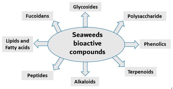
Seaweed is a popular food source rich in bioactive compounds, polysaccharides, fatty acids, peptides, proteins and vitamins. Seaweeds have several potential therapeutic activities, including anti-bacterial, anti-viral, anti-cancer, antioxidant and anti-inflammatory [
16,
17]. Seaweeds are classified into three groups based on pigment content; for example, Ochrophyta and Phaeophyceae (brown), Chlorophyta (green) and Rhodophyta (red) seaweeds containing fucoxanthin, chlorophyll A, chlorophyll B, phycocyanin and phycoerythrin [
18,
19,
20]. The most diverse are the red seaweeds, with more than 7000 species, followed by the brown and green seaweeds, with approximately 2030 and 600 species, respectively [
21]. Researchers have been isolating, purifying and screening the secondary metabolites from these organism for bioactivity in recent decades. Here, it summarize the anti-inflammatory activity of compounds isolated from various seaweed species (
Figure 1).
Figure 1. Main classes of seaweed bioactive compounds.
2.1. Anti-Inflammatory Phenolic Compounds from Seaweed
Phenolic compounds are secondary metabolites that are found in many natural extracts and are known for their antioxidant potential. Seaweed is also a rich source of various phenolic compounds, such as flavonoids and tannins, obtained by different extraction methods [
49,
50,
51]. Many categories of phenolic compounds are obtained from seaweeds [
51]. The best-studied phenolic compounds are phlorotannins, which exhibit anti-inflammatory effects in addition to other biological activities [
52]. A methanol extract of the brown seaweed
Eisenia bicyclis and its CH
2Cl
2 sub-fraction have potent anti-inflammatory effects in LPS-stimulated RAW 264.7 macrophages by inhibiting NO production [
22]. Fucosterol, purified through column chromatography from the CH
2Cl
2 sub-fraction also decreases NO production via the suppression of iNOS expression [
22]. Furthermore, the ethyl acetate sub-fraction of the methanolic extract and chromatographic sub-fractions 1-6 yielded phlorofucofuroeckol A, eckol, dieckol, phlorofucofuroeckol-A, dioxinodehydroeckol and 7-phloroeckol, which also inhibit NO production in a dose-dependent manner in LPS-stimulated macrophages [
22]. Interestingly, similar sub-fractions were extracted from an ethanolic extract and its ethyl acetate sub-fraction from another seaweed,
Ecklonia cava [
53]. These also resulted in significant decreases in the expression of IL1β, IFNγ and interferon stimulatory gene (SGI15) in the olive flounder animal model [
53]. The related phlorotannin, phlorofucofuroeckol B (PFFB), was isolated and chromatographically purified using NMR from an ethanol extract of the brown algae
Ecklonia stolonifera [
23]. PFFB suppressed the inflammatory response in LPS-stimulated BV2 microglial cells by downregulating the PGE
2, TNF-α, IL-1β and IL-6 [
23]. Furthermore, this study demonstrated that the inhibition of inflammation is via the downregulation of IκB-α/NF-κB mediated by Akt/ERK/JNK pathways [
23]. Dieckol was isolated from a methanol extract of
Ecklonia cava powder, with diethyl sub-fractionation, chromatographic purification and NMR characterization [
24]. Dieckol decreased NO production via the suppression of iNOS and COX-2 and also inhibited the generation of proinflammatory cytokines IL-1β and TNF-α through suppressing the activation of NF-κB and p38 MAPK in LPS-induced microglial cells [
24]. Furthermore, a study using commercially available dieckol revealed the inhibition of carrageenan-triggered inflammation and leukocyte infiltration and reduced pro-inflammatory cytokines (TNF-α, IL-1β, and IL-6) in a mouse model [
25]. A recent study demonstrated an increased production of NO and ROS in LPS-treated zebrafish embryos following treatment of an ethyl acetate fraction from
Ecklonia maxima, which included dieckol [
54]. Another compound, diphlorethohydroxycarmalol (DPHC), was isolated and purified from the edible brown seaweed
Ishige okamurae. DPHC suppressed the production of IL-6 via the inhibition of phosphorylation and translocation of NF-κB in LPS-stimulated RAW 264.7 macrophages [
27]. Cytokine signaling 1 (SOCS1) suppression functions as a negative feedback regulator of Janus kinase (Jak)-signal transducer and activator of transcription (STAT) signaling [
27]. DPHC downregulated STAT5 and upregulated SOCS1 in macrophages [
27]. DPHC attenuated several inflammatory symptoms (ear edema, lymph node size, serum IgE level and mast cell infiltration) in an experimental atopic dermatitis-induced inflammatory mouse model [
27]. In another study, DPHC purified from the same seaweed also reduced the expression of pro-inflammatory cytokines and suppressed muscle RING-finger protein (MuRF)-1 and muscle atrophy F-box (MAFbx)/atrgoin-1 in LPS-induced RAW 264.7 macrophages [
26]. These protein complexes are well known in muscle atrophy via NF-κB and MAPK signaling pathways in TNF-α-stimulated C2C12 myotubes [
26]. This study also showed DPHC docking in the TNFα inhibitory site in a simulation [
26]. Another interesting compound, octaphlorethol A, is a phenolic compound isolated from the ethanolic extract, purified from chromatography and further characterized by LC/MS and NMR, belonging to
Ishige foliacea, which inhibits pro-inflammatory cytokines, MAPK and NF-κB pathways in CpG oligodeoxynucleotides (CpG)-stimulated primary murine bone-marrow-derived macrophages and dendritic cells [
28].
2.2. Anti-Inflammatory Polysaccharides from Seaweed
Polysaccharides are major components of seaweed that have attracted much attention because of various health benefits [
11,
56]. Sulfated seaweed polysaccharides show significant anti-inflammatory activity in several inflammatory models [
57]. The major compounds are alginic acid and fucoidans, which have various biological effects reflecting their chemical diversity [
58]. These compounds are major anti-inflammatory components of seaweed polysaccharides. For example, brown seaweed
Sachharina japonica-derived fucoidan galactofucan demonstrated anti-inflammatory activity by reducing the production of NO and the expression of MAPK (including p38, ENK and JNK) and NF-κB (including p65 and IKKα/IKKβ) signaling pathways in endotoxin-stimulated RAW 264.7 macrophages [
29]. Similarly, sulfated fucoidan isolated from
Colpomenia sinuosa prevented oxidative stress and inflammation in the paracetamol-induced hepatic injury and inflammation rat model, as evidenced by suppressing the hepatic levels of thiobarbituric acid reactive substances, NO, iNOS, TNF-α, IL-1β and IL-6 while increasing glutathione and glutathione peroxidase enzyme activity [
30]. Fucoidan, extracted from
Fucus vesiculosus, inhibited LPS-induced inflammatory responses in RAW 264.7 macrophages and zebrafish larvae by suppressing NO and PGE2 secretion via iNOS and COX-2 inhibition as well as reducing the expression and secretion of TNF-α and IL-1β [
31]. An ethanol fraction of the hot water extract from the edible seaweed
Laminaria japonica (which contains abundant fucoidan) suppressed the production of PGE
2 and expression of MMP-9, COX-2 and pro-inflammatory cytokines in the UV-induced inflammation model of the human keratinocyte (HaCaT) cell line [
32]. Interestingly, Nagahwatta et al. purified sulfated fucoidan from the leaves of
Ecklonia maxima and demonstrated a reduction in proinflammatory cytokines such as PGE2, NO, TNFα IL6 and IL1β in RAW 264.7 macrophages [
59]. Furthermore, a recent study revealed that fucoidan had a curative effect mediated by the downregulation of the aryl hydrocarbon receptor and phosphodiesterase 4 in an ulcerative colitis rat model [
60]. More recently, sulfated polysaccharides isolated from the edible brown seaweed
Sargassum fulvellum significantly and concentration-dependently decreased the production of the inflammatory mediators NO, PGE2, TNF-α, IL-1β and IL-6, and suppressed the expression of COX-2 and iNOS in LPS-stimulated RAW 264.7 macrophages [
33]. Furthermore, these sulfated polysaccharides improve survival and decrease cell death, ROS production and NO levels in LPS-stimulated zebrafish [
33]. Another recent study showed the anti-inflammatory effect of a sulfated polysaccharide extracted from
Codium fragile. This study demonstrated the reduction in PGE2, NO, IL1β, IL6 and TNFα in LPS-induced RAW 264.7 macrophages [
61]. The sulfation of seaweed-derived low-molecular-weight fucoidans increases the potency of their anti-inflammatory properties [
62]. In contrast, the non-sulfated polysaccharide, alginic acid, from
Padina boryana, showed marked anti-inflammatory activity in particulate-matter-stimulated inflammation in human HaCaT immortalized keratinocytes and dermal fibroblasts (HDF) [
63]. Alginic acid reduced PGE
2 and COX-2 and inflammatory cytokines (IL-1β and IL-6) via the suppression of the NF-κB and MAPK pathways [
63]. Alginic acid, purified from
Sargassum wightii, demonstrated anti-inflammatory potential in adjuvant-induced arthritic rats by reducing paw edema and COX, lipoxygenase (LOX) and myeloperoxidase levels [
64]. Furthermore, it also reduced the levels of COX-2, IL-6 and TNF-α and inhibited certain key molecular mediators (such as p-p38 MAPK, P-Erk1/2 and P-JNK) of the NF-κB and MAPK pathways in Chinese fine dust (CFD)-treated HaCaT cells [
34]. β-Linked polysaccharides, including β-glucans, are known to possess immunomodulatory and anti-proliferative activities. Laminarin, a water-soluble β-glucan isolated from
Grifola frondose, reduced NO and PGE2 production and suppressed the secretion of pro-inflammatory cytokines via the downregulation of NF-κB in endotoxin-stimulated macrophages [
35].
2.3. Anti-Inflammatory Terpenoids from Seaweed
Terpenoids are the largest group of natural products with specialized secondary metabolites [
68]. These naturally occurring chemical compounds are highly diverse in chemical structure [
68]. Although many biological activities of plant-derived terpenoids have been reported, there are several marine-source-based terpenoids that have been isolated and screened for their biological activity in the last few decades [
69]. Fucosterol, epitaondiol, neorogioltriol, pacifenol, ergosterol and 7-dehydroporiferasterol, pheophytin A and apo-9′-fucoxanthinone are a few potent anti-inflammatory terpenoids isolated from various species of seaweed [
69]. For example, fucosterol, isolated from
Padina boryana, reduced particulate matter (PM)-induced inflammation in macrophages by modulating the NF-κB and MAPK pathways [
36]. It significantly suppressed the expression of inflammatory mediators such as iNOS, COX-2, pro-inflammatory cytokines and PGE2 [
36]. Epitaondiol, isolated from various seaweeds, inhibited COX pathway activity. For example, epitaondiol, isolated from
Stypopodium flabelliforme, inhibited the production of phospholipase A2 and eicosanoids (LTB4 and TXB2) and modulated the COX pathway in the in vitro isolated human neutrophils and the 12-O-Tetradecanoylphorbol-13-acetate (TPA)-induced mouse ear oedema model [
37]. Neorogioltriol, and two related diterpene neorogioldiol and O11,15-cyclo-14-bromo-14,15-dihydrorogiol-3,11-diol, were isolated from a red algae
Laurencia glandulifera [
38]. These compounds exhibit anti-inflammatory effects in M2-type macrophages via an increased expression of arginase1, MRC1, IRAK-M and the transcription factor C/EBPβ [
38]. The anti-inflammatory activity of neorogioldiol and O11,15-cyclo-14-bromo-14,15-dihydrorogiol-3,11-diol was also demonstrated in a DSS-induced colitis mice model where they reduced tissue damage and pro-inflammatory cytokine production in vivo. Ergosterol and 7-dehydroporiferasterol derivatives, isolated from
Dunaliella tertiolecta, suppressed pro-inflammatory cytokines in sheep peripheral blood mononuclear cells stimulated with LPS [
39]. Apo-9-fucoxanthinone (AF), isolated from
Sargassum muticum, showed potent anti-inflammatory activity in LPS-stimulated macrophages by suppressing the mRNA expression of several inflammatory mediators, such as iNOS, COX-2 and pro-inflammatory cytokines [
40]. These effects of AF are mainly due to the modulation of NF-κB and MPAK signaling pathways [
40].
2.4. Anti-Inflammatory Proteins and Peptides from Seaweed
Lectins, isolated from seaweed, are glycoproteins involved in cellular adhesion. Some of these lectins have anti-inflammatory activity. A 30 kDa lectin derived from
Amansia multifida has anti-inflammatory effects, reducing edema formation in a paw edema model, leukocyte migration and oxidative stress in a carrageenan-induced peritonitis model and proinflammatory cytokine (IL-1β and TNF-α) expression in a carrageenan-induced rat paw edema model [
41]. Another study demonstrated the anti-inflammatory effects of a 9kDa lectin isolated from
Bryothamnion triquetrum [
42]. This lectin inhibited the production of proinflammatory cytokines involved in the migration of neutrophils in a carrageenan-induced peritonitis mice model [
42]. These findings suggest that the anti-inflammatory effects of this lectin are mediated by the inhibition of leukocyte recruitment [
42]. In another study, a mucin-binding hololectin isolated from red marine algae demonstrated anti-nociceptive and anti-inflammatory responses, reducing abdominal writhing and the paw-licking time in carrageenan-induced peritonitis and paw edema models [
70]. Hololectin caused a similar inhibition of neutrophil migration in a peritonitis model and also suppressed paw edema, which was induced by carrageenan, dextran or serotonin [
70]. A big anti-inflammatory effect (23–44 kDa) from the green seaweed
Caulerpa cupressoides revealed a decrease in carrageenan-induced rat paw edema and neutrophil infiltration via a reduction in the expression of IL-1, IL-6, TNF-α and COX-2 [
71]. Lectins from red algae and their other potential biomedical applications have been previously summarized in the review [
72]. Only a limited number of papers reporting the anti-inflammatory activity of lectins from green seaweeds have been published. However, lectins from red and green algae have distinct structures [
73]. A recent review documented the structural diversity among various algal species [
73]. The authors classified the lectins from red and brown algae as
Oscillatoria agardhii agglutinin homolog (OAAH) and the green seaweed as
Galanthus nivalis agglutinin (GNA) [
73]. A study on the structural features of N-glycans of seaweed glycoproteins was performed on 15 economically important seaweeds (12 red and brown seaweeds, 2 green seaweeds and 1 sea grass) [
74]. Interestingly, this study revealed the absence of typical plant-based lectins with a complex structure of N-glycans (β1-2 xylosyl and α 1-3 fucosyl) in algae and seagrass [
74]. Moreover, the study showed the absence of high manose-type N-glycan (M5–M9) in the green seaweed
Ulva pertusa [
74]. A study on peptide fractions of approximately 2160 kDa from green seaweed
Ulva spp. showed it exerting immunomodulatory actions in vitro, consistent with an anti-inflammatory effect depending on Toll-like receptor 4 (TLR4) and the NFκB/p38/JNK pathway [
43].
2.5. Anti-Inflammatory Alkaloids from Seaweed
Anti-inflammatory alkaloids are commonly found in plants, but there are limited studies identifying and characterizing the activity of such compounds from seaweeds [
75]. Only limited studies have investigated the anti-inflammatory effects of seaweed alkaloids [
75]. Caulerpin is an indole alkaloid with anti-inflammatory activity isolated from several different species of seaweed [
76]. This compound is one of the main products in a study of the anti-inflammatory effect of methanolic extracts from
Caulerpa racemosa [
76]. Caulerpin reduced inflammation in DSS-induced ulcerative colitis, where it suppressed inflammatory infiltration and reduced the levels of colonic proinflammatory cytokines (IL-6, IL-17, TNF-α and IFN-γ) while increasing the expression of the anti-inflammatory cytokine IL-10 [
44]. These effects of caulerpin were accompanied by a reduction in the expression of NF-κB p65, suggesting that the anti-inflammatory effects are mediated by blocking the activation of NF-κB [
44]. Anti-inflammatory alkaloids forming red algae of the genus
Gracilaria have been identified [
77]. An aqueous extract containing polyphenols, flavonoids and ascorbic acid from
Gracilaria tenuistipitata had anti-inflammatory activity in a hepatitis C virus model [
78]. Treatment with this extract inhibited COX-2 activity, the synthesis of PGE
2, nuclear translocation of NF-κB p65 and expression of TNF-α, IL-1β and iNOS in HCV-infected cells [
78]. Fractions of a methanol extract, analyzed by mass spectroscopy, from
Gracilaria changii reduced the expression of TNF-α and IL-6 in phorbol 12-myristate 13-acetate (PMA)-stimulated U937 cells [
79]. An anti-inflammatory alkaloid, azocinyl morpholinone, isolated from
Gracilaria opuntia selectively inhibited COX-2 and 5-LOX activity and thereby reduced inflammation in a murine carrageenan-induced paw edema model [
45].
2.6. Other Anti-Inflammatory Compounds from Seaweed
3-Hydroxy-4,7-megastigmadien-9-one isolated from
Ulva pertusa markedly inhibits interleukin (IL)-12 p40, IL-6 and TNF-α cytokine production [
46]. The compound exhibits anti-inflammatory activity through the inhibition of MAPK by the dephosphorylation of ERK1/2, JNK12 and p38, as well as inhibition of the NF-κB pathway by the dephosphorylation of IκBα in CpG-stimulated bone-marrow-derived dendritic cells [
46]. This study also demonstrated that the compound downregulates the TLR9 promoter activity through AP1 and NF-κB [
46]. Floridoside is another anti-inflammatory compound derived from Laelia undulata, and inhibits the production of NO and ROS in LPS-stimulated BV2 microglial cells [
47]. This study demonstrated the floridoside-mediated downregulation of iNOS and COX-2 by blocking the phosphorylation of p38 and ERK in LPS-stimulated BV-2 cells [
47]. A non-polar extract from
Cymopolia barbata and its primary active component cymopol were analyzed for their anti-inflammatory properties [
48]. Purified cymopol upregulates the Nrf2 transcription activity and reduces the expression of proinflammatory genes such as iNOS, COX2, PGE2 and Nqo1 with an expected reduction of NO in macrophages and mouse embryonic fibroblasts in an Nrf2-dependent manner [
48]. Cymopol-containing extracts also attenuate neutrophil migration in a zebrafish tail wound model [
48] and reduce DSS-induced colitis, as measured by fecal lipocalin concentration [
48]. Further analysis of DSS-induced mice treated with a cymopol fraction and the non-polar extract through RNA-seq revealed the enrichment of mucosal-associated microbiome genera [
48]. Another compound sargachromanol G, isolated by chromatography from
Sargassum siliquastrum, reduces the expression of pro-inflammatory cytokines (TNF-α, IL-1β and IL-6) and also suppresses inflammatory markers such as NO and PGE2 production via the inhibition of iNOS and COX-2 in RAW 264.7 cells [
81]. A study of sargachromanol G on osteoclast differentiation revealed the inhibition of the receptor activator of the NF-κB ligand (RANKL)-induced activation of NF-κB by suppressing RANKL-mediated IκB-α degradation [
82]. The study further explored the mechanism of action of sargachromanol G and demonstrated the inhibition of mitogen-activated protein kinases (p38, JNK and ERK) in RANKL-stimulated RAW 264.7 macrophages [
82]. A recent study showed anti-inflammatory activity of an ethanol extract of
Sargassum siliquastrum in RAW 264.7 cells by the inhibition of NO and proinflammatory cytokines such as TNFα and IL-6 [
83]. Further studies are needed to explore these anti-inflammatory active agents responsible.
This entry is adapted from the peer-reviewed paper 10.3390/md21100524