1. Introduction
Cervical cancer is one of the most common malignancies in women worldwide and one of the deadliest forms of cancer with a high burden of disease in developing nations [
1,
2]. Infection with high-risk subtypes of the human papillomavirus is one of the most important risk factors involved in carcinogenesis [
3,
4]. Programs and health policies have influenced access to different levels of prevention. The research and development of a vaccine has been proven to significantly reduce the risk of developing cervical cancer in young women [
5] but, when diagnosed in advanced stages, the outcomes for cervical cancer remain concerning. The 5-year OS for patients with regional disease is approximately 55% [
6,
7].
Chemoradiation (CRT) followed by brachytherapy (BT) is the main treatment for locally advanced cervical cancer (LACC) [
8]. The addition of concurrent chemotherapy to external beam radiotherapy (EBRT) has improved the prognosis but treatment-related toxicity and distant recurrence remain a challenge [
9]. Since 1999, CRT has been recognized as the standard of care in LACC with the results of five randomized controlled phase III trials showing a benefit of 30% to 50% survival advantage by using cisplatin-based chemotherapy to radiation (GOG 85, GOG 120, SWOG 8797/ Intergroup 0107, RTOG 9001) [
10,
11,
12,
13,
14]. Conventional radiotherapy for cervical cancer is based on cervical examination, 3D conformal radiotherapy, and 2D intracavitary brachytherapy. Definitive CRT for early-stage cervical cancer has shown excellent local control. Compared with 2D or 3D EBRT, intensity-modulated radiation therapy (IMRT) refers to delivering clinical targeted doses using multiple beam angles and field shapes while protecting normal organs such as marrow-containing pelvic bones, bowel, rectum, and bladder [
15,
16,
17,
18]. With advances in imaging modalities, 3D-IGABT with CT or MRI has been frequently used to refine target coverage and decrease the dose to normal structures [
19].
2. From 2D to 3D Conformal Radiotherapy
In the past, the treatment of LACC with RT has been implemented with 2D EBRT using anatomical landmarks on X-rays to be able to encompass the primary disease and the potential spread. Two parallel opposed fields (AP–PA) were the basic techniques of EBRT. Later, the “four-field box” technique was introduced to achieve better conformality with four large treatment fields, with the upper border at the level of the aortic bifurcation (L3–L5 vertebral body levels; T12 level in case of extended field in case of paraaortic nodes being involved), the anterior border placed anteriorly to the pubic symphysis, and another border posteriorly outlining the sacrum at the S3/S4 level.
The lateral borders are 1.5–2cm lateral to the pelvic brim and the inferior border is the bottom of the obturator foramen. Although easy to implement in practice, geographical misses have led to reduced LC [
43]. CT invention was brought into RT planning after the 1990s, marking the evolution from 2D to 3D conformal RT. Based on the information obtained from a CT scan of the patient imaged in the same position for treatment and in a reproducible way has helped clinicians, physicists, and radiation therapists to understand concepts related to target delineation—gross tumor volume (GTV), clinical target volume (CTV), and planning target volume (PTV)—published in ICRU Report 50 and ICRU Report 62 [
44,
45]. This technique utilizes anatomical landmarks to better shape the dose distribution of the PTV with the protection of OARs with multileaf collimators (MLCs) [
46]. Three-dimensional conformal RT provides volumetric dosimetry and has the advantage of quantifying and correlating treatment outcomes and toxicities.
3. Intensity-Modulated Radiotherapy (IMRT) Volumetric Intensity-Modulated Arcs (VMAT)
IMRT is a radiotherapy technique that warrants rigorous conformation of the radiation dose to the target volume. Through IMRT, there is potential to significantly reduce long-term morbidity and improve local control. Compared with conventional EBRT, IMRT uses small beamlets with variable intensity and better conformality to 3D target volumes while minimizing the dose to critical adjacent structures. In 2002, excellent results have been reported with the initial implementation of IMRT for women with gynecological cancers.
TIME-C, Uterus-11, PARCER, INTERTECC-2, Huang et al., and Ghandi et al. are numerous major studies involving advances in radiation therapy techniques with significant lower bowel and bladder toxicity [
47,
48,
49,
50]. Emerging evidence has shifted the balance increasingly in favor of the ordinary use of IMRT. IMRT can be given via multiple static fields or in arcs, a newer radiation technique known as volumetric intensity-modulated arc radiation therapy (VMAT). Conformality, faster treatment time, and fewer monitor units are some of the main advantages offered by VMAT.
IGRT can deliver a higher dose to the primary tumor and areas at high risk for recurrence with the SIB technique without exposing adjacent normal organs to radiation.
IMRT on a routine basis faces issues such as organ movement during RT (intrafraction) and during treatment (interfraction). Jadon et al. [
51] reported that organ motion in EBRT for cervical cancer is present and the degree of movement during radiation therapy should be considered. Bladder and rectal filling influence uterine and cervical motion [
52].
As pelvic organ motion seems to be patient-specific, personalized PTV margins and adaptive image-guided radiotherapy (IGRT) have been proposed to cover accurately the target volume while enlarging the normal tissues’ protection. As IMRT planning can spare normal OARs, IMRT plans and SIB of LNs are a great strategy to reduce radiation-induced normal tissue complication probability while improving outcomes in a shorter overall treatment time.
4. Adaptive External Beam Radiotherapy
IGRT is a dynamic complex process of performing imaging before daily radiotherapy with the intent of achieving target accuracy and precision by correcting geometric and anatomic variations. IGRT techniques consist of planar or volumetric imaging to obtain tighter treatment margins. Highly conformal techniques such as IMRT and stereotactic body radiotherapy (SBRT) demand a high level of setup reproducibility to ensure that the planned dose is delivered to the interest area. Planar IGRT techniques compare 2D radiographs in the same treatment position with digitally reconstructed radiographs (DRR), images obtained from simulation. This allows accurate matching of the radiograph from simulation on bony anatomy with the radiograph from treatment. Volumetric IGRT techniques provide 3D imaging comparable with the initial simulation imaging to check position. This allows for soft tissue and bony anatomy as well (volumetric IGRT: cone-beam CT, CT on rails, and megavoltage CT imaging) [
53].
IGRT plays a central role in modern radiotherapy, especially for hypofractionation and stereotactic treatments. It can boost confidence that radiotherapy treatment is in the desired size and margins can be tailored when appropriate. Many factors can determine anatomic deformations to the tumor and OARs throughout a course of treatment:
Firstly, anatomic motion is caused by the system (e.g., musculoskeletal, gastrointestinal, genito-urinary, cardiac, and respiratory).
Secondly, the treatment-induced changes such as tumor reduction, regrowth resulting from accelerated repopulation, weight gain or loss, concomitant chemotherapy, and fibrosis of normal structures.
It can happen at three levels: offline between sessions, online immediately prior to treatment, and real-time during radiotherapy. Based on a predetermined set of scenarios, simple forms of adaptive radiotherapy apply correct measures and will define the concept of a multiadaptive image-guided radiation therapy (IGRT) plan.
In LACC, the uterus and cervix change position during treatment delivery due to variations in rectal and bladder filling and tumor shrinkage during RT [
53]. Deformable image registration for transferring anatomic contours and dose between images are described in recent practice advances for forms of adaptive radiotherapy and software tools to analyze automatic treatment planning and deformable dose summation [
54].
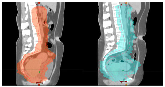
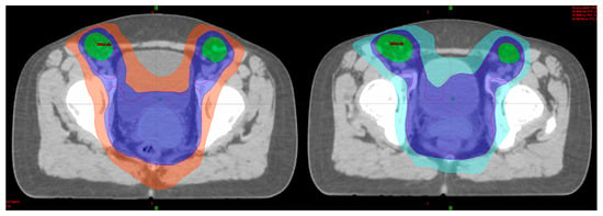
Adaptive radiotherapy is defined as a temporal adjustment of the treatment plan delivery to a patient, according to objective anatomic changes caused by weight loss, tumor shrinkage, or internal motion (Figure 1 and Figure 2).
Figure 1. Patient 1 with FIGO stage IIIC2 LACC: left image (week 1)—initial plan of EBRT and right image (week 3) during EBRT—adaptive planning (sagittal view) for tumor shrinkage.
Figure 2. Patient 1 with FIGO stage IIIC2 LACC: left image (week 1)—initial plan of EBRT and right image (week 3) during EBRT—adaptive planning (axial view) for tumor shrinkage.
A new concept of internal target volume (ITV) is generated to account for various treatment positions for LACC by performing a simulation with a full and an empty bladder and then combining the CTV to be taken into consideration for every move between these two bladder filling extremes [
55]. A margin between 3 and 7 mm (PTV) is added to the ITV to fully encompass setup and position errors. Then, volumetric IGRT is applied with cone beam CT (CBCT) to verify the position of the CTV and PTV daily prior to RT delivery. More advanced adaptive strategies have made space for a highly advanced work for treatment delivery called “plan of the day” or “online adaptive RT”. These approaches include same-day replanning and recently published review articles are available in the literature [
56].
In special scenarios, after paraaortic exploration or hernia repairs, open or laparoscopic, it is vital to use IGRT techniques to protect the wound and prevent complications or even delays in starting radiotherapy for LACC [
57].
This entry is adapted from the peer-reviewed paper 10.3390/medicina59101735