Antibody–Drug Conjugate for Cancer Treatment: History
Please note this is an old version of this entry, which may differ significantly from the current revision.
Subjects: Oncology
Contributor: , , , , , , , , , ,

Cancer, also known as malignant tumour or neoplasm, is a leading cause of death worldwide. One distinct feature from normal cells is that cancerous cells often overexpress protein on the cell membrane—for instance, the overexpression of human epidermal growth factor receptor 2. The expression of a specific protein on the cancerous cell surface acts as a marker that differentiates the normal cell and facilitates the recognition of cancerous cells. An emerging anticancer treatment, Antibody–Drug Conjugates (ADCs), utilises this unique feature to kill cancerous cells. ADCs consist of an antibody linked with a cytotoxic payload, mainly targeting the antigen found on cancerous cells. This design can increase the specificity in delivering the cytotoxin to the drug target, thus increasing the drug efficacy and reducing the side effect of cancer treatment due to off-target toxicities. 

  • cancer
  • antibody–drug conjugate
  • cytotoxin

1. Introduction

The discovery of the “Magic Bullet Theory” by a German Scientist, Paul Ehrlich, marked the targeted therapy revolution. He espoused the theory to describe his vision, where a chemical binds selectively to targeted microorganisms [1]. Ehrlich eventually realised this concept by successfully treating syphilis with a highly selective drug towards bacteria that does not harm the neighbouring healthy cells [2]. His outstanding achievement has had a significant positive impact on the healthcare system in which scientists develop drugs that could specifically target the cells in the body system.
The concept of the “magic bullet” has to some extent been realised by the development of antibody–drug conjugates (ADCs), particularly in the field of oncology. ADCs’ historical revolution has answered the crucial pharmaceutical concern of all oncologists, the need for anticancer treatment to target tumour cells efficiently along with high precision and specificity. The tremendous advancement of monoclonal antibody technology has allowed various cytotoxic agents such as cytotoxic drugs, immunotoxins, and radiopharmaceuticals to be conjugated to the bullet, resulting in high selectivity and targetability on the specific antigen expressed on cancerous cells. ADCs were proposed to reduce non-specific toxicities by altering their signalling pathways towards a therapeutic outcome or directing naturally acquired immune responses towards the tumour cells [3]. In recent years, new ADCs have been under clinical development, marking the success of this highly potential therapeutic agent in chemotherapy (Figure 1) [4]. Drago et al. (2021) have been very insightful in ADC design and maximising the potential of ADCs for cancer therapy [5].

2. Principle of Antibody–Drug Conjugate

ADC comprises a tumour antigen-specific antibody, a potent cytotoxic drug and a stable chemical linker that joins the antibody to the cytotoxic drug [10]. The physicochemical properties of respective components in an ADC are summarised in Table 1.
Table 1. Summary of the physicochemical properties of respective components in an ADC.
Components of ADC
Antibody Subunit Function
Antigen-binding Fragment (Fab) Mediate antigen recognition expressed on tumour cells.
Constant Fragment (Fc) Facilitate binding of Fab to immune cells.
Linker Type Mechanism of Drug Release
Cleavable Cleavage depends on the physiological environment.
  • Acid labile linker is cleaved at low pH, e.g., Gemtuzumab Ozogamicin.
  • Protease cleavable linker is cleaved through proteolysis, e.g., Brentuximab Vedotin.
  • Disulphide linker is cleaved by high intracellular glutathione concentrations, e.g., Mirvetuximab Soravtansine.
Non-cleavable It depends on lysosomal proteolytic degradation and requires optimal trafficking to lysosome. Thioether linker is cleaved by proteases in the cytosolic milieu, e.g., Trastuzumab Emtansine (T–DM1).
Cytotoxic Payload Target Site Examples of Payload with
Mechanism of Action
DNA
  • Calicheamicins induce double-strand DNA breaks.
  • Duocarmycin and Pyrrolobenzodiazepines (PBD) induce DNA alkylation by binding to A–T rich regions and guanine residues, respectively.
Tubulin
  • Auristatins and derivatives of Maytansine inhibit polymerisation of the microtubule and cell cycle arrest at the G2/M phase.
  • Auristatin monomethyl auristatin E (MMAE) in Brentuximab vedotin and glembatumumab vedotin, Monomethyl auristatin F (MMAF) in Depatuximab mafodotin, a derivative of Maytansine (DM1) in trastuzumab emtansine.
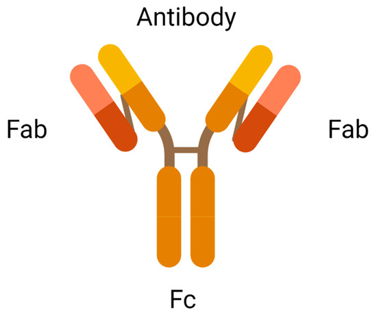
2.1. Antibody

An antibody is the most crucial element in an ADC due to its role in recognising the cancer surface marker. Therefore, an excellent antibody used in ADC should be stable during circulation in the bloodstream, less immunogenic, highly specific for the antigen, and can be internalised by the cancer cells. There are two antigen-binding fragments known as Fabs to recognise antigens expressed on cancer cells over healthy cells. A constant fragment, Fc mediates the interaction of antibodies with immune cells such as lymphocytes and B cells (Figure 2). A binding domain on the Fc receptor binds to the neonatal Fc receptor (FcRn) that regulates serum protein half-life [11]. Selection of the appropriate antibody subtype or technological engineering of the Fc domain, antigen affinity, the immune function of ADC, and biodistribution will influence the immune function of an ADC. In addition to the intrinsic cytotoxicity of drugs, the antibody on ADC mediates immune functions that are naturally acquired in patients through the activation of complement immune effector cells and signalling components, resulting in the migration of immune effector cells to the targeted sites for tumour-killing effects.
Figure 2. Antigen-binding fragments of an antibody (created with https://www.biorender.com/) (accessed on 2 December 2022).

2.2. Cytotoxic Payload

Cytotoxic payload is the effector component in ADC. The ideal candidate of an anticancer drug used in ADC should be highly potent with half-maximal inhibitory concentration (IC50) in the subnanomolar range to achieve desired therapeutic activities. Subnanomolar potency is crucial because only a few payloads can be conjugated to the targeting antibodies. Low potency of payloads results in a treatment failure. Due to the criteria of subnanomolar potency, many highly toxic drugs that were abandoned in the laboratory have now been reviewed as potential payloads in the development of ADCs. Common intracellular targets of ADC in tumour cells are DNA and Tubulin. The inhibition of these targets blocks the mitotic phase of tumour cells, slowing cell growth, and ultimately inducing apoptotic cell death [12].

2.3. Linker

The chemical linker in ADC conjoins cytotoxic payload to the antibody, stabilising the circulating ADC in the bloodstream upon administration. The functional group of linkers and the conjugation site determine the performance of ADCs in terms of circulating half-life, pharmacokinetics and pharmacodynamic profiles, and therapeutic window [14]. Disulfide, thioether, and hydrazone functional groups combine antibodies to the cytotoxic agent through intermolecular interactions. Currently, available linkers based on mechanisms of payload release are classified as cleavable or non-cleavable. Cleavable linkers depend on the physiological environment in the systemic circulation, such as pH or proteases available to release cytotoxic payload from the carrier. For example, the acid–labile linker in Gemtuzumab Ozogamicin is cleaved under low pH conditions. In contrast, protease cleavable linker in Brentuximab Vedotin is cleaved by proteases such as Cathepsin-B and plasmin. The cleavage of disulfide linkers in mirvetuximab soravtansine depends on high glutathione concentrations [15]. Non-cleavable linkers form non-reducible covalent bonds with amino acids on the antibodies. Unlike cleavable linkers, non-cleavable linkers are not affected by the physiological environment and are more stable in blood circulation [16].

2.4. Mechanism of Action of ADC

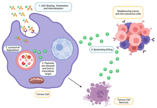
Upon intravenous administration, ADC circulates in the bloodstream and binds to an antigen explicitly expressed by targeted tumour cells (Figure 3) [18]. Following internalisation, ATP-dependent proton pumps in lysosomes to create an acidic environment to facilitate the degradation of cleavable ADCs by proteases such as plasmin and cathepsin-B, releasing cytotoxic drugs from ADC complexes [11]. In addition, lysosomal degradation, proteolytic cleavage of acid–labile linker, protease cleavable linker, and disulfide linker by the physiological environment occurs in the early or late endosomes [19]. The released cytotoxic drugs in the free circulating state resulted in the binding to intracellular targets, causing apoptosis via DNA intercalation or inhibition of microtubule polymerisation. As tumour cells lyse, free cytotoxic drugs are released and progress to bystander killing of neighbouring cancerous and non-cancerous cells via passive diffusion [20]. Apart from direct cytotoxicity, ADC-mediated effector functions include activating complement systems and infiltration of immune cells to localise at the targeted tumour via several mechanisms such as ADCP and ADCC [21].
Figure 3. Mechanism of action of ADCs at targeted tumour cells. ADCs are rapidly internalised and degraded in lysosomes upon binding to the antigen on the tumour. Payloads are then released into the cytoplasm, bind to the intracellular target and induce cell death. However, payloads can also exhibit bystander effects by diffusing out from the cells into adjacent cells (Created with https://www.biorender.com/) (accessed on 2 December 2022).

3. Antibody–Drug Conjugate Approved by FDA

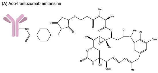
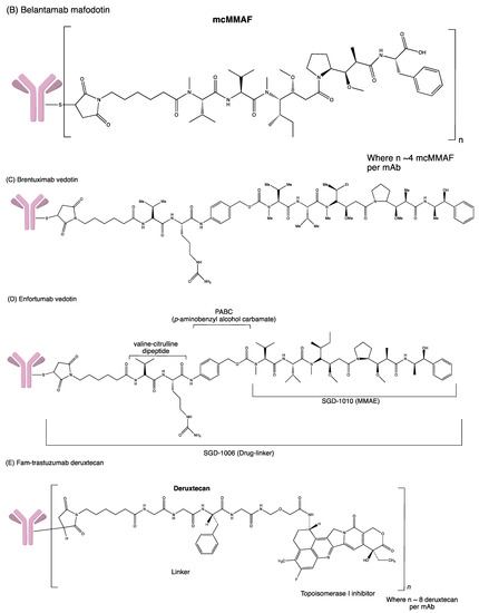
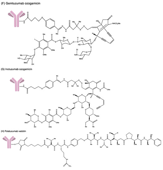
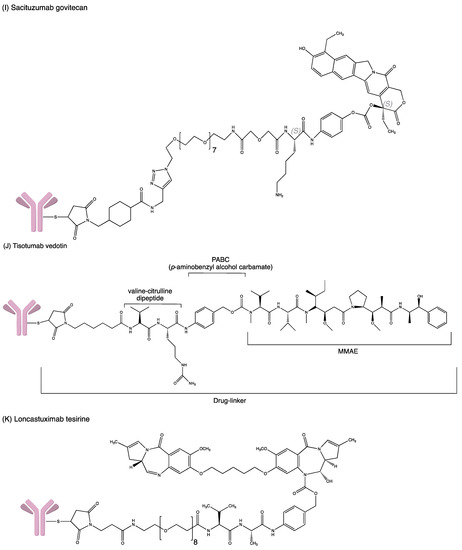
Developing an ADC is not easy as the process involves complicated bioengineering, chemistry and pharmacology. Many ADCs have recently received FDA-accelerated approval to treat severe malignancies in the hospital setting despite the challenges and hurdles (Figure 4). The FDA-approved ADCs are highlighted and updated accordingly in Table 2.
Figure 4. Structures of (A) ado-trastuzumab emtansine. (B) belantamab mafodotin. (C) brentuximab vedotin. (D) enfortumab vedotin. (E) fam-trastuzumab deruxtecan. (F) gemtuzumab ozogamicin. (G) inotuzumab ozogamicin (H) polatuzumab vedotin. (I) sacituzumab govitecan. (J) tisotumab vedotin. (K) loncastuximab tesirine.
Table 2. FDA-approved Antibody–Drug Conjugates.
ADC Antibody Target Antigen Linker Cytotoxic Payload Indication Year of Approval
Gemtuzumab ozogamicin Humanised IgG4 CD33 Acid–labile hydrazone Calicheamicin Relapse or refractory AML and newly diagnosed CD33+ AML Approved in 2000, withdrawn in 2010 but then reapproved in 2017 for relapsed/refractory malignancies

FDA approved for newly diagnosed CD33+ AML in ≥1-month paediatric patient
Brentuximab vedotin Chimeric IgG1 CD30 Protease-cleavable dipeptide MMAE HL, ALCL and different subtypes of T-cell lymphomas FDA accelerated approval in 2011

In 2018, FDA approved the treatment of previously untreated stage III-IV HL and previously untreated ALCL and other CD30+ peripheral T-cell lymphomas.
Ado-trastuzumab emtansine Humanised IgG1 HER2 SMCC DM1 Metastatic HER2+ breast cancer, previously treated with trastuzumab and a taxane and as adjuvant treatment for HER2+ early breast cancer with the residual invasive disease after neoadjuvant taxane and trastuzumab FDA approved in 2013

FDA approved for adjuvant treatment in 2019
Inotuzumab Ozogamicin Humanised IgG4 CD22 Acid–labile hydrazone Calicheamicin Relapsed or refractory B-cell precursor ALL FDA approved in February 2017
Monotherapy treatment of relapsed or refractory CD22-positive B-cell precursor EMA approved in June 2017
Polatuzumab vedotin Humanised IgG1 CD79b Cleavable dipeptide MMAE Relapsed or refractory diffuse large B-cell lymphoma FDA accelerated approval in 2019
Enfortumab vedotin Fully human IgG1 Nectin 4 Protease-cleavable dipeptide (Val–Cit) linker MMAE Locally advanced or metastatic urothelial cancer in adult patients who received prior treatment with a PD-1/L1 inhibitor and platinum-based chemotherapy in neoadjuvant/adjuvant setting FDA accelerated approved in 2019
Trastuzumab deruxtecan Humanised IgG1 HER2/ERB2 Protease-cleavable tetra-peptide (Gly–Gly–Phe–Gly) linker DXd Unresectable locally advanced or metastatic HER2+ breast cancer, previously treated with trastuzumab and a taxane, adjuvant treatment for HER2+ early breast cancer with the residual invasive disease after neoadjuvant taxane and trastuzumab FDA accelerated approved in 2019
Sacituzumab govitecan Humanised IgG1 TROP-2 Acid–labile ester (CL2 linker) SN-38 Triple-negative breast cancer, urothelial and other cancers FDA accelerated approval in April 2020 for mTNBC
FDA regular approval for in April 2021 TNBC
FDA accelerated approval in April 2021 for mUC
Belantamab mafodotin Humanised IgG1 BCMA Protease-
resistant maleimidohexanoic linker
MMAF Relapsed or refractory multiple myeloma in adults who have received at least four prior therapies FDA accelerated approval in 2020
Loncastuximab tesirine- lpyl Humanised IgG1 CD19 Valine–alanine dipeptide PDB dimer Relapsed or refractory large B-cell lymphoma FDA accelerated approval in April 2021
Tisotumab vedotin IgG1 Tissue factor (TF) mc–val–cit–PABC MMAE Recurrent or metastatic cervical cancer in patients with disease progression during or following chemotherapy FDA accelerated approval in September 2021
Moxetumomab Pasudotox - CD22 Recombinant covalently fused Pseudotox Relapsed or refractory HCL who received at least two prior systemic therapies FDA approval in September 2018
ADC: Antibody Drug Conjugate; AML: Acute myeloid leukaemia; MMAE: Monomethyl auristatin E; HL: Hodgkin lymphoma; ALCL: Anaplastic large cell lymphoma; ALL: Acute lymphoblastic leukaemia; mTNBC: metastatic triple-negative breast cancer; TNBC: metastatic triple-negative breast cancer; mUC: Metastatic Urothelial Carcinoma; PDB: Pyrrolobenzodiazepine; HCL: Hairy Cell Leukoma.

4. Antibody–Drug Conjugates under Development

4.1. Potential ADCs to Be Approved

4.1.1. AGS67E

AGS67E is a humanized IgG2 designed to target CD37. It is an ADC linked to the cytotoxic MMAE via a protease cleavable linker. AGS67E is able to exert direct proapoptotic activity and has potent cytotoxicity towards several non-Hodgkin lymphomas (NHLs) and acute myeloid leukemia (AML).
A phase I clinical trial (NCT02175433) had been conducted to assess the safety, tolerability and pharmacokinetics of AGS67E in patients with relapsed or refractory lymphoid malignancies. A total of 71 participants were enrolled in this study. AGS67E had a moderate response in 50 patients with R/R B-NHL and R/R T-NHL. The overall response rate was 22%, and the complete response rate was 14%. The most reported adverse effects were peripheral neuropathy and neutropenia. The results indicated that AGS67E showed positive effects in patients with relapsed or refractory lymphoid malignancies. Thus, further investigation of AGS67E can be continued [70].

4.1.2. Denintuzumab Mafodotin (SGN-CD19A)

Denintuzumab mafodotin (SGN-CD19A) is an ADC designed to target CD19. It is a humanised monoclonal antibody conjugated to the cytotoxic monomethyl auristatin F (MMAF) via a non-cleavable linker. Upon internalisation, MMAF will be released into the cells, which bind to the tubulin and inhibit its polymerisation. Eventually, the apoptosis of the tumour cell will occur and thus inhibit the cell growth of CD19-expressing malignancies [124].
A phase I clinical trial (NCT01786135) had been completed to assess the safety and tolerability of SGN-CD19A among 64 patients with relapsed or refractory B-lineage non-Hodgkin lymphoma (B-NHL) [83]. A total of fifty-two patients were treated every three weeks (0.5–6 mg/kg), and ten patients were treated in the q6wk schedule (3 mg/kg). In the dosing schedule, of 22 relapsed patients, seven patients had complete remission while four patients had partial remission (the complete remission rate and the objective response rate were 32 and 50%, respectively).

4.1.3. MEDI4276

MEDI4276 is an ADC consisting of a HER2-bispecific antibody that targets HER2-positive tumours. It is conjugated to AZ13599185, a tubulysin-based microtubule inhibitor via a maleimidocaproyl linker. The toxin AZ13599185 inhibits microtubule polymerization during mitosis and eventually induces cell death. MEDI4276 has potent antitumour activity in HER2-low cell lines that are refractory to Trastuzumab emtansine (T–DM1) treatment [126]. A phase I/II clinical trial (NCT02576548) had been performed to evaluate the safety, pharmacokinetics, immunogenicity, and antitumour activity of MEDI4276 in patients with select HER2-expressing advanced solid tumours.

4.1.4. Mirvetuximab Soravtansine (IMGN853)

Mirvetuximab soravtansine (IMGN853) is an immunoconjugate consisting of M9346A (humanised monoclonal antibody), linked by the cleavable disulfide to the cytotoxic DM4. The monoclonal antibody moiety of IMGN853 recognises and binds to antigen folate receptor 1 (FOLR1, folate receptor alpha). Upon the antibody–antigen interaction and internalisation, IMGN853 releases its cytotoxic payload (DM4), which binds to tubulin and disrupts the assembly of microtubules [127].
A phase III clinical trial (NCT02631876) had been conducted to access the efficacy and safety profile of mirvetuximab soravtansine versus the selected single-agent chemotherapy (Paclitaxel, Pegylated liposomal doxorubicin, Topotecan) in women with primary peritoneal cancer and/or fallopian tube cancer and platinum-resistant folate receptor alpha positive advanced epithelial ovarian cancer [128].

4.1.5. Patritumab Deruxtecan (U3-1402)

Patritumab deruxtecan (U3-1402 or HER3-DXd) is an ADC consisting of the fully humanised monoclonal antibody patritumab (known as AMG 888 or U3-1287) linked to the cytotoxic payload deruxtecan (topoisomerase I inhibitor) via a cleavable peptide linker. Patritumab specifically targets HER3 which is overexpressed on certain cancerous cells. When monoclonal antibody moiety binds to HER3 on the cell membrane, internalisation will occur, and the linker will be cleaved, releasing the payload deruxtecan. Deruxtecan induces tumour cell death through DNA damage [130].

4.1.6. Telisotuzumab Vedotin (ABBV-399)

Telisotuzumab vedotin (ABBV-399) is an ADC containing monoclonal antibody ABT-700, linked to cytotoxin MMAE (monomethyl auristatin E) via cleavable dipeptide. ABT-700 targets the c-Met receptor, a tyrosine kinase receptor that is overexpressed in patients with NSMCLC (non-small cell lung cancer). Preclinical data demonstrate that ABBV-399 can directly deliver cytotoxin to tumour cells by overexpressing the c-Met receptor [133]. A phase I clinical trial (NCT02099058) had been conducted to evaluate its pharmacokinetics, safety profile and preliminary efficacy as monotherapy compared to in combination with Erlotinib, Osimertinib, and Nivolumab in patients likely to express c-Met receptor the advanced solid tumours cell surface.

4.2. Discontinued/Terminated Clinical Trials

4.2.1. AGS-16C3F

AGS-16C3F is a novel ADC designed to target ENPP3, also known as cell-surface ectonucleotide pyrophosphatase/phosphodiesterase 3 [134]. AGS-16C3F is a fully humanised monoclonal antibody conjugated to MMAF (Monomethyl auristatin F) through a non-cleavable maleimido–caproyl linker. MMAF is an auristatin derivative, a potent microtubule disruptive agent with antineoplastic activity. Upon binding to ENPP3, internalisation occurs, followed by proteolytic cleavage of the linker to release MMAF in the cells. It will induce tumour cell apoptosis by inhibiting tubulin polymerisation and lead to G2/M phase arrest.

4.2.2. Coltuximab Ravtansine (SAR3419)

Coltuximab ravtansine, also known as SAR3419, is an ADC that targets CD19 (B-lymphocyte antigen CD19). SAR3419 is a humanised monoclonal antibody conjugated to cytotoxic DM4 (maytansinoid) through a cleavable disulfide bond. CD19 is a type 1 transmembrane glycoprotein ubiquitously expressed in B cell malignancies. Internalisation will occur upon binding to the CD19 antigen on the target cells, releasing DM4 into the cells. This will lead to microtubule disruption and cell cycle arrest and hence eventually causes cell death [135].
A phase II clinical trial (clinical trials identifier NCT01472887) has been conducted to investigate the safety and efficacy of SAR3419 in patients with diffuse large B-cell lymphoma. A total of 61 participants were recruited in this study [77]. Forty-one patients received coltuximab ravtansine, and the overall response rate was 43.9%, while overall survival, progression-free survival and median duration of response were 9.2 months, 4.4 months and 4.7 months, respectively.

4.2.3. Glembatumumab Vedotin (CDX-011)

Glembatumumab vedotin (CDX-011 or CR011-vcMMAE), an ADC designed to target glycoprotein NMB (gpNMB) that is often overexpressed in human malignant tissues, including melanoma and breast cancer. The high expression of gpNMB is associated with invasion and metastasis. CDX-011 links a CR-011, the gpNMB targeting antibody, to the MMAE (monomethyl auristatin E) through a cleavable dipeptide linker. Upon binding and internalisation of CDX-011, the release of MMAE induces cell death by disrupting the microtubules [137].

4.2.4. Lorvotuzumab Mertansine (IMGN-901)

Lorvotuzumab mertansine, also known as IMGN-901, is an ADC used to target CD56 (also known as neural cell adhesion molecule 1 or NCAM-1). IMGN-901 is a humanised monoclonal antibody conjugated to cytotoxic DM1 (mertansine, maytansinoid) via a disulfide linker. CD56 is a membrane glycoprotein expressed in malignant tissues, such as small cell lung cancer (SCLC) and leukaemia. Upon binding to the CD56 on the tumour cell surface, IMGN-901 will be internalised and cleaved to release DM1 into the cells, which causes tumour cell death by inhibiting tubulin polymerisation [138].

4.2.5. Rovalpituzumab Tesirine (Rova-T)

Rovalpituzumab Tesirine, known as Rova-T, is an ADC containing monoclonal antibody (humanised IgG1) conjugated to the cytotoxic PBD dimer via cleavable dipeptide. The antibody moiety of Rova-T selectively recognises and binds to the delta-like protein (DLL3) on the tumour cell’s surface. The dipeptide linker is cleaved upon internalisation to release the cytotoxin. Cytotoxin PBD dimer binds and induces DNA strand breakage [139]. A phase III clinical trial (NCT03033511) had been conducted to evaluate the efficacy of rovalpituzumab tesirine as maintenance therapy following first-line platinum-based chemotherapy. The result demonstrated no survival benefit of rovalpituzumab tesirine compared to placebo in treating small cell lung cancer. The overall survival in patients receiving a placebo was 9.79 months, higher than those receiving Rova-T, 8.48 months. Due to this unfavourable consequence, AbbVie officially announced discontinuing theRova-T research and development program [140].

5. Challenges of the Use of Antibody–Drug Conjugate

5.1. Toxicity of ADC

ADCs were initially designed to reduce systemic toxicities in comparison to conventional chemotherapy by targeting antibodies on highly expressed cancer biomarkers. However, toxicity remains one of the most intimidating challenges faced during the use of ADCs [27]. Chemotherapeutic drugs often cause toxicities even in the normal range of therapeutic doses [142], more so for ADCs that carry a subnanomolar range of payloads. It should be noted that ADCs cannot carry a large amount of cytotoxic payload due to the conformation and structure of the payload and antibodies. If a less potent payload is used in the development of an ADC, a higher dose is required to achieve the therapeutic effect, which may increase the risk of toxicity of the treatment. Thus, a more potent and highly super toxic agent as the cytotoxic payload is ideal for ADC designation [141]. According to [27], the issue of ADC toxicity could be improved by maximising the therapeutic index and adjusting the ADC dosage. The therapeutic index could be maximised in several ways, including integrating maytansine derivatives into ADCs, or biomarkers could be implemented to choose the correct patient population, observe initial response signals, or guide the combination therapy. Continuous monitoring should be carried out to observe the patient’s response.

5.2. Antibody & Antigen Specificity

Ideally, the antibody of an ADC should be highly specific and bind to the targeted antigen only. The purpose of having a highly specific antibody is to avoid any cross-reaction by binding to other antigens that might lead to undesirable toxicity [141]. Indeed, the targeted antigen should be highly expressed only in tumours but absent or minimally expressed in non-cancerous cells. This makes the targeted antigen tumour-specific, and the therapeutic effect can be specifically carried out on tumour cells without harming other non-cancerous cells. Sadly, most of the discovered antigens are also expressed in both cancerous and non-cancerous cells; the only difference is the level is higher in the cancerous cells than in non-cancerous cells [27].
The homogeneity or heterogeneity expression of antigen on the cell surface also determines the ADC activity. [147] stated that a homogenous antigen will conjugate at the same position in every mAb molecule, while a heterogeneous antigen will conjugate at a random position which differs in every mAb molecule. The homogenously expressed antigen is more desired in ADCs due to enhanced ADC targeting, batch-to-batch consistency, and higher desirability from a regulatory perspective [147]. A heterogeneous antigen isn’t desired in ADC; it could increase bystander killing activity. The bystander killing activity is beneficial to increase the killing rate of cancer cells but gives a poorer safety profile as the non-cancerous cells may be harmed simultaneously. Site-specific conjugation methods are developing to produce homogenous ADCs targeting just the homogenous antigen on cancer cells [147].

5.3. Immunogenicity of Antibody

Antibodies with high immunogenicity will provoke an immune response when a therapeutic agent is administered. It was desired to have an antibody with minimal immunogenicity to avoid an immune response from the body. This issue could be improved by replacing murine antibodies with humanised monoclonal antibodies to minimise the risk of immunogenicity [149]. The capability to induce receptor-mediated internalisation was also an essential parameter of the desired antibodies. The rapid internalisation of antibodies could improve ADC’s safety profile and therapeutic efficacy because it significantly decreases the possibilities of off-target release [141]. A highly stable antibody in blood circulation with a long half-life and a high molecular weight is desirable in ADC drugs. Thus, antibodies of ADC should have low immunogenicity and increased stability with rapid internalisation.

5.4. Stability of Linkers

Linkers are short bridges that bind the drug to the antibody covalently. The stability of linkers is a significant challenge faced in ADC use. In blood circulation, a stable linker will keep the drug tightly intact to the antibody and perform intercellular cleavage after reaching the targeted cell. Linker acts as a critical parameter in ADC designation as it influences the safety and efficacy of the drug in patients. An unstable linkage will degrade and release the drug before reaching the targeted cell, leading to off-target cytotoxicity. Thus, an ADC’s safety and efficacy will significantly reduce with an unstable linkage. The two main types of linkers include cleavable and non-cleavable linkers. Although non-cleavable linkage performs better than the cleavable, the most suitable linkage for each ADC will be selected mainly based on its stability with the functional group of ADC and environment [149].

5.5. Target Features of Successful ADCs

The target features of successful ADCs can be identified from several aspects. First, the toxicity of a successful ADC must be acceptable, with lower bystander killing effects to protect normal cells from being destroyed unintentionally, and the DAR should be optimized for each ADC. Second, the interaction between antigen expressed in tumours and the antibody must be highly specified to achieve a favourable on-target effect. Moreover, since ADCs consist of engineered antibodies, they may trigger an immunogenic response. Thus, a successful ADC must be safe to introduce into the human body. Finally, the stability of the linker is one of the key features of successful ADC which should only release the drug when it interacts with antigen to promise the safety and efficacy of ADC. 

6. Conclusions

Antibody–Drug Conjugate is an advanced chemotherapy that has been developed for the treatment of relapsed and/or refractory cancer. High specificity, potency and efficacy are the main features of this antibody-based therapy. Technological advancements enable the optimum design of ADC with fully humanised or chimeric antibodies, selecting clinically functional linkers and using a subnanomolar range of compounds to treat cancer patients. With the accelerated approval from FDAs, many ADCs are now benefiting cancer patients in a hospital setting. Nevertheless, there is still room for improvement for ADCs, including ADC toxicity, specificity of antibodies and antigens, the immunogenicity of antibodies, and stability of linkers.

This entry is adapted from the peer-reviewed paper 10.3390/ddc2020020

This entry is offline, you can click here to edit this entry!
Video Production Service