Cilia Distal Domain: History
Please note this is an old version of this entry, which may differ significantly from the current revision.
Contributor: , , , ,

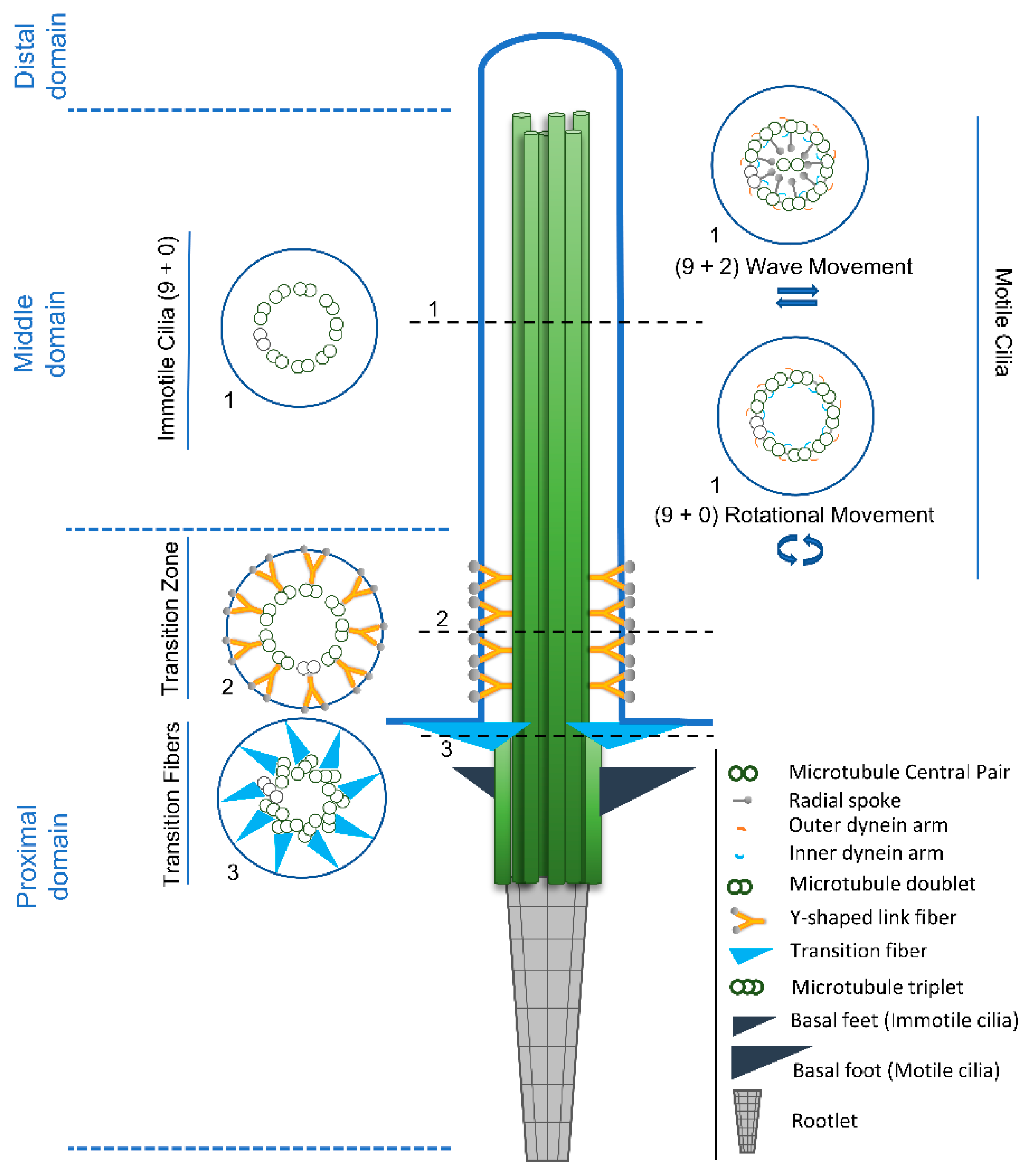
Eukaryotic cilia are microtubule-based organelles that protrude from the cell surface to fulfill sensory and motility functions. Their basic structure consists of an axoneme templated by a centriole/basal body. Striking differences in ciliary ultra-structures can be found at the ciliary base, the axoneme and the tip, not only throughout the eukaryotic tree of life, but within a single organism. Defects in cilia biogenesis and function are at the origin of human ciliopathies. This structural/functional diversity and its relationship with the etiology of these diseases is poorly understood. Some of the important events in cilia function occur at their distal domain, including cilia assembly/disassembly, IFT (intraflagellar transport) complexes’ remodeling, and signal detection/transduction. How axonemal microtubules end at this domain varies with distinct cilia types, originating different tip architectures. Additionally, they show a high degree of dynamic behavior and are able to respond to different stimuli. The existence of microtubule-capping structures (caps) in certain types of cilia contributes to this diversity. 

  • cilia
  • cilia distal domain
  • microtubule-capping structures
  • cilia structural diversity

1. Introduction

The Discovery of the Basic Cilia Structure at a Glance

Eukaryotic cilia/flagella are fascinating microtubule-based organelles that protrude from the cell surface. These organelles, composed of more than 1000 polypeptides [1][2], are complex compartments that captured the attention of cell biologists since they were first observed under a microscope. In fact, van Leeuwenhoek (1676) observing protozoa cells described them as “animalcules … with …little feet, or little legs, which were moved…” [3]. The observation of these intriguing structures, both in protozoa and in spermatozoa, led to the idea that cilia have a permanent fibrillar structure arranged with its long axis parallel to that of the cilium, and that is at the base of cilia movements by contraction [4].
The advent of electron microscopy (EM) created new opportunities to study the ciliary structure. EM observations of cilia/flagella from a number of flagellates, algae, fungi and human further showed that cilia were composed of multiple fibrils enclosed in a common matrix of protoplasm and surrounded by a membrane [5][6][7][8][9]. However, only in 1954, comparative studies by Fawcett and Porter [10] of thin sections of mollusk, amphibian, mouse and human cilia showed that these present an internal structure (axoneme) built of a ring of nine double filaments along the periphery and a central pair of single filaments. It also became clear that cilia emanate from complex elongated basal corpuscles/basal bodies from which ciliary rootlets (striated fibers) extend downward into the cytoplasm likely to anchor them [10]. A Similar arrangement of the axonemal filamentous components were observed in the cilia and flagella of plants [9], strongly suggesting that it could be conserved throughout the phylogenetic tree.
The advances on knowledge about the ciliary structure cannot be separated from structural and functional studies on the centrosome. The idea that centrioles are basically the same organelles as basal bodies was proposed in the second half of the 19th century [11]. This proposal triggered intense debate, and only much later, further EM studies permitted to establish the structural identity of basal bodies and centrioles and the confirmation that they are identical [11]. Later, it was shown that the injection of Xenopus eggs with purified basal bodies was able to replace centrioles in aster formation [12]. However, despite the body of work concerning the structure, function and biogenesis of centrosomes and basal bodies developed since, how a centriole becomes a basal body is still a process not fully understood.
The progressive improvement of preservation/fixation and negative staining techniques for EM samples, allowed detailed observations of the axoneme structures of motile cilia. This led to the description of new axonemal elements such as side arms and bridges, between the peripheral filaments of the axoneme, as well as radial spokes [13][14][15]. The first basic reconstruction of a flagellum structure from the tip to the basal body, along the axoneme, could thus be obtained, and showed the existence of structural diversity between different cilia types and species [16].
Although a detailed ciliary structure was emerging, the biochemical characterization of the ciliary compartment was lagging behind. Parallel to the characterization of cilia, elegant EM studies started to reveal other cellular structures more accurately. For example, microtubules were shown to be associated with centrioles, the mitotic apparatus and the midbody during cytokinesis [17][18][19] in diverse cell types. Their structural resemblance with the protofibrils composing the nine peripheral and central filaments of the axoneme (9+2 structure) of the human and rat sperm tails and Tetrahymena cilia [20][21] suggested a microtubule-based nature for the axoneme, and a role for these polymers in cilia movement [22]. Later, efforts to identify ciliary proteins revealed the microtubule building block tubulin and the microtubule motor protein dynein as major components of these organelles [23][24][25][26].
The existence of cilia (primary cilia), distinct from the motile cilia previously observed in protists and multiciliated tissues, were noticed in the 19th century, but it was Zimmermann (1898) that described them in mammalian cells. He designated these cilia as ‘centralgeissel’ (central flagella), showed they were associated with a pair of centrioles (the diplosome), and proposed a sensory role for them [11].
Numerous EM studies from the 1950s to 1970s using EM showed that both the centriole/basal body, and axoneme fine structures are remarkably conserved throughout the eukaryotic phylogenetic tree. This supports the idea of these being ancient structures already present in the last eukaryotic common ancestor (LECA) [27][28][29]. Furthermore, these studies also clearly show that, despite being conserved, these structures present a certain degree of structural variability. This suggests that structurally diverse cilia evolved such as to allow cells and organisms to cope with distinct environmental challenges/signals, and functional specificities or their life styles.
This important body of work led us to the present view on ciliary structure and functions. Thus, it is now well established that the basic ciliary structure consists of an axoneme made of nine radially-arranged stable microtubule doublets [30] templated by a centriole/basal body docked to the cell membrane, and covered by a specialized membrane domain (Figure 1). Typically, microtubule doublets are composed of a complete (13 protofilaments) A-tubule and an incomplete B-tubule (10 protofilaments) [31]. Additionally, a central pair of singlet microtubules can be present originating the (9+2) architecture. The relevance of cilia in human health and disease is also now unquestionable. Indeed, in vertebrates, most cells are ciliated, and multiple specialized cilia types fulfill sensory and motility functions critical for embryonic development and tissue homeostasis [32][33]. In simple terms, cilia can be classified as motile, promoting cell movement (e.g. protozoans and sperm cells) or fluid flow (e.g. trachea multicilated epithelia), or immotile (primary cilia) fulfilling mainly sensory/signaling functions (e.g. morphogenetic pathways including Notch, Wnt, Hippo, mTOR, PDGFR, GPCR, TGFβ and Hedgehog (Hh) signaling) [34]. However, a refined evaluation of cilia roles clearly showed that motile cilia can also mediate sensory functions, which is illustrated by the motile cilia in the respiratory and reproductive tracts of humans and other mammals [35]. Nevertheless, this idea was already latent in the early studies of protists and invertebrates showing the mechanoreception properties of their motile cilia [36].
Figure 1. Structural comparison between motile and primary cilia. The cilia axoneme is composed of nine pairs of microtubules (green tubules) that extend from a modified centriole named basal body. Three structural and functional cilia domains can be identified, namely the distal, the middle and the proximal domains, according to their position in relation to the basal body. Cross-section 1 shows the three most common axonemal architectures: (9+0) immotile and motile cilia, with nine microtubule doublets arranged radially; (9+2) motile cilia, with nine microtubule doublets and a central pair of microtubule singlets. Motor proteins (outer dynein arms (yellow) and inner dynein arms (blue)) together with radial spokes generate the wave-like beating of the (9+2) motile cilia; (9+0) motile cilia, with nine microtubule doublets and without central pair of microtubule singlets. In (9+0) motile cilia, motor proteins (outer dynein arms (yellow) and inner dynein arms (blue)) generate a rotational movement. Cross-sections 2 and 3 respectively show, the Y-shaped links (yellow) of the transition zone and the transition fibers (light blue), that link to the membrane creating the ciliary gate. The rootlet (grey) extends from the base of the basal body towards the nucleus and appears as long striated fibers. The basal foot (motile cilia)/basal feet (immotile cilia) (dark blue) provide additional support to the cilia. In the case of motile cilia, the basal foot aligns with the cilia beating direction.

2. CILIA: Variations of a Basic Structure to Fulfill Myriad Functions

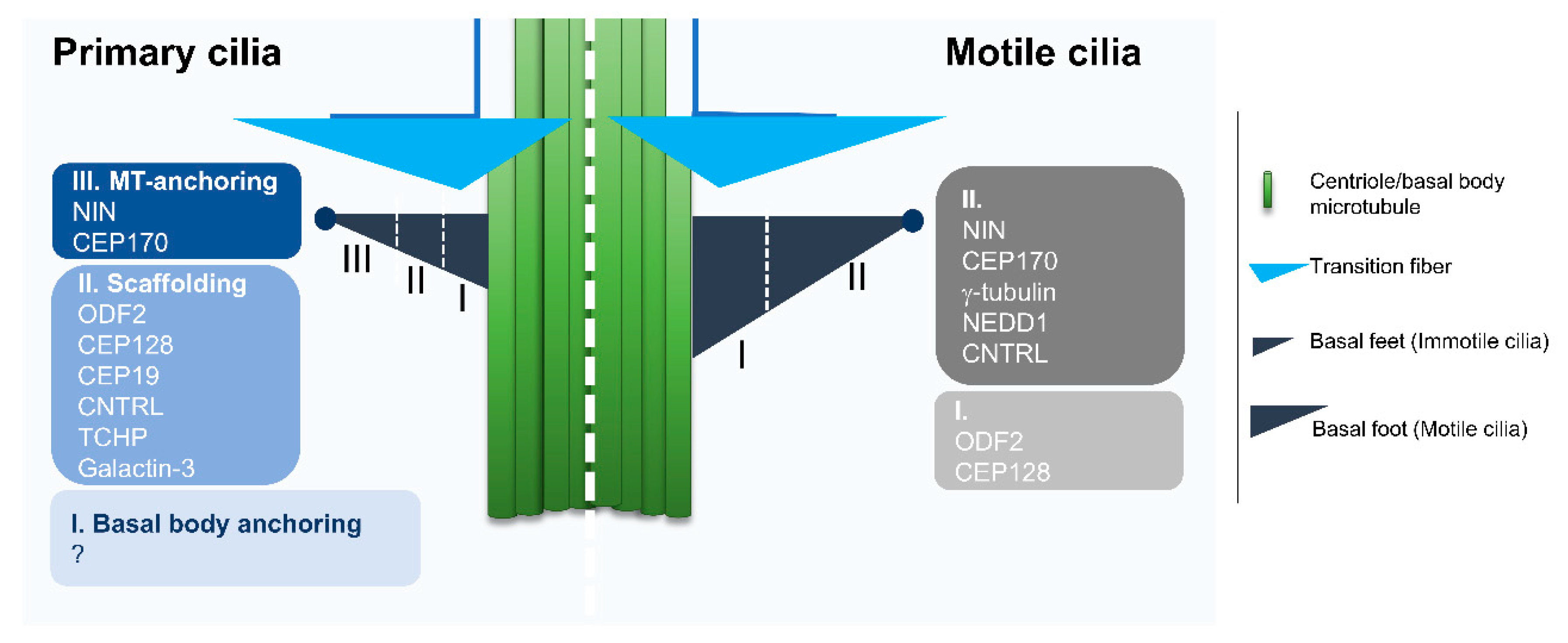
2.1. Diversity Starts at the Base: An Overview

Although the axoneme is likely the most characterized ciliary structure in multiple cilia types of many different species, cilia diversity is also reflected at the level of structures at the base (rootlets, basal feet, transition fibers, transition zone) and tip of cilia (capping structures). Concerning the ciliary base sub-structures, they can present dramatically different morphologies between organisms and even between cilia types of the same organism. In addition, they can be altogether absent, as is the case of Caenorhabditis elegans cilia, which lack transition fibers [37], and Drosophila cilia, which lack basal feet [38].
Cilia biology is intimately related to that of the centrosome, the major microtubule organizing center in animal cells. The centrosome presents two microtubule-based structures called centrioles, which are usually composed of nine microtubule triplets arranged radially. There are, however, species that present centrioles made of doublet (e.g.; certain Drosophila cells) or even singlet microtubules (e.g.; C. elegans). The two centrioles of the centrosome are asymmetric given that usually the oldest, also called mother-centriole, is decorated with sub-distal and distal appendages. These appendages have important roles in the context of cilia formation and maintenance, giving rise to basal body structures called basal feet and transition fibers, respectively (Figure 2). The mother centriole has to be converted into a basal body when ciliogenesis is triggered, and usually gives rise to primary cilia. However, motile cilia in embryonic structures like the node (mammalians) and the Kupffer’s vesicle (zebrafish) are also likely derived from the mother centriole. On the other hand, in multinucleated cells bearing multiple motile cilia, the basal bodies originate from two parallel pathways: centriole-dependent and deuterosome-dependent pathways [39].
Figure 2. Comparison of the Structural Organization and major components of basal feet from primary and motile cilia. Primary and motile cilia basal feet present different components and domain organizations as observed by super-resolution microscopy. Primary cilia have multiple basal feet whose components are organized in three distinct domains. The most distal region (III) is characterized by the presence of Ninein (NIN) and Centrosomal protein of 170 kDa (CEP170), proteins involved in microtubule (MT) anchoring and components of the centriole sub-distal appendages. The next region (II) contains proteins that are possibly involved in the scaffolding of the basal feet, namely Outer dense fiber protein 2 (ODF2). The most proximal region (I) to the basal body was named “basal body anchoring” although there was no protein assigned to this region. Motile cilia present only one basal foot, aligned with the cilia beating direction. The domain organization is simpler than that of primary cilia, corresponding, to a certain extent to a rearrangement of Regions II and III. On the most distal region of the basal foot (II), besides the proteins in common with the basal feet of primary cilia (i.e. NIN, CEP170, Centriolin (CNTRL)), there are proteins of the γ-tubulin ring complex (γ-TURC) microtubule nucleating complex (i.e. γ-tubulin and protein NEDD1). The most proximal region (I) contains proteins that are common to the primary cilia basal feet (i.e. Centrosomal protein of 128 kDa (CEP128) and ODF2) that are most likely responsible for the scaffolding (based on proteomics and super-resolution microscopy data in [40]).
Despite the origin of the centriole/basal body, there are several structures that can be observed at the base of most eukaryotic cilia that are poorly characterized but fulfill important roles, and reflect vertebrate cilia ultra-structural diversity (Figure 1). One of them is called the rootlet, which extends from the base of the basal body towards the nucleus and appears as long striated fibers. Rootletin has been described as the protein that organizes rootlets, but other components may yet need identification [41][42][43]. Ciliary rootlets seem to provide physical support to the ciliary structure [42], but they have been proposed to play additional roles like the transport of cargo to the ciliary base [44]. Interestingly, rootletin assembles rootlets of different lengths in different cell types, suggesting that these structures may fulfill cilia-type specific roles [41], likely in a cross-talk with other cytoskeletal components. In addition, there seem to exist inter-species diversity in ciliary rootlet structures. Indeed, Fawcett and Porter [10] recognized early, through EM studies, that the rootlets’ structure is more prominent in the cells of mollusks, less well developed in amphibia and differentially developed in mammalian. For example, ciliated epithelial cells of the rabbit trachea contain no rootlet structures, contrary to those of other mammalian species [45].
Basal feet also provide support to the cilium and acquire a prominent role in the context of motile cilia. Indeed, basal feet are critical to orient basal bodies in multiciliated cells such that there is efficient coordinated ciliary beating in the same direction, which is achieved through the basal foot role in organizing microtubules [46][47]. This is reminiscent of the role of the mother centriole sub-distal appendages, which anchor microtubules to the centrosome. In primary cilia, basal feet are important in determining the exposure of the cilium to the external environment. Indeed, the loss of sub-distal appendages/basal feet leads to the full exposure of the usually partially submerged primary cilia of human RPE-1 cells, which greatly impacts cilia-dependent signaling [48]. Typically, it has been assumed that the composition of basal feet is the same as that of the sub-distal appendages, which is supported by a few studies [48]. However, it is possible that during the mother centriole to basal body conversion the protein composition changes, something that still warrants investigation. Moreover, basal feet appear dramatically different in motile cilia. Indeed, whereas basal feet are present in multiple units in the context of primary cilia, motile cilia present one prominent basal foot (Figure 2). These two types of basal feet share components, but further studies are needed to determine their protein composition in the context of different cilia types. Additionally, it will be important to understand how the same molecules organize such different basal feet in multiple cell types. Of note, recent studies using super-resolution microscopy showed slightly different relative positions of basal feet components in primary versus motile mammalian cilia (Figure 2) [40].
The transition fibers anchor the basal body to the cell membrane. Importantly, these fibers, together with the ciliary transition zone present at the proximal end of the axoneme, function as a ciliary gate. This gate is absolutely key to determine the protein and lipid composition of the cilium compartment. In agreement, the ciliary gate is a ciliopathy hotspot with many components of the transition zone and transition fibers being coded by ciliopathy genes. Extensive reviews on the ciliary gate have been published recently and the researchers would like to direct the readers to them [49][50]. Different transition zone architectures can be found throughout the eukaryotic tree of life or even in the same organism reflecting eukaryotic cilia functional diversity [38][51][52].
In most multiciliated cells found in the epithelia of vertebrates and other organisms, motile cilia beat in a synchronized fashion, presenting a typical (9+2) pattern. However, monociliated cells bearing (9+2) motile cilia can be found in the pronepheric duct of zebrafish [53][54]. In the case of primary/immotile cilia they are usually found alone in each cell, but there are some exceptions [55][56].
Although the (9+0) axonemal structure has been mainly associated with primary cilia this pattern is not exclusive of this cilia type. Indeed, (9+0) motile cilia are present in embryonic structures like the mouse node or the kupfer’s vesicle in the zebrafish (reviewed in [57]), and in the gametes of protists, flat worms, annelids, and eels [58]. Along the same line, the (9+2) pattern is also present in immotile cilia, as is the case of human/mouse olfactory cilia, and the cilium in mechanosensory hair cells of the inner ear. In fact, motility requires the presence of axoneme-associated structures, such as outer (ODA) and inner dynein arms (IDA), the nexin-dynein complex, which regulates the activity of the dynein arms, and the radial spokes [32][33] (see Figure 1), but not necessarily the presence of the central pair of microtubules [59]. However, there are evidences that the central apparatus is involved in determining the bending plane of cilia movement, and therefore related to the type of movement that cilia are able to promote. The (9+2) motile cilia of ciliated epithelia and sperm flagella, present a rhythmic waving or beating motion, whereas the (9+0) nodal cilia move in a rotational manner and produce a leftward fluid flow (nodal flow) [60]. The ODA and IDA are essential for ciliary motility since they generate the force that causes microtubule doublet to slide. This produces a helical beat that can be modified by the inter-doublet links and the interactions between the central pair projections and the radial spokes [61]. ODA’s function is mainly to control the ciliary beat frequency by changing the velocity of doublet sliding, without changing the beat type [62]. On the other hand, IDA, in conjunction with the radial spokes and the central pair complex, control the bend amplitude and the form of the beat [63]. In Chlamydomonas, ODA are composed of three dynein heavy chains while in metazoans they possess only two [31][64]. IDA are less studied, and in Chlamydomonas, they present seven major different heavy chain dyneins and at least three minor heavy chain dyneins [65]. The complexity of dynein arms confers, in many organisms, the ability to change their ciliary beat pattern in response to specific environmental signals. This picture may be more complex since besides the structural units of ODA and IDA are highly conserved throughout evolution [66], genomic studies show diversification of dynein genes with specific phylogenetic distribution which may be related to species specific functions [67]. Radial spokes regulate dynein activity, acting as mechano-chemical transducers between the central apparatus and the outer microtubule doublets. These T-shaped structures are composed of at least 23 distinct proteins in Chlamydomonas and are periodically repeated throughout the axoneme. Most organisms contain three radial spokes in each repeat unit (e.g.; Tetrahymena, Trypanosoma, sea urchins, mammals [52][68][69] while Chlamydomonas and Sarcophaga only possesses two complete and one incomplete repeat units [70][71][72][73][74]. Even though the morphology of radial spokes is conserved, their components have diverged evolutionarily. For example, Ciona radial spokes have only one RSP4/6 protein, whereas Tetrahymena has three [75].
The structural diversity in the middle domain beyond its impact in cilia motility is probably related with other cilia-specific functions. Recently, a new axonemal microtubule organization was observed in C. elegans cilia cephalic male (CEM) neurons, in which the cilia middle regions doublet microtubules are splayed to form complete A- and B-tubule singlets while remaining attached at their proximal and distal ends [76]. Variations to the (9+0) and (9+2) structures can be found throughout the eukaryotic tree of life. Most often, they consist either of extra structures surrounding the (9+2) axoneme (usually in sperm cells), or in the absence of one of the central microtubules [16][52][77][78]. This illustrates how this domain is poorly structurally/functionally characterized in diverse cilia types.

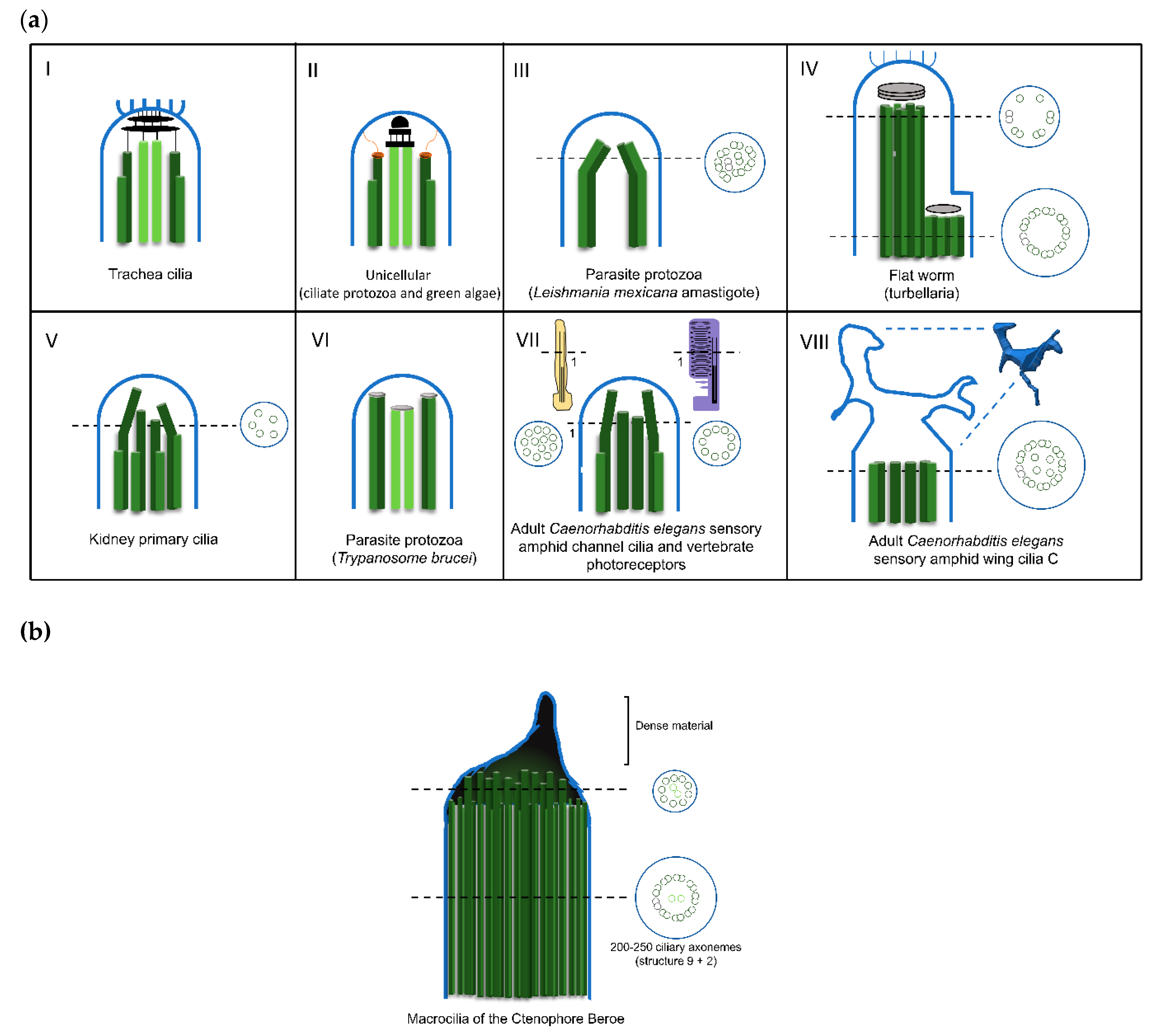
2.2. The Amazing Architecture Diversity of the Cilia Distal Domain: Cilia Tips Segments and Caps

In recent years, the cilium compartment started to be regarded as being composed of distinct domains that are functionally and structurally specialized (Figure 1). Here the researchers will focus the attention of how the distal ciliary domain (the tip or tip segment) is structurally organized and may present complex capping microtubule structures (caps).
The first cross sections from the transition region from the axoneme middle segment to the tip of motile cilia/flagella from a variety of organisms (e.g.; mammalian sperm [79][80]; invertebrate sperm [10][81]; gill cilia [51], ciliate protozoa [82][83]; flagellate protozoa [14]; fungal zoospores [84] and unicellular algae [85]) showed that, in general, cilia/flagella tend to narrow towards their most distal end, presenting a blunt or slightly pointed tip. This region is characterized by a progressive alteration and disorganization of the normal microtubule (9+2) pattern. Although variations can be identified, in general the peripheral microtubule doublets tend to become singlets, usually through the loss of the B-tubule. Frequently, these singlets preserve their radial organization, but their number decreases towards the distal end of the cilium/flagellum. However, this organization is completely lost when all the doublets are converted into singlets, and sometimes the central pair becomes indistinguishable. Still, in certain ciliates, as well as in Chlamydomonas, the central pair is observable until the end, and terminates in complex cap structures [15][86][87]. In addition, in most cilia from different origins (e.g.; rumen protozoa [82]; gill cilia [88] and Euplotes cilia [83][89]), amorphous electron dense material was observed between the microtubule central pair and the membrane, and the lumen of the singlets was electron-opaque just before the cilium/flagellum ends [15].
In fact, more detailed studies of thin sectioned tips of Chlamydomonas flagella showed that the central pair microtubules were inserted into a cap (central microtubule cap) [15][90] which was attached to the distal tip of the flagellar membrane. The cap is composed of two plates oriented perpendicular to the long microtubule axis, that seem to be attached to each other by small filaments and a spherical bead of approximately 50 nm in diameter. At the distal end, the bead links the plates to the membrane. Also, the A-tubules of the outer doublet microtubules of the flagella terminate in a pair of filaments that were designated as distal filaments [90]. These structures are attached to the membrane and to a plug-like structure that is inserted into the tip of the A-tubules. A comparison between the Chlamydomonas flagella tip structures and those of Tetrahymena and Aequipecten irridians bay scallop gill cilia showed that, these present similar structures linking the ends of both central pair and outer doublet microtubules to flagellar membranes (Figure 3aII) [87]. Moreover, in Tetrahymena, the central microtubule cap is bound to the ends of the two central microtubules by plug structures similar to those observed in A-tubules [91].
Figure 3. Diversity of cilia distal domain architectures. (a) Schematic representation of representative cilia tip structures. (I) In trachea cilia, the axoneme structure is (9+2) and at the tip the A-tubules and central pair are bound to a central cap that is attached to the ciliary crown, a structure constituted by a cluster of fibrils that project from the membrane to the outside of the cilia tip. (II) In the ciliate protozoa (Tetrahymena) and green alga (Chlamydomonas), the axoneme structure is (9+2). At the tip, the A-tubules are attached to the membrane by distal filaments that, in their proximal ends, form a plug which inserts into A-tubule lumen, while the central microtubule pair is bound to the central cap. (III). In the parasite protozoa Leishmania mexicana amastigote, the axoneme structure is (9+0). Distally, the 9-fold symmetry is broken by microtubule doublets progressively occupying a more central position. (IV) In the flat worm turbellarian the cilia present a typical (9+2) axonemal pattern through the main part of its length. Near the tip, this pattern changes to one where the microtubule central pair and doublets 1, 2, 3, 8 and 9 terminate in a distal cap that is attached to a ciliary crown, whereas doublets 4 to 7 end at a dense material of a proximal secondary cap. Therefore, the tip is asymmetric. (V) In the kidney primary cilia, the axoneme is (9+0) and progressively, along the axoneme, microtubule doublets suffer a displacement towards the center and the axoneme narrows towards the tip. In the distal segment, doublets are converted into singlets through the loss of the B-tubule. (VI). In the parasite protozoa Trypanosome brucei, the tip is (9+2) but the microtubule central pair is slightly shorter. Microtubule doublets and central pair present a dense material at the ends. (VII) In the adult Caenorhabditis elegans sensory amphid channel cilia (yellow) the axoneme is (9+a few singlets), and the microtubules maintain a radial organization and in vertebrate photoreceptors (purple) the axoneme is (9+0). However, both tips present only microtubule singlets. The differences can be observed at the cross section (VIII). In the adult Caenorhabditis elegans’ sensory amphid wing cilia C, the middle region of the axoneme contains both doublets and singlets that “splay apart” laterally and end together ~0.5 μm below the distal membrane. The tip presents a membranous fan-like structure. (b) Macrocilia of the ctenophore Beroe. Macrocilia present hundreds of ciliary axonemes with a (9+2) structure surrounded by a common membrane, with a giant capping structure. At the tip, doublet microtubules end and only singlet microtubules remain, linked together by electron-dense material. Central microtubule pairs are often still recognizable at this level. Schemes were based on: [45][86][92][93][94][95][96].
In the parasite protozoan Trypanosoma, motility is achieved through the undulating behavior of a single (9+2) flagellum. The analysis of the flagella tip of different promastigote Trypanosomatida species (i.e.; Crithidia deanei; Herpetomonas megaseliae, Trypanosoma brucei and Leishmania major) showed that these (9+2) flagella present a blunt end where two regions of dense material are detected (Figure 3aVI). One is essentially associated with the central pair of microtubules, whereas the other connects to the doublets [97]. Depending on the species, this dense material may show a ring shape or adopt a more disc-like structure [97]. On the other hand, the intracellular amastigote stages of Leishmania parasites possess a short cilium with an interesting partial structure of (9+0). In these cilia, 1 or 2 doublets are subject to displacement from the 9-ring organization throughout the axoneme, generating a variable architecture towards the cilia tip, where no organized structures are observed (Figure 3aIII) [92]. Moreover, connections between the microtubule doublets and the ciliary membrane were observed throughout the majority of the cilium length [92]. Due to the fact that these cilia are found in close contact with the membrane of the parasitophorous vacuole inside host cells, it was proposed that they act as a sensory organelle with important functions in host-parasite interactions and signaling [92].
In mammalian epithelial cilia, as the ones lining the trachea, oviducts, and bronchioles the central microtubules’ cap is amorphous in appearance. Additionally, instead of being directly linked to the cilia membrane like in protozoa, it links to a ciliary crown, a structure constituted by a cluster of fibrils emanating from the ciliary membrane at the very tip of the organelle (Figure 3aI) [93][98][99][100]. In these cilia, the plugs of A- and central pair microtubules end in a plate of electron-dense amorphous material, and five layered discs are found in the space between the membrane and the amorphous plate [93][98][99]. The crown fibrils seem to be linked to the plates through the membrane, given that they are resistant to treatments with detergents that affect it. A similar structure, with slight differences, was described in the cilia of other vertebrates such as, chicken, rabbits, frog embryos, adult frog palates [101], Anuran seminal vesicles [102], mouse thymic cysts [100] and the haptocilia of Acoel turbellarians [86][103]. On the other hand, bovine tracheal cilia lack ciliary crowns, being the dense cap linked to membrane through thin filaments, resembling the way the central cap is linked to the membrane in protozoan cilia [104]. Interestingly, Dirksen and Satir (1972) [98] noticed that the cilia crown was absent from the mouse developing oviduct and respiratory tract cilia [93], appearing only when the growth is complete. This strongly suggests that the cilia tip structure varies throughout the different stages of ciliary maturation and between tissues.
Contrary to the observations in protozoan cilia, in tracheal, oviduct, and frog palate motile cilia, the plugs are linked to the central microtubule cap instead of the membrane [104]. It is noteworthy that, in the Tetrahymena oral apparatus cilia, the distal filaments also link the outer doublets to the central microtubule cap [87] being an exception relatively to the remainder cilia of this organism. This indicates that the tip structures present not only a degree of structural variation from species to species, between different tissues and developmental stages of an organism, but also within a single-cell organism.
Asymmetrical cap structures were found in certain amphibian cilia, as well as in Acoel turbellarians (flatworms) (Figure 3aIV) [103][105]. For example, palate cilia in the frog Bombina orientalis end in one large cap, linked to the membrane and to doublet microtubules number 4 to 7, and a smaller cap that is connected to doublets number 1, 2, 3, 8, and 9 and the two central microtubules. A plug structure inserted into the lumen of each microtubule attaches the A-microtubules to the caps [105]. In the case of Acoel turbellarians, the adhesive plate on the ventral and posterior tail surface possesses cilia with a typical (9+2) axonemal pattern through the main part of its length. However, near the tip the microtubule central pair and doublets 1, 2, 3, 8 and 9 terminate in a distal cap, whereas doublets 4 to 7 end at a dense material of a proximal secondary cap [86][103]. The distal cap has 2 to 4 laminar structures, and possesses attached filaments that resemble the fibrils of the ciliary crown of the mammalian tracheal cilia cap. Interestingly, some of the doublets end as a single A-microtubule, whereas for others, the A- and B-microtubules terminate in the cap. These cilia are motile, display slow, irregular beating with an irregular bending and most probably possess an adhesion role, allowing the animal to stick to the substrate [86]. Noteworthy is the fact that in both organisms the same groups of microtubules are linked to distinct caps but the orientation of the asymmetric structure, in respect to the cilia stroke are different [105].
Different cap structures, essentially composed of amorphous electron-dense material distributed around the distal ends of the doublet and central microtubules, were identified in cilia from different organisms. The lips of the ctenophore Beroe possess multiple rows of motile ciliary axonemes surrounded by a common membrane, and these are thought to help the organism spread its lips over preys, pushing them into the stomach cavity [94]. These multiple axonemes present a giant capping structure at the tip, formed by extensions of the A- and central-pair microtubules, which are bound together by amorphous material (Figure 3b) [94]. The sensory cilium of the chordotonal sensillum in the American cockroach antenna pedicel, possesses an axoneme with a (9+0) pattern throughout its length. At its dilated the tip, the doublets become singlets and terminate in an electron-dense spheroid [106]. It was proposed that these cilia may be involved in mechanoelectric transduction, when vibrations are applied to the sensillum [106]. Additionally, dense material was observed at the distal ends of the axonemal microtubules in stiff non-motile sensory cilia of the bivalvia Lima hians [107].
Concerning the ultrastructure of primary cilia distal tips, not much data is available, but their tips seem to lack cap structures or even electron-dense material (Figure 3aV) [108]. In cultured kidney IMCD3 cells the doublet microtubules suffer a displacement, distorting the (9+0) arrangement towards the tip. Moreover, these cilia narrow towards the tip where a few doublets and singlets are still detected [92]. However, in many of these cilia, the organization pattern of the microtubule doublets can be extremely variable along their length, and many of them present connections between the doublets and the ciliary membrane throughout the axoneme. In certain cases, these structural details associated with their specialized functions may overcome the necessity to have a cap structure.
This analysis of how different cilia organize their distal segment suggests that during evolution, eukaryotic cilia have acquired or modified ciliary capping structures in order to fulfill specific roles in different cilia types and this is reflected by their multiple structural modalities. Even single cell organisms present different functional cilia/flagella and these multiple ciliary tip-organizations and associated structures seem to be related to specific ciliary roles. In the case of motile cilia, it has been proposed that those involved in cell propulsion, for example in ciliates and flagellates, present tip cap structures that evolved to allow beating and locomotion in water. These caps will probably help to maintain cilia structure integrity continuously under mechanical stress caused by beating, still accommodating the possibility of dynamic regulation of cilia length in response to stimuli. On the other hand, the specialized structures (ciliary crown) found in the majority of vertebrate ciliated epithelia, likely allow for efficient interaction with ovules and movement of viscous mucus and lubricants required for tissue homeostasis. These processes, compared to cellular movement, will also require different force generation. The crown fibrils probably increase adhesion efficiency creating a larger surface and allowing a better adaptation to interaction with random shapes. Still, exceptions, like that of bovines limit this view. Interestingly, similar structures are found in cilia of Acoel turbellarians, which appear to be involved in sticking the organism to a substrate. It is also possible that these cilia structures may have evolved from early organisms for which cilia adhesion properties to particles/preys or substrates would favor fitness. It is tempting to hypothesize that the crown fibrils may be able to transduce signals from adhesion interactions to the cilia tip. If this is true these signals could trigger local modifications in microtubule dynamics and increase/decrease in a few nanometers the length of the distal segment which would adapt stroke to the physical/chemical features of the media. Supporting this idea is the fact that motor cilia are able to change size in response to environmental stimuli [109]. The biochemical characterization of distinct cilia types in different organisms and tissues will definitely help elucidate the functions of these diverse ciliary capping structures. However, despite the limited information on the molecular composition of the ciliary surface, it is interesting that cilia tips present adhesive properties illustrated by the ability of virus and bacteria to adhere to cilia [110]. In fact, cationic ferritin binds preferentially to cilia crown, showing the presence of anionic sites in these localizations [111].
The asymmetry of some cap structures was proposed to be related with the definition of the cilia stroke orientation, but no constant pattern was found in the different examples described so far. In the case of the Acoel turbellarians the accentuated asymmetry creates a small thinner segment in the tip that will also allow a better interaction with substrates with random shapes which may create opportunities to better sense the substrate and adhere. In the case of frog palate cilia asymmetry is probably important to help define the direction of the stroke. These cilia propel mucus and particles in it over of the oro-pharynx epithelium in the posterior direction and down the oesophagus [112]. Interestingly, they are shorter than cilia generating water propulsion and present relatively small difference in height between the effective stroke and the recovery stroke which seems to help the mucus movement. Additionally, these cilia do not present a high coordination showing intermittent activity, which suggests that cilia motility is regulated by reflex control dependent upon mechanical stimulation. Dentler [45] suggested that cap structures may cause modification in the patterns of microtubule sliding. This modification would cause a twist along the axoneme or create an effective stroke with a nonplanar bend. Alternatively, the microtubules may stretch or contract during sliding with the purpose of accommodating the restriction of sliding by the cap. Therefore, different cap structures may contribute to different cilia bending patterns.
Finally, sensory cilia seem not to have complex structures at their tips but in certain cases amorphous material can be observed. Whatever cilia are essentially motile or sensory the analysis of their distal cilia compartments shows that in the majority of the cases the microtubule pattern of the axoneme of nine doublets tends to be progressively converted in singlets by disappearance of the B-tubules. Thus, most cilia before ending in a cap+plugs or dense electron material or not showing any visible structure present a short segment of singlets.

2.3. The Distal Domain of Cilia: Components and Functions

The existence of cap structures displaying complex architectures in several cilia types, poses several questions concerning the structural/functional compatibility of these structures with the events known to occur at the cilium distal domain. For example, studies in Chlamydomonas have shown that in growing axonemes the precursor proteins, including tubulin heterodimers, are transported by the IFT machinery to the tip where the assembly occurs [113][114][115][116]. Chlamydomonas flagellar resorption also seems to occur by disassembly of axonemal microtubules from the tip [90][117]. Furthermore, the central microtubule cap is maintained linked to the microtubule central pair both during cilia recovery and disassembly (resorption) [90]. Although the distal filaments linked to the plugs seem to be more loosely attached to the outer doublet microtubules, these structures stay linked to the microtubules during their elongation [87][90]. In tracheal cilia, the ciliary crown is preserved on cilia even when the cilia membrane is removed using triton X-100, which strongly supports these structures being tightly attached to the microtubules [93]. This suggests that even though caps are clearly structurally different between organisms such as Chlamydomonas and vertebrates, these may have similar composition and functional properties.
As previously mentioned, in Chlamydomonas mature and capped flagella, tubulin assembles and disassembles continuously at their distal end, indicating that the tips are dynamic structures [118]. Also, in the C. elegans sensory cilium, microtubule plus ends lying at the distal (A-tubule) and middle (B-tubule) segment tips exchange tubulin subunits and these localized dynamics are necessary to maintain proper axoneme structure [119]. Supporting this view is the observation that Tetrahymena tubulin-knockout cells, at ∼26 h post-mating, lacked most microtubules and had a dramatically reduced cilia number. Additionally, among the remaining (pre-existing) cilia, many had splayed tips, likely due to the lack of new tubulin subunits’ addition, of resulting in tip instability [120].
Moreover, the dynamics of cilia distal segments allows changing the length of this ciliary domain in response to external stimuli, like the absence of sensory signal transduction, and hypo- or hyper-osmotic challenges [95][121]. Interestingly, the tubulin diffusion rate is slower at the cilia tip than at other ciliary regions, which suggests that tubulin interacts with other proteins at the cilia cap [122]. Therefore, these structures may regulate tubulin turn-over, as well as that of other cilia components at the tip domain.
The distal compartment is also where IFT particle trains, transported by kinesin II in most organisms [123][124][125], release their cargo. In addition, they undergo a reorganization process that includes the split apart of the IFT complexes and their mixing with other complex components from other IFT trains. This includes the switch of motor protein so that the trains are transported to the ciliary base by dynein 2 [126][127][128][129]. Therefore, the tip, in parallel with the ciliary base, is one of the critical points in the regulation of IFT association with their cargo. This regulation will be in a constant interplay with the mechanisms involved in cell cycle regulation and cell responses to environmental changes [130].
How the complex cap structures accommodate the dynamic behavior of the overall cilia tip region is even more puzzling, knowing that they assemble early during cilia recovery after deciliation, at least in Chlamydomonas, Tetrahymena, Trypanosoma and amphibian ciliated epithelia [87][96][131] while the cilium is still growing. In addition, and most importantly, some axonemal components, such as the axonemal dynein 22S complex [132] and the radial spoke complexes are preassembled in the cell body before their transport and integration into the growing cilia [133].
How the complex cap structures are assembled is almost unknown. Studies in Tetrahymena and frog ciliated epithelia strongly support the idea that the assembly of the cap is a multi-step process, in which structures of different sizes and shapes are brought together before the axoneme appears fully capped, and are recruited early during cilia assembly [131][134]. Atomic Force Microscopy (AFM) in tapping mode, with resolution at the nanometer range, and with minimum sample manipulation, was used to follow the first steps of Tetrahymena cilia assembly [134]. This method allowed the observation that this process requires the transient assembly of structures, composed of three components that are placed asymmetrically on an early elongating axoneme wall [134]. These structures are visible in small uncapped axonemes ranging from 210–460 nm in height. In these small axonemes, the microtubule central pair was never observed, which is probably due to the fact that these singlet microtubules have delayed polymerization compared to those forming the outer doublets, and thus are not visible on the growing tip. In fact, this seems to be the case, as a more recent study showed that the central pair, capped by the ciliary tip complex, can be over a micrometer shorter than the B-tubules [135]. Growing axonemes with ∼1 μm of length present a collar-like structure of 200 nm in thickness that is probably an incomplete cap at their distal tips. The complete cap seems to be assembled afterwards, upon the arrival of additional structures also visible on the small cilium wall. Similarly, Portman et al. [131] described that, in the cilia of palate epithelial cells of the frog Bombina, the cap assembly process begins with the formation of a disk-shaped plate during the first µm of growth. In fact, electron-dense material, similar in appearance to the capping structures, was observed between the microtubule tips and the ciliary membrane in small cilia of 500 nm. The Tetrahymena ciliary cap seems to be fully formed by the time cilia reach ∼1.5 µm in length, whereas in the frog it seems to be completed after the first 2 μm, when they become indistinguishable from the caps of fully mature cilia.
The role of tip structures isolated from rabbit trachea was investigated in vitro, and their binding to the ciliary microtubules is sufficient to block the addition of tubulin subunits [136]. This led to the proposal, by Dentler and LeCluyse, that the regulation of axoneme microtubule polymerization depends on how caps and plugs associate with their tips [136]. Furthermore, these authors proposed a mechanism where a momentary release of the plugs from the microtubule walls would allow tubulin addition to the microtubule growing tips. This is supported by the observation that the attachment of caps and plugs to microtubules seems to vary throughout the process of Chlamydomonas flagella regeneration. During this process, the plugs seem to become more tightly attached to A-tubules as the flagellum reaches its full length [87], which may inhibit microtubule polymerization. Although the cap prevents the addition of tubulin heterodimers to central microtubules in vitro, uncapped microtubules present in the same preparation were able to nucleate and polymerize [89]. This led Dentler to propose that the central microtubule cap is a nucleation center for the assembly of these microtubules that grow by their proximal ends [87]. This suggests that tip structures may regulate microtubule assembly and cilia length during cilia/flagella regeneration. In Tetrahymena reciliation studies, small cilia between 1.5 µm (with a complete cap) and the full-length of 7 μm (average size of mature cilia) showing different stages of cap assembly were rarely observed. In contrast, smaller growing axonemes were frequently detected, which suggests that Tetrahymena cilia present a first stage of growing that occurs at a relatively slower pace until they reach 1.0 ∼ 1.5 µm, which is coincident with cap assembly. The capped axonemes then lengthen at a faster rate, which strongly supports the idea that the cap modifies the axoneme microtubule polymerization rate. This hypothesis is fully supported by the recent work that shows that Tetrahymena cilia present fast and slow growth phases, and that the slow elongation phase is a period of continued maturation process of the tip region [135]. As suggested by Portman et al. [131] it is conceivable that during cilia biogenesis, the assembly of capping structures may structurally stabilize the growing microtubules by maintaining them straight, which would facilitate their polymerization and/or assist in the correct placement of proteins like tubulin, dyneins and radial spokes.
In C. elegans, IFT transport delivers distinct tubulin isotypes to the ends of axonemal microtubules of sensory cilia, where they become differentially localized. For example, the β-tubulin isotype TBB-4, localizes all along the cilium, whereas the α −tubulin TBA-5 isotype concentrates at the distal cilia singlets [119]. Similarly, cilia-specific tubulin isoforms were found in mammals [137][138]. The occurrence of specific tubulin isotypes at the cilia tip may contribute to confer specific properties to the microtubules in this ciliary domain, which in turn will be recognized by specific structural/regulatory proteins that travel to and/or are resident there. On the other hand, tubulin functional diversity is also conferred by different post-translational modifications. Thus, it is possible that caps promote post-translational modifications of microtubules, creating codes distinct from those of the microtubules in the middle axoneme section. This idea is supported by the fact that some ciliary post-translational modifications such as glycylation in ependymal motile cilia are only detected after the cilia reaches a certain length, which may be related to maturation and full cap assembly [139].
The structural and functional dynamics of the cilia tip is probably associated with the requirement of specific cilia tubulin isotypes and post-translational isoforms in order to achieve correct microtubule spatial organization, with strictly regulated microtubule dynamics and associated turn-over. Furthermore, this should maintain a confined space in close association with the cilia membrane, and keep the ability to respond to external stimuli. In this scenario, the cilia tip may require specific microtubule regulators that will operate at this domain in association with caps structures, when present.

2.4. What Can We Learn about the Functions of the Distal Domain from Proteins Residing There?

Compared with other ciliary domains, not much is known about the structural proteins of the tip, probably because the lability of these structures hinders their biochemical characterization [90][140][141]. Concerning the microtubule capping structures, the first cap component to be identified was a Tetrahymena protein of 97 kDa that was detected using autoantibodies of patients with CREST (Calcinosis/Raynaud’s phenomenon/Esophageal dysmotility/Sclerodactyly/Telangiectasis), as kinetochores markers [141]. These first results strongly supported the hypothesis that caps and kinetochores may share similar regulatory mechanisms for microtubule polymerization/depolymerization. Still, the identity of the Tetrahymena protein(s) recognized by these sera remains unknown. From this research to the present, a number of proteins that accumulate/reside at the ciliary tip have been identified and can be clustered in distinct functional groups.

This entry is adapted from the peer-reviewed paper 10.3390/cells8020160

References

  1. Waters, A.M.; Beales, P.L. Ciliopathies: An expanding disease spectrum. Pediatr. Nephrol. 2011, 26, 1039–1056.
  2. Ostrowski, L.E.; Blackburn, K.; Radde, K.M.; Moyer, M.B.; Schlatzer, D.M.; Moseley, A.; Boucher, R.C. A proteomic analysis of human cilia: Identification of novel components. Mol. Cell Proteom. 2002, 1, 451–465.
  3. Haimo, L.T.; Rosenbaum, J.L. Cilia, flagella, and microtubules. J. Cell Biol. 1981, 91, 125s–130s.
  4. Dellinger, O.P. The cilium as a key to the structure of contractile protoplasm. J. Morphol. 1909, 20, 171–210.
  5. Schmitt, F.O.; Hall, C.E.; Jakus, M.A. The Ultrastructure of Protoplasmic Fibrils. Biol. Symp. 1943, 10, 261–276.
  6. Harvey, E.B.; Anderson, T.F. The Spermatozoon And Fertilization Membrane Of Arbacia Punctulata As Shown By The Electron Microscope. Biol. Bull. 1943, 85, 151–156.
  7. Longest, P.M. Structure of the cilia in Ectocarpus mitchellae and Codium decorticatum. J. Elisha Mitchell Sci. Soc. (Chapel Hill, NC) 1946, 62, 249–252.
  8. Jakus, M.A.; Hall, C.E. Electron microscope observations of the trichocysts and cilia of Paramecium. Biol. Bull. 1946, 91, 141–144.
  9. Manton, I.; Clarke, B. Electron Microscope Observations on the Spermatozoid of Fucus. Nature 1950, 166, 973–974.
  10. Fawcett, D.W.; Porter, K.R. A study of the fine structure of ciliated epithelia. J. Morphol. 1954, 94, 221–281.
  11. Bloodgood, R.A. From Central to Rudimentary to Primary: The History of an Underappreciated Organelle Whose Time Has Come.The Primary Cilium. In Methods in Cell Biology; Academic Press: Cambridge, MA, USA, 2009.
  12. Heidemann, S.R.; Kirschner, M.W. Aster formation in eggs of Xenopus laevis. Induction by isolated basal bodies. J. Cell Biol. 1975, 67, 105–117.
  13. Afzelius, B. Electron microscopy of the sperm tail; results obtained with a new fixative. J. Biophys. Biochem. Cytol. 1959, 5, 269–278.
  14. Gibbons, I.R.; Grimstone, A.V. On flagellar structure in certain flagellates. J. Biophys. Biochem. Cytol. 1960, 7, 697–716.
  15. Ringo, D.L. Flagellar motion and fine structure of the flagellar apparatus in Chlamydomonas. J. Cell Biol. 1967, 33, 543–571.
  16. Satir, P. STUDIES ON CILIA: II. Examination of the Distal Region of the Ciliary Shaft and the Role of the Filaments in Motility. J. Cell Biol. 1965, 26, 805–834.
  17. Bessis, M.; Nomarski, G.; Thiery, J.P.; Breton-Gorius, J. . Rev. Hematol. 13, 249–270.
  18. Ledbetter, M.C.; Porter, K.R. A “Microtubule” In Plant Cell Fine Structure. J. Cell Biol. 1963, 19, 239–250.
  19. Slautterback, D.B. Cytoplasmic Microtubules. I. Hydra. J. Cell Biol. 1963, 18, 367–388.
  20. Watson, M.R.; Hopkins, J.M. Isolated cilia from Tetrahymena pyriformis. Exp. Cell Res. 1962, 28, 280–295.
  21. Pease, D.C. The Ultrastructure Of Flagellar Fibrils. J. Cell Biol. 1963, 18, 313–326.
  22. Ledbetter, M.C.; Porter, K.R. Morphology of Microtubules of Plant Cell. Science 1964, 144, 872–874.
  23. Stephens, R.E.; Renaud, F.L.; Gibbons, I.R.; Stevens, R.E. Guanine nucleotide associated with the protein of the outer fibers of flagella and cilia. Science 1967, 156, 1606–1608.
  24. Borisy, G.G.; Taylor, E.W. The mechanism of action of colchicine. Binding of colchincine-3H to cellular protein. J. Cell Biol. 1967, 34, 525–533.
  25. Shelanski, M.L.; Taylor, E.W. Isolation of a protein subunit from microtubules. J. Cell Biol. 1967, 34, 549–554.
  26. Gibbons, I.R.; Rowe, A.J. Dynein: A Protein with Adenosine Triphosphatase Activity from Cilia. Science 1965, 149, 424–426.
  27. Cavalier-Smith, T. The phagotrophic origin of eukaryotes and phylogenetic classification of Protozoa. Int. J. Syst. Evol. Microbiol. 2002, 52, 297–354.
  28. Carvalho-Santos, Z.; Azimzadeh, J.; Pereira-Leal, J.B.; Bettencourt-Dias, M. Tracing the origins of centrioles, cilia, and flagella. J. Cell Biol. 2011, 194, 165–175.
  29. Mitchell, D.R. The Evolution of Eukaryotic Cilia and Flagella as Motile and Sensory Organelles. In Eukaryotic Membranes and Cytoskeleton; Springer New York: New York, NY, USA, 2007; pp. 130–140.
  30. Sharma, N.; Kosan, Z.A.; Stallworth, J.E.; Berbari, N.F.; Yoder, B.K. Soluble levels of cytosolic tubulin regulate ciliary length control. Mol. Biol. Cell 2011, 22, 806–816.
  31. Nicastro, D.; Schwartz, C.; Pierson, J.; Gaudette, R.; Porter, M.E.; McIntosh, J.R. The Molecular Architecture of Axonemes Revealed by Cryoelectron Tomography. Science 2006, 313, 944–948.
  32. Goetz, S.C.; Anderson, K.V. The primary cilium: A signalling centre during vertebrate development. Nat. Rev. Genet. 2010, 11, 331–344.
  33. Satir, P.; Christensen, S.T. Overview of Structure and Function of Mammalian Cilia. Annu Rev. Physiol. 2007, 69, 377–400.
  34. Wheway, G.; Nazlamova, L.; Hancock, J.T. Signaling through the Primary Cilium. Front. Cell Dev. Biol. 2018, 6, 8.
  35. Bloodgood, R.A. Sensory reception is an attribute of both primary cilia and motile cilia. J. Cell Sci. 2010, 123, 505–509.
  36. Spirostomum, I.; Author, S.; Jennings, H.S. Studies on Reactions to Stimuli in Unicellular Organisms. III Reactions to Localized Stimuli. Am. Nat. 1899, 33, 373–389.
  37. Nechipurenko, I.V.; Berciu, C.; Sengupta, P.; Nicastro, D. Centriolar remodeling underlies basal body maturation during ciliogenesis in Caenorhabditis elegans. Elife 2017, 6.
  38. Jana, S.C.; Mendonça, S.; Machado, P.; Werner, S.; Rocha, J.; Pereira, A.; Maiato, H.; Bettencourt-Dias, M. Differential regulation of transition zone and centriole proteins contributes to ciliary base diversity. Nat. Cell Biol. 2018, 20, 928–941.
  39. Shahid, U.; Singh, P. Emerging Picture of Deuterosome-Dependent Centriole Amplification in MCCs. Cells 2018, 7, 152.
  40. Nguyen, Q. P. H., Liu, Z., Albulescu, A., Ouyang, H., Zlock, L., Coyaud, E., Laurent, E., Finkbeiner, W., Moraes, T. J., Raught, B., & Mennella, V. (2020). Comparative Super-Resolution Mapping of Basal Feet Reveals a Modular but Distinct Architecture in Primary and Motile Cilia. Developmental cell, 55(2), 209–223.e7. https://doi.org/10.1016/j.devcel.2020.09.015
  41. Chen, J.V.; Kao, L.-R.; Jana, S.C.; Sivan-Loukianova, E.; Mendonça, S.; Cabrera, O.A.; Singh, P.; Cabernard, C.; Eberl, D.F.; Bettencourt-Dias, M.; Megraw, T.L.; et al. Rootletin organizes the ciliary rootlet to achieve neuron sensory function in Drosophila. J. Cell Biol. 2015, 211, 435–453.
  42. Yang, J.; Gao, J.; Adamian, M.; Wen, X.-H.; Pawlyk, B.; Zhang, L.; Sanderson, M.J.; Zuo, J.; Makino, C.L.; Li, T. The Ciliary Rootlet Maintains Long-Term Stability of Sensory Cilia. Mol. Cell Biol. 2005, 25, 4129–4137.
  43. Mohan, S.; Timbers, T.A.; Kennedy, J.; Blacque, O.E.; Leroux, M.R. Striated Rootlet and Nonfilamentous Forms of Rootletin Maintain Ciliary Function. Curr. Biol. 2013, 23, 2016–2022.
  44. Gilliam, J.C.; Chang, J.T.; Sandoval, I.M.; Zhang, Y.; Li, T.; Pittler, S.J.; Chiu, W.; Wensel, T.G. Three-Dimensional Architecture of the Rod Sensory Cilium and Its Disruption in Retinal Neurodegeneration. Cell 2012, 151, 1029–1041.
  45. Dentler, W.L. Cilia and Flagella. In Cytology and Cell Physiology; Elsevier: Amsterdam, The Netherlands, 1987; pp. 391–456.
  46. Clare, D.K.; Magescas, J.; Piolot, T.; Dumoux, M.; Vesque, C.; Pichard, E.; Dang, T.; Poirier, F. Basal foot MTOC organizes pillar MTs required for coordination of beating cilia. Nat. Commun. 2014, 5, 4888.
  47. Kunimoto, K.; Yamazaki, Y.; Nishida, T.; Shinohara, K.; Ishikawa, H.; Hasegawa, T.; Okanoue, T.; Hamada, H.; Noda, T.; Tamura, A.; et al. Coordinated Ciliary Beating Requires Odf2-Mediated Polarization of Basal Bodies via Basal Feet. Cell 2012, 148, 189–200.
  48. Mazo, G.; Soplop, N.; Wang, W.-J.; Uryu, K.; Tsou, M.-F.B. Spatial Control of Primary Ciliogenesis by Subdistal Appendages Alters Sensation-Associated Properties of Cilia. Dev. Cell. 2016, 39, 424–437.
  49. Reiter, J.F.; Leroux, M.R. Genes and molecular pathways underpinning ciliopathies. Nat. Rev. Mol. Cell Biol. 2017, 18, 533–547.
  50. Gonçalves, J.; Pelletier, L. The Ciliary Transition Zone: Finding the Pieces and Assembling the Gate. Mol. Cell. 2017, 40, 243–253.
  51. Gibbons, I.R. The relationship between the fine structure and direction of beat in gill cilia of a lamellibranch mollusc. J. Biophys. Biochem. Cytol. 1961, 11, 179–205.
  52. Gibbons, I.R. Cilia and flagella of eukaryotes. J. Cell Biol. 1981, 91, 107s–124s.
  53. Inaba, K. Sperm flagella: Comparative and phylogenetic perspectives of protein components. Mol. Hum. Reprod. 2011, 17, 524–538.
  54. Kramer-Zucker, A.G.; Olale, F.; Haycraft, C.J.; Yoder, B.K.; Schier, A.F.; Drummond, I.A. Cilia-driven fluid flow in the zebrafish pronephros, brain and Kupffer’s vesicle is required for normal organogenesis. Development 2005, 132, 1907–1921.
  55. Bird, S.D.; Legge, M.; Walker, R.J. Cultured peritoneal mesothelial cells exhibit apical primary cilia. Cell Biol. Int. 2004, 28, 79–92.
  56. Moran, D.T.; Rowley, J.C.; Jafek, B.W.; Lovell, M.A. The fine structure of the olfactory mucosa in man. J. Neurocytol. 1982, 11, 721–746.
  57. Babu, D.; Roy, S. Left-right asymmetry: Cilia stir up new surprises in the node. Open Biol. 2013, 3, 130052.
  58. Goldstein, S. Motility of the 6+0 flagellum of Lecudina tuzetae. Cell Motil. Cytoskeleton 1982, 2, 369–383.
  59. Afzelius, B. Cilia-related diseases. J. Pathol. 2004, 204, 470–477.
  60. Nonaka, S.; Tanaka, Y.; Okada, Y.; Takeda, S.; Harada, A.; Kanai, Y.; Kido, M.; Hirokawa, N. Randomization of left-right asymmetry due to loss of nodal cilia generating leftward flow of extraembryonic fluid in mice lacking KIF3B motor protein. Cell 1998, 95, 829–837.
  61. Okada, Y.; Takeda, S.; Tanaka, Y.; Belmonte, J.-C.I.; Hirokawa, N. Mechanism of Nodal Flow: A Conserved Symmetry Breaking Event in Left-Right Axis Determination. Cell 2005, 121, 633–644.
  62. Christensen, S.T.; Guerra, C.; Wada, Y.; Valentin, T.; Angeletti, R.H.; Satir, P.; Hamasaki, T. A Regulatory Light Chain of Ciliary Outer Arm Dynein in Tetrahymena thermophila. J. Biol. Chem. 2001, 276, 20048–20054.
  63. Habermacher, G.; Sale, W.S. Regulation of flagellar dynein by phosphorylation of a 138-kD inner arm dynein intermediate chain. J. Cell Biol. 1997, 136, 167–176.
  64. Nicastro, D.; McIntosh, J.R.; Baumeister, W. 3D structure of eukaryotic flagella in a quiescent state revealed by cryo-electron tomography. Proc. Natl. Acad. Sci. USA 2005, 102, 15889–15894.
  65. Viswanadha, R.; Sale, W.S.; Porter, M.E. Ciliary Motility: Regulation of Axonemal Dynein Motors. Cold Spring Harb. Perspect. Biol. 2017, 9, a018325.
  66. Inaba, K. Molecular basis of sperm flagellar axonemes: Structural and evolutionary aspects. Ann. N. Y. Acad. Sci. 2007, 1101, 506–526.
  67. Kollmar, M. Fine-Tuning Motile Cilia and Flagella: Evolution of the Dynein Motor Proteins from Plants to Humans at High Resolution. Mol. Biol. Evol. 2016, 33, 3249–3267.
  68. Chasey, D. Further observations on the ultrastructure of cilia from Tetrahymena pyriformis. Exp. Cell Res. 1972, 74, 471–479.
  69. Olson, G.E.; Linck, R.W. Observations of the structural components of flagellar axonemes and central pair microtubules from rat sperm. J. Ultrastruct. Res. 1977, 61, 21–43.
  70. Hopkins, J.M. Subsidiary Components of the Flagella of Chlamydomonas Reinhardii. J. Cell Sci. 1970, 7.
  71. Warner, F.D. New observations on flagellar fine structure. The relationship between matrix structure and the microtubule component of the axoneme. J. Cell Biol. 1970, 47, 159–182.
  72. Barber, C.F.; Heuser, T.; Carbajal-González, B.I.; Botchkarev, V.V.; Nicastro, D. Three-dimensional structure of the radial spokes reveals heterogeneity and interactions with dyneins in Chlamydomonas flagella. Mol. Biol. Cell 2012, 23, 111–120.
  73. Pigino, G.; Bui, K.H.; Maheshwari, A.; Lupetti, P.; Diener, D.; Ishikawa, T. Cryoelectron tomography of radial spokes in cilia and flagella. J. Cell Biol. 2011, 195, 673–687.
  74. Pigino, G.; Ishikawa, T. Axonemal radial spokes: 3D structure, function and assembly. Bioarchitecture 2012, 2, 50–58.
  75. Zhu, X.; Liu, Y.; Yang, P. Radial Spokes—A Snapshot of the Motility Regulation, Assembly, and Evolution of Cilia and Flagella. Cold Spring Harb. Perspect. Biol. 2017, 9, a028126.
  76. Silva, M.; Morsci, N.; Nguyen, K.C.Q.; Rizvi, A.; Rongo, C.; Hall, D.H.; Barr, M.M. Cell-Specific α-Tubulin Isotype Regulates Ciliary Microtubule Ultrastructure, Intraflagellar Transport, and Extracellular Vesicle Biology. Curr. Biol. 2017, 27, 968–980.
  77. Phillips, D.M. Insect sperm: Their structure and morphogenesis. J. Cell Biol. 1970, 44, 243–277.
  78. Dallai, R.; Lupetti, P.; Mencarelli, C. Unusual axonemes of hexapod spermatozoa. Int. Rev. Cytol. 2006, 254, 45–99.
  79. Fawcett, D.W.; Iton, S. The fine structure of bat spermatozoa. Am. J. Anat. 1965, 116, 567–609.
  80. Fawcett, D.W. The anatomy of the mammalian spermatozoon with particular reference to the guinea pig. Z Zellforsch Mikrosk Anat. 1965, 67, 279–296.
  81. Silveira, M.; Porter, K.R. The spermatozoids of Flatworms and their microtubular systems. Protoplasma 1964, 59, 240–265.
  82. Roth, L.E.; Shigenaka, Y. The structure and formation of cilia and filaments in rumen protozoa. J. Cell Biol. 1964, 20, 249–270.
  83. Roth, L.E. Aspects of ciliary fine structure in Euplotes patella. J. Biophys. Biochem. Cytol. 1956, 2, 235–240.
  84. Koch, W.J. Studies of the Motile Cells of Chytrids. I. Electron Microscope Observations of the Flagellum, Blepharoplast and Rhizoplast. Am. J. Bot. 1956, 43, 811.
  85. Manton, I. Electron microscopical observations on a very small flagellate: The problem of Chromulina pusilla Butcher. J. Mar. Biol. Assoc. UK 1959, 38, 319.
  86. Tyler, S. An Adhesive Function for Modified Cilia in an Interstitial Turbellarian. Acta Zool. 1973, 54, 139–151.
  87. Dentler, W.L. Structures linking the tips of ciliary and flagellar microtubules to the membrane. J. Cell Sci. 1980, 42, 207–220.
  88. Satir, P. Studies on cilia. 3. Further studies on the cilium tip and a "sliding filament" model of ciliary motility. J. Cell Biol. 1968, 39, 77–94.
  89. Dentler, W.L. Microtubule-Membrane Interactions in Cilia and Flagella. Int. Rev. Cytol. 1981, 72, 1–47.
  90. Dentler, W.L.; Rosenbaum, J.L. Flagellar elongation and shortening in Chlamydomonas. III. structures attached to the tips of flagellar microtubules and their relationship to the directionality of flagellar microtubule assembly. J. Cell Biol. 1977, 74, 747–759.
  91. Dentler, W.L. Attachment of the cap to the central microtubules of Tetrahymena cilia. J. Cell Sci. 1984, 66, 167–173.
  92. Gluenz, E.; Höög, J.L.; Smith, A.E.; Dawe, H.R.; Shaw, M.K.; Gull, K. Beyond 9+0: Noncanonical axoneme structures characterize sensory cilia from protists to humans. FASEB J. 2010, 24, 3117–3121.
  93. Kuhn, C.; Engleman, W. The structure of the tips of mammalian respiratory cilia. Cell Tissue Res. 1978, 186, 491–498.
  94. Tamm, S.L.; Tamm, S. Visualization Of Changes In Ciliary Tip Configuration Caused By Sliding Displacement Of Microtubules In Macrocilia Of The Ctenophore Beroe. J. Cell Sci. 1985, 79, 161–179.
  95. Mukhopadhyay, S.; Lu, Y.; Shaham, S.; Sengupta, P. Sensory Signaling-Dependent Remodeling of Olfactory Cilia Architecture in C. elegans. Dev. Cell. 2008, 14, 762–774.
  96. Varga, V.; Moreira-Leite, F.; Portman, N.; Gull, K. Protein diversity in discrete structures at the distal tip of the trypanosome flagellum. Proc. Natl. Acad. Sci. USA 2017, 114, E6546–E6555.
  97. Woolley, D.; Gadelha, C.; Gull, K. Evidence for a sliding-resistance at the tip of the trypanosome flagellum. Cell Motil. Cytoskeleton 2006, 63, 741–746.
  98. Dirksen, E.R.; Satir, P. Ciliary activity in the mouse oviduct as studied by transmission and scanning electron microscopy. Tissue Cell. 1972, 4, 389–403.
  99. Jeffery, P.K.; Reid, L. New observations of rat airway epithelium: A quantitative and electron microscopic study. J. Anat. 1975, 120, 295–320.
  100. Cordier, A.C. Ultrastructure of the cilia of thymic cysts in “nude” mice. Anat Rec. 1975, 181, 227–249.
  101. Dentler, W.L. Linkages between Microtubules and Membranes in Cilia and Flagella. In Ciliary and Flagellar Membranes; Springer US: Boston, MA, USA, 1990; pp. 31–64.
  102. Chailley, B.; N’Diaye, A.; Boisvieux-Ulrich, E.; Sandoz, D. Comparative study of the distribution of fuzzy coat, lectin receptors, and intramembrane particles of the ciliary membrane. Eur. J. Cell Biol. 1981, 25, 300–307.
  103. Tyler, S. Distinctive features of cilia in metazoans and their significance for systematics. Tissue Cell. 1979, 11, 385–400.
  104. Dentler, W.L.; LeCluyse, E.L. Microtubule capping structures at the tips of tracheal cilia: Evidence for their firm attachment during ciliary bend formation and the restriction of microtubule sliding. Cell Motil. 1982, 2, 549–572.
  105. LeCluyse, E.L.; Dentler, W.L. Asymmetrical microtubule capping structures in frog palate cilia. J. Ultrastruct. Res. 1984, 86, 75–85.
  106. Toh, Y.; Yokohari, F. Structure of the antennal chordotonal sensilla of the American cockroach. J. Ultrastruct. Res. 1985, 90, 124–134.
  107. Owen, G.; McCrae, J.M. Sensory Cell/Gland Cell Complexes Associated with the Pallial Tentacles of the Bivalve Lima hians (Gmelin), with a Note on Specialized Cilia on the Pallial Curtains. Phil. Trans. R. Soc. B 1979, 287, 45–62.
  108. Pedersen, L.B.; Schrøder, J.M.; Satir, P.; Christensen, S.T. The Ciliary Cytoskeleton. In Comprehensive Physiology; John Wiley & Sons, Inc.: Hoboken, NJ, USA, 2012; pp. 779–803.
  109. Goodenough, U.W. Tipping of flagellar agglutinins by gametes of Chlamydomonas reinhardtii. Cell Motil. Cytoskeleton 1993, 25, 179–189.
  110. Tuomanen, E. Adjuncts to the therapy of bacterial meningitis. Pediatr. Infect. Dis. J. 1990, 9.
  111. Anderson, R.G.; Hein, C.E. Distribution of anionic sites on the oviduct ciliary membrane. J. Cell Biol. 1977, 72, 482–492.
  112. Aiello, E.; Sleigh, M. Ciliary function of the frog oro-pharyngeal epithelium. Cell Tissue Res. 1977, 178, 267–278.
  113. Johnson, K.A.; Rosenbaum, J.L. Polarity of flagellar assembly in Chlamydomonas. J. Cell Biol. 1992, 119, 1605–1611.
  114. Witman, G.B. The site of in vivo assembly of flagellar microtubules. Ann. N. Y. Acad. Sci. 1975, 253, 178–191.
  115. Rosenbaum, J.L.; Carlson, K. Cilia regeneration in Tetrahymena and its inhibition by colchicine. J. Cell Biol. 1969, 40, 415–425.
  116. Rosenbaum, J.L.; Moulder, J.E.; Ringo, D.L. Flagellar elongation and shortening in Chlamydomonas. The use of cycloheximide and colchicine to study the synthesis and assembly of flagellar proteins. J. Cell Biol. 1969, 41, 600–619.
  117. Johnson, U.G.; Porter, K.R. Fine structure of cell division in Chlamydomonas reinhardi. Basal bodies and microtubules. J. Cell Biol. 1968, 38, 403–425.
  118. Marshall, W.F.; Rosenbaum, J.L. Intraflagellar transport balances continuous turnover of outer doublet microtubules. J. Cell Biol. 2001, 155, 405–414.
  119. Hao, L.; Thein, M.; Brust-Mascher, I.; Civelekoglu-Scholey, G.; Lu, Y.; Acar, S.; Prevo, B.; Shaham, S.; Scholey, J.M. Intraflagellar transport delivers tubulin isotypes to sensory cilium middle and distal segments. Nat. Cell Biol. 2011, 13, 790–798.
  120. Seixas, C.; Cruto, T.; Tavares, A.; Gaertig, J.; Soares, H. CCTα and CCTδ Chaperonin Subunits Are Essential and Required for Cilia Assembly and Maintenance in Tetrahymena. PLoS ONE 2010, 5, e10704.
  121. Rich, D.R.; Clark, A.L. Chondrocyte primary cilia shorten in response to osmotic challenge and are sites for endocytosis. Osteoarthr. Cartil. 2012, 20, 923–930.
  122. Craft, J.M.; Harris, J.A.; Hyman, S.; Kner, P.; Lechtreck, K.F. Tubulin transport by IFT is upregulated during ciliary growth by a cilium-autonomous mechanism. J. Cell Biol. 2015, 208, 223–237.
  123. Prevo, B.; Mangeol, P.; Oswald, F.; Scholey, J.M.; Peterman, E.J.G. Functional differentiation of cooperating kinesin-2 motors orchestrates cargo import and transport in C. elegans cilia. Nat. Cell Biol. 2015, 17, 1536–1545.
  124. Kozminski, K.G.; Beech, P.L.; Rosenbaum, J.L. The Chlamydomonas kinesin-like protein FLA10 is involved in motility associated with the flagellar membrane. J. Cell Biol. 1995, 131, 1517–1527.
  125. Snow, J.J.; Ou, G.; Gunnarson, A.L.; Walker, M.R.S.; Zhou, H.M.; Brust-Mascher, I.; Scholey, J.M. Two anterograde intraflagellar transport motors cooperate to build sensory cilia on C. elegans neurons. Nat. Cell Biol. 2004, 6, 1109–1113.
  126. Pazour, G.J.; Dickert, B.L.; Witman, G.B. The DHC1b (DHC2) isoform of cytoplasmic dynein is required for flagellar assembly. J. Cell Biol. 1999, 144, 473–481.
  127. Porter, M.E.; Bower, R.; Knott, J.A.; Byrd, P.; Dentler, W. Cytoplasmic Dynein Heavy Chain 1b Is Required for Flagellar Assembly in Chlamydomonas. Mol. Biol. Cell 1999, 10, 693–712.
  128. Signor, D.; Wedaman, K.P.; Orozco, J.T.; Dwyer, N.D.; Bargmann, C.I.; Rose, L.S.; Signor, D.; Scholey, J.M. Role of a class DHC1b dynein in retrograde transport of IFT motors and IFT raft particles along cilia, but not dendrites, in chemosensory neurons of living Caenorhabditis elegans. J. Cell Biol. 1999, 147, 519–530.
  129. Chien, A.; Shih, S.M.; Bower, R.; Tritschler, D.; Porter, M.E.; Yildiz, A. Dynamics of the IFT machinery at the ciliary tip. Elife 2017, 6.
  130. Pedersen, L.B.; Rosenbaum, J.L. Chapter Two Intraflagellar Transport (IFT). Curr. Top. Dev. Biol. 2008, 85, 23–61.
  131. Portman, R.W.; LeCluyse, E.L.; Dentler, W.L. Development of microtubule capping structures in ciliated epithelial cells. J. Cell Sci. 1987, 87, 85–94.
  132. Fok, A.K.; Wang, H.; Katayama, A.; Aihara, M.S.; Allen, R.D. 22S axonemal dynein is preassembled and functional prior to being transported to and attached on the axonemes. Cell Motil. Cytoskeleton 1994, 29, 215–224.
  133. Qin, H.; Diener, D.R.; Geimer, S.; Cole, D.G.; Rosenbaum, J.L. Intraflagellar transport (IFT) cargo. J. Cell Biol. 2004, 164, 255–266.
  134. Seixas, C.; Gonçalves, J.; Melo, L.V.; Soares, H. Tetrahymena Cilia Cap is Built in a Multi-step Process: A Study by Atomic Force Microscopy. Protist 2017, 168, 697–717.
  135. Reynolds, M.J.; Phetruen, T.; Fisher, R.L.; Chen, K.; Pentecost, B.T.; Gomez, G.; Ounjai, P.; Sui, H. The Developmental Process of the Growing Motile Ciliary Tip Region. Sci. Rep. 2018, 8, 7977.
  136. Dentler, W.L.; LeCluyse, E.L. The effects of structures attached to the tips of tracheal ciliary microtubules on the nucleation of microtubule assembly in vitro. Prog. Clin. Biol. Res. 1982, 80, 13–18.
  137. Péchart, I.; Kann, M.L.; Levilliers, N.; Bré, M.H.; Fouquet, J.P. Composition and organization of tubulin isoforms reveals a variety of axonemal models. Biol. cell. 1999, 91, 685–697.
  138. Jensen-Smith, H.C.; Ludueña, R.F.; Hallworth, R. Requirement for the βI and βIV tubulin isotypes in mammalian cilia. Cell Motil. Cytoskeleton 2003, 55, 213–220.
  139. Bosch Grau, M.; Gonzalez Curto, G.; Rocha, C.; Magiera, M.M.; Marques Sousa, P.; Giordano, T.; Spassky, N.; Janke, C. Tubulin glycylases and glutamylases have distinct functions in stabilization and motility of ependymal cilia. J. Cell Biol. 2013, 202, 441–451.
  140. Fisch, C.; Dupuis-Williams, P. Ultrastructure of Cilia and Flagella - Back to the Future! Biol. Cell. 2011, 103, 249–270.
  141. Miller, J.M.; Wang, W.; Balczon, R.; Dentler, W.L. Ciliary Microtubule Capping Structures Contain a Mammalian Kinetochore Antigen. J. Cell Biol. 1990, 110, 703–714.
More
This entry is offline, you can click here to edit this entry!
Video Production Service