Your browser does not fully support modern features. Please upgrade for a smoother experience.
Please note this is an old version of this entry, which may differ significantly from the current revision.
Subjects:
Oncology
Colorectal cancer (CRC), encompassing cancers of the colon and rectum, is one of the most prevalent malignancies worldwide and a leading cause of cancer-related death. Long non-coding RNAs (lncRNAs) are RNA molecules that exceed 200 nucleotides in length and are characterized by their lack of classical protein-coding capacity. In CRC, lncRNAs are known to notably modulate fundamental cellular mechanisms and reciprocal interactions, thereby shaping the immune landscape of the TME.
- colorectal cancers (CRCs)
- immune cells
- tumor environment (TME)
- long non-coding RNAs (lncRNAs)
1. Introduction
Colorectal cancer (CRC), encompassing cancers of the colon and rectum, is one of the most prevalent malignancies worldwide and a leading cause of cancer-related death [1]. This complex disease, arising from a blend of genetic and environmental factors, manifests as a substantial challenge in oncology due to late-stage diagnosis and resistance to conventional therapies. Understanding the intricate web of CRC requires a deep exploration of its underlying mechanisms, including the significant role of inflammation.
Inflammation is pivotal in CRC pathogenesis, from the inception of aberrant crypt foci to advanced metastatic disease [2,3,4,5]. It not only fuels the initiation and progression of CRC but also fundamentally shapes the tumor microenvironment and the immune response to the tumor [6]. This intertwining of inflammation and cancer has unveiled new frontiers in treatment, including the potential role of long non-coding RNAs (lncRNAs).
Recent evidence has revealed lncRNAs, non-coding RNAs exceeding 200 nucleotides in length, as key players in various biological processes, including inflammation and cancer [7]. Dysregulated lncRNAs are increasingly being recognized as significant contributors to the pathogenesis of CRC immune evasion, and the response to therapy. Moreover, lncRNAs appear to be intricately connected to the inflammatory processes driving CRC, marking them as potential predictive biomarkers for treatment response [8].
The interest in lncRNAs extends to their potential role in enhancing the efficacy of existing treatments, such as immune checkpoint blockade therapies like anti-PD-1/PD-L1 [9,10]. While these therapies have shown promise, their effectiveness varies between individuals, and many patients remain unresponsive. The exploration of lncRNAs in this context might unveil novel avenues for identifying the patients who are most likely to benefit from these immunotherapies and open doors for innovative therapeutic targets [11].
2. The Fundamentals of Long Non-Coding RNAs
Long non-coding RNAs (lncRNAs) are RNA molecules that exceed 200 nucleotides in length and are characterized by their lack of classical protein-coding capacity. These critical biomolecules have recently been brought to the forefront of molecular biology due to their essential roles in regulating several biological processes such as gene expression, cellular differentiation, and development [11].
To further elucidate the intriguing nature of lncRNAs, it is vital to understand their genomic classification, which can provide insight into their nature and, to some extent, their function. As shown in Table 1, lncRNAs can be classified according to the genomic locations of their genes.
Table 1. The classification of long non-coding RNAs (lncRNAs) according to the genomic locations of Their genes.
| Classification | Description |
|---|---|
| Sense lncRNAs | Transcribed from the same strand as a protein-coding gene and may overlap entirely or partially with the gene [12]. |
| Antisense lncRNAs | Transcribed from the opposite strand of a protein-coding gene and may overlap with exons or introns [13]. |
| Intronic lncRNAs | Located within the introns of a protein-coding gene but transcribed independently [14]. |
| Intergenic lncRNAs | Situated between protein-coding genes and do not overlap with them. Also known as long intergenic non-coding RNAs (lincRNAs) [15]. |
| Bidirectional lncRNAs | Transcribed in close proximity to a protein-coding gene but in the opposite direction [16]. |
| Enhancer lncRNAs (eRNAs) | Associated with enhancer regions and may regulate the activity of enhancers, influencing gene expression [17]. |
This classification is valuable for understanding potential regulatory roles and interactions within cellular processes. While some lncRNAs may function in close conjunction with neighboring protein-coding genes, others might exert influence over more distant genomic regions or engage in complex interactions that are not solely dictated by their location [12]. Further research is needed to fully grasp the diverse functions of lncRNAs, considering factors such as cell type, developmental stage, and post-transcriptional modifications.
Expanding upon their roles, lncRNAs also play significant roles in various cellular processes, including gene expression regulation, RNA interactions, RNA–protein interactions, structural roles, and signaling regulation. An overview of these functions and mechanisms is presented in Table 2.
Table 2. Overview of long non-coding RNA (lncRNA) functions and mechanisms in cellular processes.
| Broad Function | Specific Mechanism | Description |
|---|---|---|
| Gene Expression Regulation | Transcriptional Control | Involves the activation/repression of transcription, enhancer activity, RNA polymerase interference, chromatin remodeling, histone modification, and DNA methylation [18,19,20]. |
| Post-transcriptional Control | Includes the regulation of splicing, mRNA stability, and translation [21,22]. | |
| RNA Interactions | miRNA Sponging | lncRNAs may sequester miRNAs away from their target mRNAs [23]. |
| RNA-RNA Interactions | Includes base pairing with other RNAs, affecting function or stability [24]. | |
| RNA–Protein Interactions | Scaffolding and Sequestration | lncRNAs can act as scaffolds for protein complexes or sequester proteins away from functional locations. May overlap with gene expression regulation and RNA interactions [25,26]. |
| Structural Roles | Nuclear Architecture | Contributes to the organization of nuclear structures. May have indirect effects on gene regulation [27]. |
| Signaling Regulation | Pathway Modulation | Involves interactions with signaling molecules or pathway components, potentially impacting various cellular processes, including gene expression, growth, and stress [28,29]. |
The multifaceted nature of lncRNAs and the context-dependent specificity of their functions may lead to real-world scenarios in which the complexities extend beyond this categorization [18,19,20,21,22,23,24,25,26,27,28,29].
Shifting the focus to the broader context of oncogenesis, lncRNAs modulate key cellular processes like proliferation, apoptosis, migration, and invasion. They can potentially instigate the transformation of normal cells into malignant ones [30,31,32]. Moreover, lncRNAs significantly shape the anti-tumor immune landscape by interacting with DNA, RNA, and proteins and by modulating the expression of immune response genes [33].
The mechanisms through which lncRNAs function are as diverse as their roles. For instance, lncRNAs like HOTAIR can promote or hinder the assembly of transcriptional machinery at gene promoters, interacting with the Polycomb Repressive Complex 2 (PRC2) and thereby silencing target genes [34]. Certain lncRNAs also influence post-transcriptional processes, such as mRNA splicing, stability, and translation with MALAT1 [35,36,37]. Furthermore, lncRNAs can function as molecular scaffolds or as molecular decoys, like the lncRNA GAS5 does [38,39].
Delving into their role in cellular communication, lncRNAs play a critical role within the tumor microenvironment. They can be encapsulated into extracellular vesicles such as exosomes and secreted from cells, altering various cellular functions inside recipient cells. This intercellular lncRNA exchange has been associated with aspects of tumor biology like immune modulation, angiogenesis, metastasis, and therapy resistance [40].
In conclusion, this chapter provides general information about this versatile and complex class of biomolecules. The insights laid out will serve as a foundation for the forthcoming discussion on the role of lncRNAs in processes associated with oncogenesis in colorectal cancer. The text is intended to offer a comprehensive yet concise overview, setting the stage for a deeper exploration of the topic.
It would be valuable to mention the role of the sources of information when investigating issues connected with lncRNAs. Various lncRNA–disease association data resources have played an instrumental role in capturing associations with diseases, including CRC. LncRNADisease, now upgraded to LncRNADisease v2.0, includes associations with 529 diseases [41]. Lnc2Cancer is a manually curated database focusing specifically on lncRNA–cancer associations [42], while MNDR covers a wide array of ncRNA–disease associations in mammals [43]. Additionally, resources like LNCipedia [44], lncRNAWiki [45], and lncRNome [46] offer sequence and annotation information, complementing the understanding of the attributes of lncRNAs. Comprehensive platforms such as LncTarD 2.0 have further enhanced the capacity to discern the properties of cancer stem cells and potential clinical applications [47]. Community collaboration is a salient feature, exemplified by databases like lncRNAWiki, which integrate data from various sources [45].
3. Interplay of lncRNAs and Consensus Molecular Subtypes (CMSs) in CRC
The Consensus Molecular Subtypes (CMSs) of CRC offer an invaluable framework for delving into the intricate molecular landscape of this heterogeneous disease [48,49,50]. The distinct biological profiles of CMS1 (MSI immune), CMS2 (canonical), CMS3 (metabolic), and CMS4 (mesenchymal) each carry implications for diagnosis, prognosis, and therapeutic intervention (Table 3).
Table 3. Typical molecular genetic alterations associated with the CMSs of CRC.
| CMS Subtype | Typical Molecular Genetic Alterations |
|---|---|
| CMS1 (MSI Immune) | High microsatellite instability (MSI-H), DNA mismatch repair (MMR) deficiency, hypermutated phenotype, and a high neoantigen load. |
| CMS2 (Canonical) | Chromosomal instability, a high level of somatic copy number alterations, the activation of the WNT and MYC signaling pathways, and mutations in APC and TP53. |
| CMS3 (Metabolic) | Microsatellite stable (MSS), metabolic dysregulation, KRAS mutations, and involvement in the PI3K/AKT signaling pathway and possibly others that affect metabolism, such as CDK2 signaling. |
| CMS4 (Mesenchymal) | Stromal invasion and involvement in the RAS/MAPK, Rb/E2F, CDK8/β-catenin, and Raf/ERK pathways. There is a focus on the epithelial-to-mesenchymal transition (EMT) and the regulation of pathways related to cell growth and migration. |
The CMS1 (MSI immune) subtype, characterized by high microsatellite instability (MSI-H), showcases a hypermutated phenotype that often incites robust immune responses. Within this subtype, both lncRNAs, HOTAIR and LINK-A, are involved in oncogenic activities, focusing on immune activation and hypermutation. HOTAIR, an antisense lncRNA, regulates gene expression by interacting with the Polycomb Repressive Complex 2 (PRC2) and LSD1, modulating H3K27 methylation and thereby affecting gene silencing. This has consequences for PTEN methylation and pathways like PI3K/p-AKT/p-MDM2/p53, and PI3K/AKT/mTOR in tumorigenesis [51,52,53,54]. LINK-A, an intergenic lncRNA, modulates pathways, attenuating PKA activity on TRIM71 and causing AKT hyperactivation, leading to tumorigenesis [55,56,57,58] (Table 4).
Table 4. Interactions and functions of lncRNAs in CRC CMSs.
| lncRNA | CMS Subtype | Main Characteristics | References |
|---|---|---|---|
| HOTAIR | CMS1 (MSI Immune) | Antisense lncRNA. Gene expression regulation. Oncogenic. Interacts with PRC2 and LSD1 to modulate H3K27 methylation, affecting gene silencing. Consequences: PTEN methylation, PI3K/p-AKT/p-MDM2/p53, and PI3K/AKT/mTOR pathways in tumorigenesis; regulates ASTN1, PCDHA1, and MUC5AC in metastasis. | [49,50,53,54] |
| LINK-A (LINC01139) | CMS1 (MSI Immune) | Intergenic lncRNA. Pathway modulation. Oncogenic. Facilitates crosstalk between the PIP3 and GPCR pathways, attenuating PKA activity on TRIM71, leading to the degradation of PLC and tumor suppressors Rb and p53. Directly binds to phosphatidylcholine, AKT, and PIP3, causing AKT hyperactivation and tumorigenesis. | [55,56,57,58] |
| CCAT1 | CMS2 (Canonical) | Intergenic lncRNA. Nuclear architecture; scaffolding. Oncogenic. Mediates chromosome looping with CTCF, affecting c-Myc promoter and promoting c-Myc expression. Acts as a ceRNA; serves as a scaffold for epigenetic complexes, with chromosome looping central to interaction. | [59,60] |
| CRNDE | CMS2 (Canonical) | Intergenic lncRNA. miRNA sponging; pathway modulation; transcriptional control. Oncogenic. Molecular sponge for miRNAs; promotes cell growth. Activates/inhibits the Wnt/β-catenin, PI3K/AKT/mTOR, Ras/MAPK, and Notch1 signaling pathways. Binds to EZH2. | [61,62,63] |
| lncRNA-ATB | CMS3 (Metabolic) | Intergenic lncRNA. miRNA sponging. Oncogenic. Interacts with miR-141-3p and miR-200c, influencing the CDK2 pathway, affecting EMT process, and contributing to cancer progression. | [64,65,66,67] |
| RP11-462C24.1 (RPL34-DT) | CMS3 (Metabolic) | Intergenic lncRNA. Pathway modulation; transcriptional control. Oncosuppressive. Upregulates HSP70; inhibits the PI3K/AKT signaling pathway. | [68,69] |
| H19 | CMS4 (Mesenchymal) | Intergenic lncRNA. RNA interactions; pathway modulation. Oncogenic. Promotes CRC progression by targeting RB with miR-675, sponging miR-200a and miR-138, leading to HMGA2 upregulation. Activates the RAS/MAPK, Rb/E2F, CDK8/β-catenin, and Raf/ERK pathways. | [70,71,72,73,74] |
| lincRNA-p21 (TP53COR1) | CMS4 (Mesenchymal) | Intergenic lncRNA. RNA–RNA interactions; RNA–protein interactions. Oncosuppressive. Interacts with the JUNB and CTNNB1 mRNAs, reducing translation. Antagonism via HuR. mTOR/lincRNA-p21 involved in carcinogenesis, progression, metastasis. Part of the p53 network. | [75,76,77,78] |
The CMS2 (canonical) subtype is marked by chromosomal instability and WNT and MYC signaling and is associated with the lncRNAs CCAT1 and CRNDE. These lncRNAs in this subtype are engaged in oncogenic mechanisms, such as miRNA sponging and pathway modulation. CCAT1, an intergenic lncRNA, might interact with EZH2 to mediate chromosome looping with CTCF, affecting the c-Myc promoter and leading to gene silencing [59,60]. CRNDE, another intergenic lncRNA, is involved in the Wnt/β-catenin and other pathways, promoting cell growth [61,62,63] (Table 4).
The CMS3 (metabolic) subtype, distinguished by metabolic dysregulation and prevalent KRAS mutations, includes lncRNAs like lncRNA-ATB and RP11-462C24.1. Both lncRNAs in this subtype are intergenic and contribute to metabolic reprogramming, either through oncogenic or oncosuppressive functions. lncRNA-ATB may induce the epithelial-to-mesenchymal transition (EMT) by sponging miR-200 family members, while RP11-462C24.1 is associated with oxidative phosphorylation regulation and the upregulation of HSP70 [64,65,66,67,68,69] (Table 4).
In the CMS4 (mesenchymal) subtype, which is known for TGF-β activation, stromal invasion, and angiogenesis, the lncRNAs H19 and lincRNA-p21 are vital. These lncRNAs are linked with EMT, angiogenesis, inflammation, and matrix remodeling. H19 can upregulate HMGA2 by sponging the let-7 microRNA, and lincRNA-p21 can induce EMT in response to TGF-β, augmenting the invasive and metastatic potential of cancer cells [70,71,72,73,74,75,76,78] (Table 4).
Interestingly, these lncRNAs not only play a variety of roles in cellular processes but also demonstrate subtype specificity in CRC. For instance, HOTAIR’s association with CMS1 and its potential role in immune evasion, or CCAT1′s correlation with CMS2 and its influence on cell proliferation and invasion, underline the versatility and specificity of lncRNAs. lncRNAs are emerging as potential therapeutic targets. Their distinct expression in different CMS subtypes could be exploited to develop personalized therapeutic strategies, such as inhibiting overexpressed lncRNAs like HOTAIR in CMS1 or CCAT1 in CMS2. Furthermore, lncRNAs like H19 in CMS4 have been associated with a poor prognosis, suggesting their potential utility as diagnostic or prognostic biomarkers [6].
Another fascinating aspect of lncRNAs is their influence on drug resistance in CRC, making them a crucial focus for improving treatment outcomes. Additionally, lncRNAs have been found to interact with microRNAs, influencing their function and contributing to complex regulatory networks. For instance, the interaction of H19 with the let-7 miRNA in CMS4 deregulates its targets, promoting the epithelial-to-mesenchymal transition and stemness [79]. As the field of lncRNA research continues to develop, it offers promising new possibilities for understanding the molecular intricacies of CRC subtypes and refining therapeutic strategies to enhance patient outcomes.
4. The Role of Immunity and Inflammation in CRC Tumor Stroma
LncRNAs have emerged as pivotal regulatory elements across a broad spectrum of biological processes. Their roles are especially conspicuous within the intricate orchestration of the tumor microenvironment (TME), a crucial aspect of cancer’s multifaceted architecture. In CRC, lncRNAs are known to notably modulate fundamental cellular mechanisms and reciprocal interactions, thereby shaping the immune landscape of the TME. Consequently, elucidating the influence of lncRNA-guided processes on inflammation, immune responses, and metabolic reprogramming within the TME is crucial. This section sheds light on the main orchestrators involved in shaping the immunoregulatory milieu surrounding tumor cells (Figure 1).
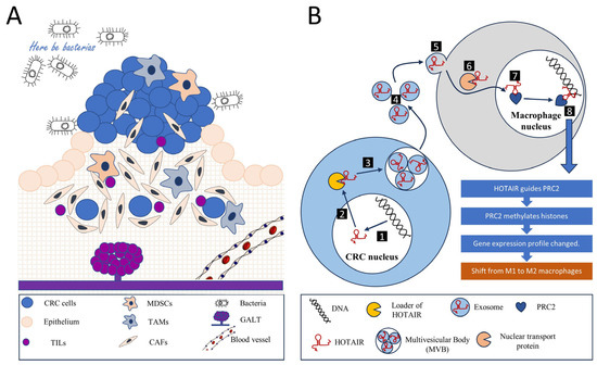
Figure 1. Tumor microenvironment and intercellular communications. (A) A colorectal cancer tumor is a complex, multicellular, and multi-dimensional entity. The tumor microenvironment encompasses not only tumor cells originating from the intestinal epithelium but also various components that help regulate metabolism, nutrition, and immune responses. These components include an extracellular matrix, a diverse population of tumor-infiltrating lymphocytes (TILs), myeloid-derived suppressor cells (MDSCs), tumor-associated macrophages (TAMs), and cancer-associated fibroblasts (CAFs), along with blood and lymphatic vessels. Another characteristic aspect of colorectal cancer is the presence of gut-associated lymphoid tissue (GALT). Additionally, bacteria play an essential role within the microenvironment. All these components interact to maintain a balance, thereby establishing a conducive environment for tumor growth. Among other forms of interaction, intercellular communication involving long non-coding RNAs (lncRNAs) is noteworthy. (B) The signaling mechanism of the long non-coding RNA (lncRNA) HOTAIR can serve as an example of intercellular communication through an lncRNA. In the nucleus of the tumor cell, the transcription and primary modification of the HOTAIR gene RNA occur (1), after which the mature lncRNA HOTAIR moves to the cytoplasm (2). Here, via a loader mechanism, HOTAIR is incorporated into a multivesicular body (MVB) where exosome particles loaded with HOTAIR are formed (3). These exosomes are then released into the extracellular environment (4). Their content is subsequently absorbed by macrophages via pinocytosis (5). Nuclear transport proteins guide HOTAIR into the macrophage’s nucleus (6), where this RNA interacts with the Polycomb Repressive Complex 2 (PRC2) chromatin remodeling complex (7). The HOTAIR-PRC2 complex identifies specific genomic regions (8), leading the PRC2 complex to methylate histones and silence a series of genes, thereby altering the gene expression profile of the macrophage. This results in a switch in macrophage polarization from M1 (anti-tumor) to M2 (pro-tumor), helping to sustain an environment conducive to tumor growth.
Inflammation plays a pivotal role in the TME of CRC. Cancer-associated fibroblasts (CAFs) and tumor-associated macrophages (TAMs) significantly contribute to this process, secreting pro-inflammatory cytokines such as interleukin-6 (IL-6) and tumor necrosis factor-alpha (TNF-α). Paradoxically, these cytokines can foster chronic inflammation, potentially promoting tumor progression [80]. Meanwhile, the ensuing inflammation can stimulate the recruitment and activation of immune cells capable of eliminating tumor cells.
The tumor stroma in CRC fosters the establishment of an immunosuppressive environment through various mechanisms. Notably, stromal cells, chiefly CAFs, secrete immunosuppressive factors such as transforming growth factor-beta (TGF-β) and interleukin-10 (IL-10), which inhibit T cell activity while enhancing regulatory T cell (Treg) functions [81,82]. Similarly, TAMs secrete factors like TGF-β, IL-10, and programmed cell death ligand-1 (PD-L1), collectively inhibiting the function of cytotoxic T cells and promoting the activity of Tregs [83,84].
Moreover, chronic exposure to tumor antigens and inflammatory signals in the TME can drive a state of T cell exhaustion which is characterized by diminished effector functions and the persistent expression of inhibitory receptors, including programmed cell death protein-1 (PD-1) and cytotoxic T-lymphocyte-associated protein 4 (CTLA-4). This state renders T cells less effective in eliminating cancer cells, thus facilitating immune evasion [85].
Another significant immunosuppressive mechanism in the TME involves the recruitment of regulatory immune cells. The tumor stroma can attract immunosuppressive cell types such as myeloid-derived suppressor cells (MDSCs) and Tregs, which impede the activity of cytotoxic T cells and natural killer (NK) cells, further facilitating tumor immune evasion [86,87].
Metabolic reprogramming is a defining feature of the TME. Both tumor cells and stromal cells can reshape the metabolic landscape of the TME, leading to conditions like hypoxia and nutrient deprivation. These conditions can adversely affect immune cell function and survival. For instance, tumor cells and CAFs can deplete vital nutrients like glucose and amino acids, thereby inhibiting T cell function [86].
CAFs, as primary stromal cells, play a crucial role in remodeling the extracellular matrix (ECM). This remodeling can construct a physical barrier that hinders immune cell infiltration and access to tumor cells. Therefore, understanding these mechanisms and interactions is vital for devising therapeutic strategies to counteract the immunosuppressive TME and boost antitumor immunity in CRC.
Within CRC, the TME can induce various forms of cell polarization to enhance the immunosuppressive state, thereby aiding the tumor in immune evasion. Macrophages within the TME often adopt an M2 polarization state known as “alternatively activated”. These M2 TAMs foster tissue repair, angiogenesis, and immune suppression by producing anti-inflammatory cytokines such as IL-10 and TGF-β and expressing high levels of immune checkpoint molecules like PD-L1, thereby inhibiting T cell function [87].
Conventional CD4+ T cells within the TME can be polarized into Tregs, identified via the expression of the transcription factor FOXP3 [83]. Tregs suppress the immune response by inhibiting the function of cytotoxic CD8+ T cells and other immune cells. Additionally, chronic exposure to antigens within the TME can instigate T cell exhaustion, which is characterized by upregulated inhibitory receptors like PD-1 and CTLA-4 and diminished effector functions [88].
MDSCs, a heterogeneous group of immature myeloid cells, can suppress the function of T cells through multiple mechanisms, including the production of immunosuppressive factors like arginase-1, nitric oxide, and reactive oxygen species [89,90]. Tumor-induced factors can also render dendritic cells (DCs) tolerogenic, diminishing their ability to activate T cells and potentially inducing Tregs or anergic T cells. These cellular polarization processes assist the colorectal tumor in establishing an immunosuppressive environment, thus contributing to immune evasion [91]. However, these processes also provide potential targets for therapeutic intervention, such as strategies aimed at reprogramming TAMs toward an M1 (pro-inflammatory and tumoricidal) phenotype, inhibiting Tregs, or reversing T cell exhaustion.
This entry is adapted from the peer-reviewed paper 10.3390/biomedicines11092411
This entry is offline, you can click here to edit this entry!