Rabbits as Animal Models for Anti-Tick Vaccine Development: History
Please note this is an old version of this entry, which may differ significantly from the current revision.

Studies evaluating candidate tick-derived proteins as anti-tick vaccines in natural hosts have been limited due to high costs. To overcome this problem, animal models are used in immunization tests. The most commonly used rabbit breeds were New Zealand (73.8%), Japanese white (19%), Californians (4.8%) and Flemish lop-eared (2.4%) rabbits. Anti-tick vaccines efficacy resulted in up to 99.9%. Haemaphysalis longicornis (17.9%) and Ornithodoros moubata (12.8%) were the most common tick models in vaccination trials. Experiments with rabbits have revealed that some proteins (CoAQP, OeAQP, OeAQP1, Bm86, GST-Hl, 64TRP, serpins and voraxin) can induce immune responses against various tick species. 

  • antigen
  • humoral and adaptive response
  • immunization
  • rabbit
  • tick

1. Introduction

Ticks are obligate blood-sucking ectoparasites that parasitize a large number of terrestrial and semi-terrestrial vertebrates, including humans [1,2,3]. Although they have been considered cosmopolitan parasites, most tick species are restricted to specific habitats, especially in tropical and subtropical regions [4,5]. Ticks transmit a wide variety of pathogens, being the second most important vectors of pathogens affecting humans, and the main vector in domestic and wild animals [6,7].
Traditional methods to control these arthropods are mainly based on the use of synthetic acaricides [8,9,10]. However, the application of these products has disadvantages, including the selection of resistant tick populations, environmental contamination, and residues in products of animal origin such as milk and meat [11].
These issues raise the need to develop alternative control methods, including the selection of parasite-resistant breeds [12,13]; biological control using entomopathogenic fungi (Metarhizium spp., Beauveria spp.) [14,15]; entomopathogenic nematodes (Heterorhabditidae and Steinernematidae) [16,17]; regulator ants (Solenopsis germinata, S. saevissima, Camponotus rengira, and Ectatomma quadridens) [18,19]; pesticides [20,21]; and immunological control through the application of anti-tick vaccines [22,23,24].
The evaluation of tick vaccines in natural hosts has limitations, mainly due to the high costs of maintaining and using farm or wild animals in experiments. For this reason, animal models such as hamsters, guinea pigs, and rabbits are commonly used [34,35,36]. These animals have been used as models for basic and applied research, not only to test immune responses generated by anti-tick vaccines, but also to study resistance to chemical acaricides and tick-borne pathogen infection under laboratory conditions [37,38,39,40].
The use of hamsters, guinea pigs, and rabbits in tick vaccination experiments generally has low maintenance costs, minimal space requirements, short reproductive cycles and larger numbers of pups produced per year compared to some natural hosts [41,42,43]. However, there are distinct benefits and disadvantages to each of these models. For instance, the use of hamsters is limited by low blood volume, compared to the use of guinea pigs and rabbits [44,45]. On the other hand, guinea pigs have thick skin, which makes blood collection relatively difficult, sometimes even requiring anesthetic techniques to collect small volumes, in contrast to rabbits, which do not require anesthetic techniques for blood collection [46].
Another limitation in experimental animal models is the number of ticks that can be used when performing the infestation. Studies in rabbits have reported that these animals can support a higher burden of adult ticks [23,47] compared to mice, hamsters or guinea pigs [48,49]. Interestingly, the rabbit model was the first animal model used in several immunological studies and was crucial, for example, in the development of Louis Pasteur’s rabies vaccine in 1881 [50]. In 1976, the World Health Organization (WHO) [51] highlighted rabbits as among the most important laboratory animals for the study of different diseases [51,52,53,54]. The most common breeds of laboratory rabbits are derived from the European rabbit (Oryctolagus cuniculus) [55]. 
Laboratory rabbits have proven to be the most suitable and accessible hosts for all life-stages of various tick species during infestation and vaccination experiments [41,58]. This is because it has several advantages over the use of laboratory mice and rats, such as: (i) a longer life span than mice and rats [59]; (ii) a larger body size (up to four times larger than rats); (iii) higher blood volume, cell and tissue samples [60]; (iv) the production of copious antiserum [51,56]; and (v) easy maintenance and breeding [56].
Moreover, it is evident that rabbit-based experiments are more cost-effective when comparing trials conducted using large animals such as bovines. Various factors contribute to the overall costs, including animal prices, the extended maintenance period, a higher demand for feed, as well as the size and complexity of the animal facilities. Bovines require a greater amount of physical space and specialized infrastructure, along with large feed quantities. As a result, more demanding waste management systems are necessary for bovine experiments.

2. Vaccination in Rabbits

Rabbits are currently used as a model organism in anti-tick vaccines assays against ticks of the genera Amblyomma, Dermacentor, Hyalomma, Haemaphysalis, Ixodes, Ornithodoros, and Rhipicephalus (Figure 1) [23,35,63,64,65,66].
Figure 1. Tick-derived proteins evaluated in tick vaccination trials using rabbits as an animal model.

2.1. Haemaphysalis spp.

The tick Haemaphysalis longicornis tick is native to east Asia, with sparse distribution in Australia, New Zealand, and the U.S. [67,68]. It has a three-host life cycle, infesting cattle, and wild animals such as ungulates, lagomorphs, carnivores, and birds [69,70]. Immunological studies have shown different immunogenic proteins with the potential to develop a vaccine against H. longicornis from China and Japan. Japanese white rabbit and New Zealand breeds were mostly used in the infestation experiments.

2.2. Ornithodoros spp.

Ornithodoros erraticus and Ornithodoros moubata are nidicolous and endophilic argasid ticks that are widely distributed in different regions [72,73,74,75], and can intermittently feed on various vertebrates such as birds and canines [76,77]. Eight tick-derived proteins were evaluated for the development of vaccines against O. erraticus and O. moubata using rabbits as an animal model. Oleaga et al. tested the O. moubata ferritin 2 orthologues in New Zealand white rabbits, obtaining 71% efficacy for OmFer2, which corresponded to a decreased egg-hatching rate and in the subsequent number of emerging O. moubata larvae [78]. On the other hand, Pérez-Sánchez’s research group tested the immune response against aquaporin, showing moderate vaccine efficacy against O. erraticus [35].

2.3. Rhipicephalus spp.

Rhipicephalus appendiculatus, Rhipicephalus microplus, and Rhipicephalus sanguineus s.l. are medically important ixodid ticks of the genus Rhipicephalus [81]. Rhipicephalus appendiculatus is distributed in central, eastern, and southeastern Africa [82,83]. Rhipicephalus microplus and R. sanguineus s.l. are cosmopolitan ticks, distributed in the tropical and subtropical regions of the globe [6,84]. They present monoxene (R. microplus) and hetorexone (R. sanguineus s.l. and R. appendiculatus) lifecycles, preferring domestic hosts such as bovines, canines, and some wild animals, respectively. They feed on humans as incidental hosts [84,85].
The voraxin α homologue of the R. appendiculatus tick was used to immunize Japanese white rabbits, which resulted in a reduction in the weight of ticks, followed by a 50% reduction in egg mass [86]. On the other hand, a different study determined the vaccinal efficacy of rGST in New Zealand white rabbits, showing that rGST caused a reduction in the number of female R. sanguineus s.l. infestations [28].

2.4. Ixodes spp.

Ixodes ricinus and Ixodes scapularis are ixodid ticks that are characterized by a heteroxenous life cycle, and infest cattle, deer, dogs, and a wide variety of vertebrates, including humans [89,90,91]. The nymphal stage is most frequently responsible for transmitting pathogens to humans [92,93]. Of the 265 species of Ixodes, 55 are distributed in the neotropical regions of the planet [5]; however, I. ricinus and I. scapularis can be found only in the northern hemisphere [89]. Vaccination studies against I. ricinus and I. scapularis using the New Zealand rabbit breed were reported in the U.S., Spain, and the Netherlands (Figure 4) [48,94,95].
Figure 4. Geographical distribution of studies using rabbits as animal models to test anti-tick vaccines. Parts of the figures were drawn by using pictures from Servier Medical Art: http://smart.servier.com/ (accessed on 18 May 2023).

2.5. Dermacentor spp.

Dermacentor marginatus is an ixodid tick that has a heteroxenous life cycle and a variety of hosts including canines, horses, and humans [97,98]. It is a tick with a cosmopolitan distribution, present mainly in the Nearctic, Palearctic, and Neotropic ecozones of the planet [99,100,101]. In the search for proteins for the development of a vaccine against D. marginatus, the New Zealand white rabbit was used as an animal model in infestations and vaccination experiments. A study infested New Zealand breed rabbits with D. marginatus after administering the last dose of the immunogen of GST, recording moderate vaccine efficacy against D. marginatus (Figure 5) [30].
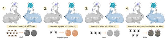
Figure 5. Comparison of different models of tick infestation in rabbits: 1. larval-stage tick infestation; 2. nymphal-stage tick infestation; 3. adult-stage tick infestation; and 4. nymphal- and adult-stage tick infestation. Parts of the figures were drawn by using pictures from Servier Medical Art: http://smart.servier.com/ (accessed on 21 February 2023).

3. Summary

To date, 57 tick-derived proteins have been evaluated as potential anti-tick vaccines by studying the immunogenic responses generated using rabbits as an experimental model. Rabbit models for anti-tick vaccination trials have allowed for a better understanding of the physiological mechanisms of ticks infesting mammal hosts. For example, a study of the serpins HLS1, rHLS2, rSerpin, and RmS-17 in rabbits stimulated an immune response that affected the prolonged duration of feeding, increased mortality, and reduced oviposition in ticks like H. longicornis and R. microplus [47,64,87,102].
Globally, the use of rabbits has provided novel evidence on a vaccine based on salivary glycine-rich proteins in various medically important tick species. According to the findings obtained by Zhou et al., using rabbits immunized with the glycine-rich protein RH50, the protein was only expressed in the salivary glands of partially fed ticks and not in the salivary glands of unfed ticks or in the midgut, fat body, or ovary of partially fed ticks, in contrast to what was previously reported for p29 and Bm86 proteins [63,88,121].
Rabbits have been used as an immunization model to evaluate immunological responses to a given antigen (Q38, Bm86, GST, serpins and voraxin) against different tick species. For example, high vaccine efficacy against both I. ricinus and D. reticulatus was obtained with the chimeric protein Q38 containing subolesin/akirin [39].
Similarly, experiments on rabbits using voraxin α, a protein derived from the male tick and transferred to the female through copulation to stimulate female blood-feeding [104], have yielded vaccine efficiency by reducing feeding times in Amblyomma hebraeum. There is an amino acid sequence similarity between the voraxin α of A. hebraeum (85%) and that of D. variabilis (92%) and R. appendiculatus (85%) [86]. The immunization results could therefore potentially be similar, making this protein a good multispecies vaccine candidate.
The use of rabbits as animal models in the discovery of anti-tick molecules has been fundamental in enabling the testing of these molecules before inoculation of the natural hosts. It was verified that rabbits present an immune response similar to that of the natural hosts. For example, the use of the ferritin 2 protein to immunize rabbits infested with I. ricinus (IrFER2) yielded an efficiency of 98%, while the efficiency of the same protein used in bovines infested with R. microplus and R. annulatus (RmFER2) was 64% and 72%, respectively [96]. 
The immune responses generated by the different proteins studied in rabbits could vary depending on the challenges of ticks in immature or mature life stages. For example, the response generated by the p29 and HL34 proteins in the life stages of larvae, nymphs, and adults of H. longicornis fed on immunized rabbits suggests that these proteins may be involved in mediating key physiological functions in the tick [63,71]. Although mature and immature ticks commonly express native p29, their sensitivities to rabbit immune responses against rp29 appear to be different [63], while the native HL34 is expressed in both immature (larvae and nymphs) and adult ticks. It is thus likely that immunity against rHL34 is directed towards immature and mature ticks [71].
Additionally, studies on rabbits have allowed us to broaden our knowledge about the “exposed” and “hidden” antigens of anti-tick proteins. For example, it was reported that HLS1 acts on the expression of hidden antigens, inhibiting the secretion of rHLS1 in rabbits during feeding [64]. Also, 64TRP isoforms were characterized as “dual-acting” anti-tick proteins against R. sanguineus s.l. and I. ricinus; they target both “exposed” and “hidden” antigens, preventing attachment, and feeding by affecting the feeding site, as well as cross-reacting with ‘hidden’ midgut antigens, resulting in the death of engorged ticks [48].
Results obtained from the study of the tick saliva proteome have shown a variety of proteins that protect ticks against host immune responses and antihemostatic mechanisms [131,132,133,134,135,136]. This is because, during hematophagy, tick salivary glands undergo remarkable growth and differentiation, accompanied by a significant increase in the synthesis of different proteins [137]. Tirloni et al. identified 187 tick and 68 bovine proteins in the saliva proteome of R. microplus, demonstrating that R. microplus saliva is rich in hemolipoproteins, lipocalins, peptidase inhibitors, antimicrobial peptides, glycine, and maintenance proteins [133]. These proteins, together with pharmacological bioactive lipids, can counteract the host’s defenses and hemostatic mechanisms [131,138], while the host physiological systems can trigger changes in the feeding activity of ticks [139] by stimulating proteins to limit host defense mechanisms [140].

This entry is adapted from the peer-reviewed paper 10.3390/pathogens12091117

This entry is offline, you can click here to edit this entry!
ScholarVision Creations