Polysomnography in the Diagnosis of Sleep-Disordered Breathing: History
Please note this is an old version of this entry, which may differ significantly from the current revision.
Subjects: Pediatrics
Contributor: , , , , , ,

Obstructive sleep-disordered breathing (SDB) is a spectrum of different clinical conditions distinguished by upper-airway dysfunction during sleep with snoring and/or increased respiratory effort due to increased upper airway resistance and pharyngeal collapsibility. Diagnostic methods for SDB in children involve a combination of clinical assessment, medical history evaluation, questionnaires, and objective measurements. Polysomnography (PSG) is the diagnostic gold standard. It records activity of brain and tibial and submental muscles, heart rhythm, eye movements, oximetry, oronasal airflow, abdominal and chest movements, body position. Despite its accuracy, it is a time-consuming and expensive tool.

  • sleep-disordered breathing
  • overnight polysomnography
  • diagnosis
  • children
  • respiratory events

1. Introduction

Obstructive sleep-disordered breathing (SDB) is a spectrum of different clinical conditions distinguished by upper-airway dysfunction during sleep with snoring and/or increased respiratory effort due to increased upper airway resistance and pharyngeal collapsibility  [1]. Obstructive SDB, ranging from primary snoring to obstructive sleep apnoea syndrome (OSAS), has variable severity of intermittent upper airway obstruction that leads to both sleep fragmentation and changes in gas exchange with night-time symptoms, daytime symptoms, and long-term deleterious health effects [2][3][4]. For this reason, it has been assumed that early recognition of SDB and its timely resolution through adequate treatment are necessary to reverse or prevent consequences such as impaired cognitive development, behavioural problems, poor academic performance, increased risk of cardiovascular complications, reduced quality of life, and increased health care use [2][3][4].
SDB occurs in all paediatric ages, from newborn to adolescent, and the prevalence varies depending on the targeted populations studied and the methodology and diagnostic criteria used: the paediatric general prevalence of OSAS is estimated at about 1–5%, with the most predominate age occurring at 2–8 years, while the prevalence of primary snoring is reported to be approximately 7.5%, although it has been reported to be as high as 30% in some published studies [5][6][7][8].
Some clinical conditions such as Down’s Syndrome (35% to 70%) [9], Prader–Willi syndrome (93%) [10], cerebral palsy (18%) [11], mitochondrial diseases (11%) [12], obesity (up to 60%) have a higher prevalence [13].
Despite the high prevalence and the need for early recognition of SDB to stem the large spectrum of adverse consequences, the availability and the accessibility of diagnostic services remain a limitation. The American Academy of Sleep Medicine recommends, as part of a routine physical exam, inquiring whether the child or adolescent snores and/or presents signs or symptoms of SDB [14]. In case of an affirmative answer, the clinicians should refer patients to a physician with expertise in paediatric sleep or to a paediatric sleep laboratory to perform a sleep study. Furthermore, high-risk children (i.e., children with tonsillar and adenoid hypertrophy, craniofacial anomalies, genetic syndrome [15]) or children with comorbidities should be referred to a sleep physician regularly.
Overnight polysomnography (oPSG) is the “gold standard” for the diagnosis of SDB, but it is an expensive and time-consuming tool and can be challenging to undertake in young children. Alternative diagnostic methods should be considered if oPSG is not available [1].

2. Polysomnography

Overnight polysomnography (oPSG) is considered the current gold standard method for diagnosis of childhood SDB [1][14]. The oPSG monitors continuously and simultaneously sleep stages and breathing functions allowing one to correlate the sleep architecture information and cortical arousal events with respiratory events. It is performed using the standard recordings of following physiologic functions: brain activity by frontal, central and occipital electroencephalography (EEG) channels, eye movements by bilateral electrooculography (EOG), muscle (tibial and submental) activity by electromyography (EMG), heart rhythm by 2-lead electrocardiography (ECG), peripheral pulse oximetry, oronasal airflow measurement using thermal sensor thermistor and nasal pressure transducer, abdominal and chest wall movements by respiratory inductance plethysmography, and body position by automated detection methods or observation. EEG, EOG, and EMG are required for accurate sleep staging. Video recording is often used to assist in sleep staging [16]. EEG and EMG allow one to record the arousals from sleep associated with SDB in childhood [17].
Regarding respiratory events, to identify apnoea/hypopnoea during sleep study in children, the use of an oronasal thermal airflow sensor and/or nasal pressure transducer to monitor airflow is recommended. When these signals are not functioning or not reliable, one of the following alternative apnoea/hypopnoea sensors are suggested: respiratory inductance plethysmography sum (RIPsum), respiratory inductance plethysmography flow (RIPflow), and dual respiratory inductance plethysmography (RIP) belt signals [18]. For monitoring respiratory effort, the Task Force of the American Academy of Sleep Medicine upholds the oesophageal manometry or the calibrated or uncalibrated dual thoracoabdominal RIP belts [18]. Oesophageal pressure measurement is widely considered the very accurate tool for diagnosing the upper airway resistance syndrome (UARS), an SDB characterised by arousals (observed during oPSG) without oxygen desaturations, apnoeas, or hypopneas but with the increase in respiratory effort. The abnormal decrements in oesophageal pressure (Pes) measured which oesophageal manometry reflects intrathoracic pressure and increased inspiratory effort [19].
The pulse oximetry with a maximum acceptable signal averaging time of ≤3 s at a heart rate of 80 beats per minutes should be used for detection of oxygen saturation. One of the parameters analysed in oPSG is the Apnoea Hypopnea Index (AHI). It is commonly used to classify OSAS severity: mild OSAS is defined by an AHI score ranging from 1 to 5, moderate OSAS falls within the AHI range of 5 to 10, and finally, severe OSAS is characterized by an AHI score greater than 10 [1].
The oPSG should be carried out by a sleep laboratory with experience in children’s healthcare. Children should be housed in a friendly environment with a parent sleeping next to them. The assessment of oPSG recording requires trained paediatricians and expert technicians to ensure the high quality of recordings, and to assure that the PSG performance, scoring, and interpretation are appropriate for the age and condition of the child [14]. The oPSG is highly costly in terms of human resources, time-consumption, and actual costs. It requires a high expertise equipment with a high staff-to-patient ratio (one to one). A single monitoring can take 8 to 10 h to record and 3 to 4 h to analyse. The determination of the arterial partial pressure of carbon dioxide (PCO2) is necessary to the diagnosis of alveolar hypoventilation. Given the difficulty of drawing an arterial sample during sleep, PCO2 recording can be performed by means of the end-tidal PCO2 (PETCO2) or transcutaneous PCO2 (PTCCO2) [18] (Figure 1 and Figure 2). However, the monitoring of PCO2 is not performed on a routine basis due to the lack of simple, cheap, and reliable CO2 monitors [20]. Unlike for the adult population, paediatric PSG results should not be corroborated over several nights; however, tests should be repeated if results are not conclusive [14].
Figure 1. Overnight transcutaneous CO2 (TcCO2) trend of a 7-year-old female affected by Rett Syndrome. (a) The green trace is the measured TcCO2 while the blue trace is the drift corrected TcCO2; they follow the same pattern over the time. The monitoring shows a hypoventilation pattern (mean TcCO2 57.2 mmHg, time spent with TcCO2 > 50 mmHg 91%). The peaks in TcCO2 correlated with (b) oxygen desaturation events and (c) the increase of the heart rate. An improvement in the pattern is observed between 06:00 and 08:00 a.m., as the patient wakes up.
Figure 2. Overnight End-Tidal CO2 (PetCO2) of a 12-year-old male with Duchenne muscular dystrophy. In the initial phase, there is an oxygen saturation without evident desaturation with an PetCO2 between 40 and 45 between 20 and 22 breaths. After a few hours, the heart rate decreases and phasic oxygen desaturations appear with an ETCO2 that significantly increases with peaks > 50 mmHg while the respiratory rate decreases; such findings are characteristic of sleep apnoea and alveolar hypoventilation.

This entry is adapted from the peer-reviewed paper 10.3390/children10081331

References

  1. Kaditis, A.G.; Alonso Alvarez, M.L.; Boudewyns, A.; Alexopoulos, E.I.; Ersu, R.; Joosten, K.; Larramona, H.; Miano, S.; Narang, I.; Trang, H.; et al. Obstructive Sleep Disordered Breathing in 2- to 18-Year-Old Children: Diagnosis and Management. Eur. Respir. J. 2016, 47, 69–94.
  2. Amin, R.; Somers, V.K.; McConnell, K.; Willging, P.; Myer, C.; Sherman, M.; McPhail, G.; Morgenthal, A.; Fenchel, M.; Bean, J.; et al. Activity-Adjusted 24-Hour Ambulatory Blood Pressure and Cardiac Remodeling in Children with Sleep Disordered Breathing. Hypertension 2008, 51, 84–91.
  3. Lee, P.-C.; Hwang, B.; Soong, W.-J.; Meng, C.C.L. The Specific Characteristics in Children with Obstructive Sleep Apnea and Cor Pulmonale. Sci. World J. 2012, 2012, 757283.
  4. Bourke, R.; Anderson, V.; Yang, J.S.C.; Jackman, A.R.; Killedar, A.; Nixon, G.M.; Davey, M.J.; Walker, A.M.; Trinder, J.; Horne, R.S.C. Cognitive and Academic Functions Are Impaired in Children with All Severities of Sleep-Disordered Breathing. Sleep Med. 2011, 12, 489–496.
  5. Lumeng, J.C.; Chervin, R.D. Epidemiology of Pediatric Obstructive Sleep Apnea. Proc. Am. Thorac. Soc. 2008, 5, 242–252.
  6. Fund, N.; Green, A.; Chodick, G.; Orin, M.; Koren, G.; Shalev, V.; Dagan, Y. The Epidemiology of Sleep Disorders in Israel: Results from a Population-Wide Study. Sleep Med. 2020, 67, 120–127.
  7. Kara, C.O.; Ergin, H.; Koçak, G.; Kiliç, I.; Yurdakul, M. Prevalence of Tonsillar Hypertrophy and Associated Oropharyngeal Symptoms in Primary School Children in Denizli, Turkey. Int. J. Pediatr. Otorhinolaryngol. 2002, 66, 175–179.
  8. Castronovo, V.; Zucconi, M.; Nosetti, L.; Marazzini, C.; Hensley, M.; Veglia, F.; Nespoli, L.; Ferini-Strambi, L. Prevalence of Habitual Snoring and Sleep-Disordered Breathing in Preschool-Aged Children in an Italian Community. J. Pediatr. 2003, 142, 377–382.
  9. Borrelli, M.; Corcione, A.; Rongo, R.; Cantone, E.; Scala, I.; Bruzzese, D.; Martina, S.; Strisciuglio, P.; Michelotti, A.; Santamaria, F. Obstructive Sleep Apnoea in Children with Down Syndrome: A Multidisciplinary Approach. J. Pers. Med. 2022, 13, 71.
  10. Canora, A.; Franzese, A.; Mozzillo, E.; Fattorusso, V.; Bocchino, M.; Sanduzzi, A. Severe Obstructive Sleep Disorders in Prader-Willi Syndrome Patients in Southern Italy. Eur. J. Pediatr. 2018, 177, 1367–1370.
  11. Koyuncu, E.; Türkkani, M.H.; Sarikaya, F.G.; Özgirgin, N. Sleep Disordered Breathing in Children with Cerebral Palsy. Sleep Med. 2017, 30, 146–150.
  12. Ramezani, R.J.; Stacpoole, P.W. Sleep Disorders Associated with Primary Mitochondrial Diseases. J. Clin. Sleep Med. 2014, 10, 1233–1239.
  13. Verhulst, S.L.; Van Gaal, L.; De Backer, W.; Desager, K. The Prevalence, Anatomical Correlates and Treatment of Sleep-Disordered Breathing in Obese Children and Adolescents. Sleep Med. Rev. 2008, 12, 339–346.
  14. Marcus, C.L.; Brooks, L.J.; Draper, K.A.; Gozal, D.; Halbower, A.C.; Jones, J.; Schechter, M.S.; Sheldon, S.H.; Spruyt, K.; Ward, S.D.; et al. Diagnosis and Management of Childhood Obstructive Sleep Apnea Syndrome. Pediatrics 2012, 130, 576–584.
  15. Xu, Z.; Wu, Y.; Tai, J.; Feng, G.; Ge, W.; Zheng, L.; Zhou, Z.; Ni, X. Risk Factors of Obstructive Sleep Apnea Syndrome in Children. J. Otolaryngol. Head Neck Surg. 2020, 49, 11.
  16. Grigg-Damberger, M.; Gozal, D.; Marcus, C.L.; Quan, S.F.; Rosen, C.L.; Chervin, R.D.; Wise, M.; Picchietti, D.L.; Sheldon, S.H.; Iber, C. The Visual Scoring of Sleep and Arousal in Infants and Children. J. Clin. Sleep Med. 2007, 3, 201–240.
  17. Scholle, S.; Zwacka, G. Arousals and Obstructive Sleep Apnea Syndrome in Children. Clin. Neurophysiol. 2001, 112, 984–991.
  18. Berry, R.B.; Budhiraja, R.; Gottlieb, D.J.; Gozal, D.; Iber, C.; Kapur, V.K.; Marcus, C.L.; Mehra, R.; Parthasarathy, S.; Quan, S.F.; et al. Rules for Scoring Respiratory Events in Sleep: Update of the 2007 AASM Manual for the Scoring of Sleep and Associated Events. Deliberations of the Sleep Apnea Definitions Task Force of the American Academy of Sleep Medicine. J. Clin. Sleep Med. 2012, 8, 597–619.
  19. Guilleminault, C.; Poyares, D.; Palombini, L.; Koester, U.; Pelin, Z.; Black, J. Variability of Respiratory Effort in Relation to Sleep Stages in Normal Controls and Upper Airway Resistance Syndrome Patients. Sleep Med. 2001, 2, 397–405.
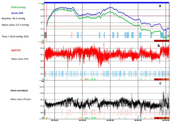
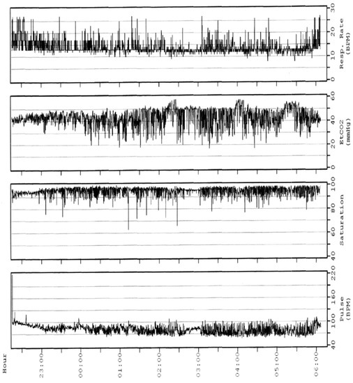
  20. Amaddeo, A.; Fauroux, B. Oxygen and Carbon Dioxide Monitoring during Sleep. Paediatr. Respir. Rev. 2016, 20, 42–44.
More
This entry is offline, you can click here to edit this entry!
Video Production Service