Anthranilic Acid and Beyond: History
Please note this is an old version of this entry, which may differ significantly from the current revision.

Anthranilic acid, a kynurenine metabolite produced by host and microbe alike, is a bioactive compound with potential systemic neurological effects. Though in the past much research has focused on the kynurenine pathway at large, the role of anthranilic acid in the gut–brain axis is emerging as an important piece of the puzzle. Anthranilic acid is a direct metabolic product of kynurenine digestion in humans and microbes and represents an alternate branch to two other immediate products (i.e., 3-hydroxykynurenine and kynurenic acid). Kynurenine is hydrolyzed to anthranilic acid and L-alanine with the help of kynureninase. With the transfer of an amino group, kynurenine becomes the alternate product kynurenic acid, and the addition of an oxygen by kynurenine-3-monoxygenase turns kynurenine into 3-hydroxykynurenine. Anthranilic acid, through non-specific hydroxylation, becomes 3-hydroxyanthranilic acid, which is the precursor for quinolinic acid, picolinic acid, and the terminal product NAD+. The ratio of anthranilic acid to the downstream 3-hydroxyanthranilic acid has been suggested as one potential biomarker for both neurological and physiological disorders. 

  • anthranilic acid
  • microbiome
  • gut–brain axis
  • metabolites

1. Human Production of Anthranilic Acid and Derivatives

The production of anthranilic acid by humans is integral to this research since it supports the concept of co-production between the host and the microbiome of a bioactive compound. Though the gut microbiome contributes many important and biologically relevant metabolites, humans make their own contributions to systemic metabolism. Many host tissues and cell types, including neurons and macrophages, have the enzymatic repertoire to support most or all of the kynurenine pathway [1][2][3]. There are various drivers of the kynurenine pathway in vivo and many factors that exert control over the divergence into the many branches and resultant metabolites.
In vitro studies have shown the kynurenine pathway in multiple human cell types can be induced and specifically altered by interferon-gamma [4]. Macrophages in particular were driven towards the production of 3-hydroxyanthranilic acid from L-tryptophan via interferon-gamma activation [4]. A recent review has further highlighted the interplay of immune markers and regulation of the kynurenine pathway, stating that key to this relationship is the indoleamine 2,3-dioxygenase enzyme (IDO) [5]. IDO appears to participate in crosstalk with markers of inflammation and though IDO plays a secondary role in pushing TRP towards kynurenine and further down, kynurenine-metabolites under homeostatic conditions, the onset of inflammation drives IDO activity [5]. Pro-inflammatory cytokines, IL6, TNF-alpha, and IL-4, drive the expression of IDO through shared signaling pathways, like through the activation of AHRs [6]. IDO expression creates an environment that suppresses the immune response through the production of neurotoxic metabolites and depletion of the central immune-mediating amino acid Trp [7]. Multiple studies have also revealed that IDO is highly induced in colonic tissue and mucosa of patients with IBD and Crohn’s diseases, both diseases that are marked by chronic inflammatory states in the gut and potential microbial interference, but for which the underlying taxa and mechanisms are currently unknown [8][9]. Inflammation is often correlated, if not implicated, with neurological disorders and declining health. The tight partnership between IDO activity and inflammatory markers may provide one piece of the metabolic–neurologic activity relationship puzzle. Tryptophan 2,3-dioxygenase (TDO), like IDO, is a rate-limiting enzyme that controls conversion of Trp into the early metabolites of the kynurenine pathway [10]. Whereas IDO can be found throughout host tissues, it is primarily expressed in the liver, where 90% of kynurenine metabolites are produced [11]. Hormonal cascades in the host, such as those induced by stress, have been shown to drive TDO activity in the liver and cause a resultant increase in circulating kynurenine metabolites [11].
Hepatic circulation and utilization of Trp metabolites are not limited to the kynurenine pathway. Tryptamine, produced from the decarboxylation of Trp by gut microbes, attenuates pro-inflammatory cytokine production in the liver [12]. Skatole is another Trp derivative, specifically a product of the indole pathway, that is produced in the gut by microbes but potentially bioactive in the liver. One study found that patients without liver disease (hepatic encephalopathy) had no detectable serum skatole, but those with signs of liver disease had detectable skatole levels between 0 and 442 nmol/L [13]. This study did not test the directionality of skatole levels and so did not postulate whether skatole was a causative agent of disease or a byproduct [13]. Skatole may be toxic at high concentrations in the body, but a recent study showed skatole may attenuate hyperlipidemic conditions of the liver [14]. Trp metabolites produced by microbes in the gut, like skatole and tryptamine, find their way to the liver through hepatic circulation and once localized, can either attenuate or exacerbate disease conditions. p-Cresol, a product of amino acid fermentation and [15] mentioned previously in relation to autism spectrum disorders, is another metabolite that is derived from microbial activity that enters hepatic circulation. Modification of p-cresol in the liver gives rise to uremic toxins and can intensify renal disease as well as promote systemic inflammation [15]. Microbial compounds in the gut that make their way to the liver through hepatic circulation can then be further modified by the host, altering catabolite activity. 
There are internal controls of metabolic-related enzymatic activity in the liver, and the original limiting factor for the kynurenine pathway is ultimately the availability of the primary substrate, Trp. As Trp is an essential amino acid only acquired from diet and microbial metabolism, the availability of Trp in the gut is a necessary starting point for the estimation of circulating secondary metabolites. An 8-week-long high-fat diet in mice resulted in a measurable perturbation of gut taxa and subsequently significantly altered the gut, serum, and liver metabolome [12]. The gut microbial community has been shown to be a key driver of metabolism and so it cannot be discounted in the discussion around circulating kynurenine metabolites like anthranilic acid. Despite our understanding of the human contribution to circulating anthranilic acid compounds, it is not always possible to disentangle host and microbe metabolisms from one another. The inability to fully dissociate the production of anthranilic acid complicates the already challenging problem of tracing this metabolite’s biological activity in the host. Just as is it important to contextualize anthranilic acid research with the potential metabolic contributions of the humans being studied, so too is it important to understand microbial contributions to the shared metabolic pool.

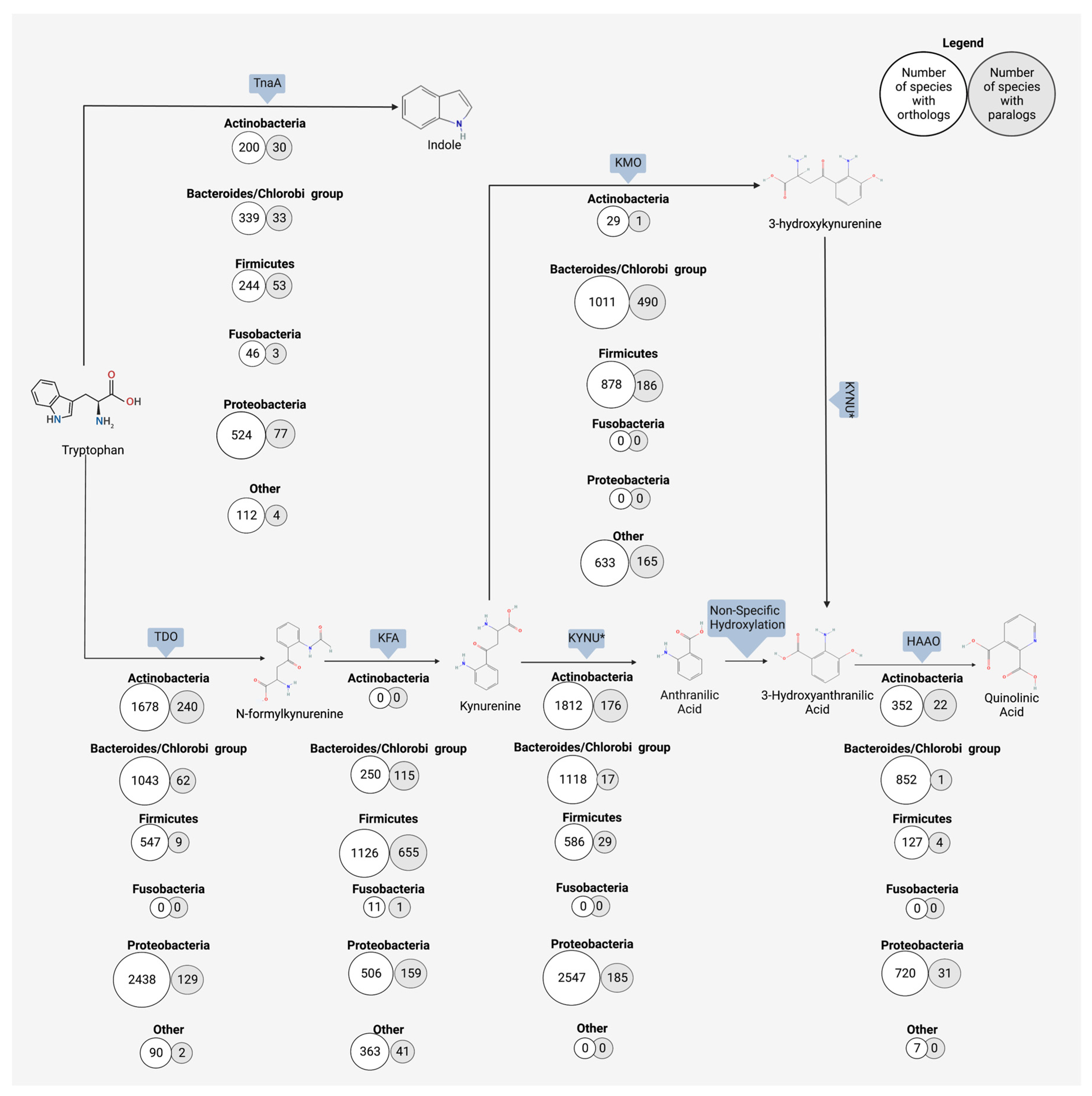
2. Bacterial Production of Anthranilic Acid and Derivatives

The gut microbiome has the functional capacity to produce anthranilic acid and downstream metabolites from the breakdown of kynurenine, and ultimately from the original source of Trp. Research on microbial production of anthranilic acid and its derivatives mostly focuses on industrially applicable microbes, which produce anthranilic acid in large quantities Anthranilic acid, and its derivatives, are used in the pharmaceutical industry for a myriad of applications, including as antimicrobials and as a potential regulator of diabetic symptoms [16]. In the food industry, the anthranilic acid derivative, methyl-anthranilate, is used as a flavor compound to impart a grape essence to candies and other food items [17]. Many microbes, though it may vary by species, have the enzymatic repertoire to perform either all or part of the kynurenine pathway [18]. A search using the OrthoDB database revealed six major bacterial phyla contain organisms with the kynureninase gene (KYNU), which catalyzes the conversion of kynurenine to anthranilic acid (Figure 1) [19]. Anthranilic acid to 3-hydroxyanthranilic acid is a non-specific hydroxylation step, but the conversion of 3-hydroxyanthranilic acid to subsequent quinolinic acid relies on the expression of 3-hydroxyanthranilate 3,4-diozygenase (HAAO). Five major phyla contain organisms with the HAAO gene, including the gut-dominating Firmicutes and Bacteroidetes (Figure 1) [19]. The microbial production of anthranilic compounds in the gut is an important, though understudied, area of research on Trp-related neuroactive compounds.
Figure 1. Orthologous and paralogous genes encoding enzymes for the microbial metabolism of tryptophan towards the kynurenine and indole pathways. The number of phyla containing orthologs of the microbial enzyme responsible for each reaction was determined through a search using the OrthoDB database. Enzymes or non-specific reaction types are indicated by rectangular shapes above each phyla list. White circles indicate the number of species in the taxa containing orthologs and gray circles represent the number of species containing paralogs in that phyla. Six chosen phyla are shown, and all other phyla with hits are grouped into “other”. Indole is derived from tryptophan digestion by tryptophanase (TnaA). Down an alternate route from indole, kynurenine can be produced from tryptophan with a two-step process involving tyrptophan 2,3-dioxygenase (TDO) and kynurenine formamidase (KFA). The breakdown of the resulting kynurenine to anthranilic acid by microbes is catalyzed by kynureninase (KNYU). KYNU also catalyzes the reaction of 3-hydroxykynurenine to 3-hydroxyanthranilic acid; this overlap in activity is denoted by an asterisk (*). Anthranilic acid is converted to 3-hydroxyanthranilic acid via non-specific hydroxylation. 3-hydroxyanthranilic acid can then be converted to multiple downstream metabolites, such as picolinic acid (not shown) or quinolinic acid (shown here). The production of quinolinic acid from 3-hydroxyanthranilic acid is catalyzed by 3-hydroxyanthranilate 3,4-dioxygenase (HAAO).
Studies that focus on the production of anthranilic acid by gut microbiota many times rely on a more universal approach to this analysis, highlighting the production in the gut by broad functional groupings and not by a specific taxon. The difficulty of teasing apart microbes and their metabolic products in the gut is in large part due to the complex interactions between microbes and the present diversity [18], which is coupled to a very high genomic diversity of microbiome members [20][21]. One point that is indisputable, however, is that the gut microbiota plays a major role in shifting the availability of free Trp within the host. Germ-free mice have altered free Trp levels compared to inoculated mice and show increased kynurenine pathway metabolites [22][23]. The gut microbiota at large has the capacity to support the kynurenine pathway in the gut. Previous work demonstrated that the genomes of gut bacteria contain the necessary enzyme homologs to produce anthranilic acid and kynureninase, as well as other enzymes responsible for producing neuroactive metabolites in this pathway [18][24]. The strain or species that have the higher or lower metabolic capacity for Trp turnover are largely missing from the literature in this area.
The ability to produce anthranilic acid or other Trp-related neuroactive metabolites may be species specific. Metabolic production is also context specific, as the host dietary intake and the resulting concentrations of these shared metabolic products shifts the equilibrium for both microbe and host. Human pathogen Psuedomonas can produce multiple components of the kynurenine pathway [25]. More relevant to this review, however, is that P. aeruginosa produces anthranilate and then uses it as a precursor to several virulence factors [25]. Kurnasov and colleagues characterized the anthranilate pathway in prokaryotes by conducting a genome comparison and searching for three enzymes, tryptophan 2,3-dioxygenase, kynurenine formamidase, and kynureninase, which together produce a eukaryotic-like anthranilate pathway [26]. Among others, Burkholderia fungorum, Bacillus cereus, Ralstonia solanacearum, and Ralstonia metallidurans were found to encode for all three anthranilate-related enzymes [26]. Another study looking at cold stress in pigs and its effect on microbiome diversity and activity identified anthranilic acid as one of the few metabolites that increased under cold stress conditions [27]. Prevotella UCG-003 was revealed to be the most strongly correlated microbe with this cold stress-induced increase in anthranilic acid [27]. It is well defined that the gut microbiota is an important contributor to the production of neuroactive compounds from Trp.

3. Neurological Outcomes of Anthranilic Acid

Research on the mechanistic role of anthranilic acid in neurological diseases has been limited up until now. Many studies focus primarily on the use of anthranilic acid and its derivatives as biomarkers [28], but do not necessarily drill down to determine the causative or protective effects of these compounds on host systems. While there is a place for studying these bioactive metabolites from the perspective of diagnostic potential, future therapeutic development will inevitably rest squarely on a mechanistic understanding of metabolic bioactivity. Current literature suggests that anthranilic acid leads a double life; the metabolite appears to exert neuroprotective effects in some cases while displaying neurotoxic effects in others [29][30]. The potential neurological activity of anthranilic acid in vivo is complex and at times conflicting. Anthranilic acid resides at the increasingly explored intersection of diet, microbe, and host neuroactivity, and a more thorough understanding of anthranilic acid’s bioactivity is important for future use as a biomarker or therapeutic target.
Anthranilic acid is often seen as part of the neurotoxic branch of the kynurenine pathway, a branch that includes quinolinic acid and hydroxykynurenine [31][32]. Elevated serum levels of anthranilic acid have been measured in people with schizophrenia and in those diagnosed with Parkinson’s disease [28][33][34]. Oxenkrug and colleagues showed circulating anthranilic acid levels in patients with schizophrenia were two times higher than those of the control population, confirming the results of a previous rat model study [28]. Though the study could not attribute the elevated anthranilic acid to anything specific, such as increased enzymatic activity in the pathway, the authors did affirm that the increase was not correlated with anti-psychotic drug intake [28]. Adding another layer of complexity to this puzzle is the difference seen between the sexes. One study found that increased circulating anthranilic acid in schizophrenic subjects was not only related to neurological diagnosis, but also correlated with being female [33]. Females in the study had plasma anthranilic acid levels 27% greater than those of their male counterparts, all of whom had a schizophrenic diagnosis [33]. It is possible this difference could be explained by sex hormones. For example, estrogen can inhibit some enzymes of the kynurenine pathway, or potentially by the deficiency of these enzymes resulting from other multifaceted signaling cascades that differ between biologically male and female subjects [35][36]. The impact of estrogen on other signaling pathways has already been shown with another host and microbe-derived neurotransmitter, dopamine. Previous work established estrogen affects dopamine activity, typically boosting synthesis and release, in both animal models and follow-up human studies [37]. Gut bacteria, including E. coli, some Bacillus species, Klebsiella pneuomoniae, and Staphylococcus aureus, have been reported to produce dopamine from tyrosine [38]. Though primarily thought of as a mammal’s molecule, dopamine may be produced by bacteria as a quorum-sensing molecule, increasing motility and biofilm formation, or as a growth-enhancing factor, through improved iron regulation [38]. The interplay between the hormone estrogen, neurotransmitter dopamine, and microbial production of these bioactive compounds is already established in the literature, so it is not surprising to see emerging literature suggesting a hormone–metabolite crosstalk in the context of other microbially produced bioactive metabolites like kynurenine.
Beyond schizophrenia, other neurological conditions (e.g., chronic migraines) have been correlated to the levels of circulating anthranilic acid. A study on chronic migraines in humans tracked the serum levels of multiple kynurenine metabolites and found a 339% increase in the concentration of anthranilic acid in those suffering from chronic migraines [39]. In tandem with that increase, the same study showed a 63% decrease in the downstream metabolite 3-hydroxyanthranilic acid [39]. Importantly, the study excluded subjects with neurological comorbidities, suggesting that this marked an increase in anthranilic acid was specific to migraines and not due to a known underlying cause. It is possible that the raised anthranilic acid level is a response to neural activity and simply marks the diagnosis of chronic migraines. More interestingly, the authors of this study propose a combination of N-methyl D-aspartate (NMDA) receptor overactivity in migraines and the reduction of neuroprotectant kynurenic acid, which correlates with an increase in anthranilic acid, which together contribute to migraine symptoms [39]. This same research group also demonstrated concurring results in patients with a similar diagnosis of repeated cluster headaches. Those with cluster headaches had a 54% increase in serum anthranilic acid and a 54% decrease in 3-hydroxyanthranilic acid as compared to the healthy control patients [40]. Taken together, these studies indicate anthranilic acid is certainly a part of headache-related diagnoses. It remains to be determined if this metabolite is a causative or a responsive biomarker.
Evaluation of depression’s metabolic roots has indicated that there are correlating Trp metabolite concentrations. In one study on circulating anthranilic acid in patients diagnosed with depression, anthranilic acid was found to be inversely correlated to the severity of depression, though this finding was dependent on sex [41]. As was indicated in the case of schizophrenia and biological sex, anthranilic acid levels associated with depression appeared more pronounced in women as compared to men in the same study [41]. This study was careful to exclude medication as a factor. Previous work has noted a difference in circulating kynurenine metabolites between medicated and non-medicated subjects [42], making medication an additional confounding factor for gut–brain axis research. A study detailing the onset of depression in patients undergoing treatment for Hepatitis C showed a decrease in Trp availability with a corresponding increase in anthranilic acid circulation [43]. During this 24-week longitudinal study, subjects with increased anthranilic acid levels were significantly more likely to be diagnosed with major depressive disorder [43]. The role of anthranilic acid as a biomarker, or even as a cause, of depression disorders is an ongoing area of research with promising findings for the development of potentially more targeted treatments. There are, however, conflicting studies which show no correlation or weak correlation between depressive symptoms and anthranilic acid serum concentrations [44]. As human studies are often rife with confounding factors, this conflict amongst studies comes as no surprise. Biological sex [33][41] and medication [42] can affect the availability of Trp metabolites and are examples of factors that are not always accounted for but can play significant roles in determining the outcomes of these studies. To obtain meaningful and reproducible results, it is imperative that mechanistic animal model approaches are employed for a more concrete resolution to the involvement of anthranilic acid in depressive disorders.
Contrasting to predicted neurotoxic properties, studies have suggested that anthranilic acid derivatives may have neuroprotective bioactivity [29][45]. Decreased 3-hydroxyanthranilic acid levels in stroke patients, along with the corresponding increase in precursor anthranilic acid, had a negative effect on health outcomes for recovering patients [46]. The inverse, increased 3-hydroxyanthranilic acid levels, is correlated with more signs of recovery in patients that suffered from a stroke [46]. 3-hydroxyanthranilic acid is a redox-active Trp metabolite. The increased levels of this compound in circulation may have a potent anti-inflammatory effect, stemming from both this redox activity and through the redirecting of anthranilic acid towards a more beneficial chemical structure [29][46]. In vitro work with 3-hydroxyanthranilic acid has shown this compound to modulate the pro-inflammatory activities of macrophages, ultimately reducing generalized inflammation [47]. Inflammation plays a known part in many neurological disorders, as highlighted by a recent review [48]. Altogether, this indicates the immunomodulatory activities, and specifically inflammation-regulating activities, of anthranilic acid derivatives which are key parts of this metabolite–host neurological link and a specific disease or etiology.
Research on Trp metabolites and their relation to neurological outcomes is ongoing and has resulted in an abundance of broad descriptive studies for Trp compound relationships in patients with neurological symptoms. Though examples have been identified for strong correlations between anthranilic acid and multiple disease states, not all neurological conditions are showing the same trend. Studies on multiple sclerosis in children and Huntington’s disease showed no or weak correlations with anthranilic acid [49][50]. A recent meta-analysis investigating the presence of Trp-associated compounds across multiple neurological studies showed specific diseases have a weak correlation with anthranilic acid [51]. Results from human–microbiome studies can sometimes be conflicting, underscoring the complexity of host health and metabolic activity research. However, the observation of the Trp and kynurenine metabolic pathway dysregulation as related to host health is well-supported in the literature, but the lack of correlation in cases means this neurological relationship is more complex than a single molecule or single enzymatic reaction. Instead, the relationship for a suite of metabolites to host neurology is likely owed to something more specific regarding the branching and balance (i.e., ‘push/pull’) of these metabolic pathways that are interconnected between the Trp concentration and the co-metabolism of the microbiome and the host.
The key to understanding this apparent paradox between neuroprotective and neurotoxic activity may be in part due to those metabolic pathways which are not linear. These pathways are dynamic networks with many forces directing the movement of metabolism between the arms of complex interconnecting routes and shunts. Anthranilic acid alters the availability of its own primary precursor, kynurenine, and in so doing changes the compound equilibrium. Anthranilic acid is an inhibitor of 3-hydroxyanthranilic acid oxidase (3-HAO) enzymatic activity, the inhibition of which results in decreased production of quinolinic and picolinic acid from 3-hydroxyantranilic acid [45]. Iron is likewise able to inhibit 2-HAO enzymatic activity [52]. Interestingly anthranilic acid can readily chelate this circulating iron, resulting in a complex interaction where iron may indirectly increase anthranilic acid concentrations through 3-HAO inhibition and the increased concentration may then sequester that very same iron [45][52].
In pharmaceutical applications, anthranilic acid and related compounds have exhibited anti-inflammatory properties [16][53]. In a study on rats modeling rheumatoid arthritis, a positive dose-dependent response to synthetic anthranilic acid derivative N-(3′,4′-dimethoxycinnamonyl) anthranilic acid (3,4-DAA) was observed [53]. 3,4-DAA is structurally highly similar with 3-hydroxyanthranilic acid and 3-hydroxykynurenic acid. Arthritic rats received a daily dose of 200, 300, or 400 mg/kg, and all dose groups displayed reduced inflammation via immunomodulatory activity [53]. Endogenous anthranilic acid is also suggested to have these same anti-inflammatory properties [45][54]. 3-hydroxyanthranilic acid also has illustrated anti-inflammatory and antioxidant properties. Such properties are partially derived from the metabolite’s induction of hemooxygenase-1, which is involved in the control of inflammation [29]. As metabolomics work is never black and white, focusing on a single metabolite alone does not provide a clear picture of potential bioactivity. Instead, it is necessary to examine the interplay of network flux, protein regulation, and gene expression, all in combination with an understanding of the host–microbe interplay.
Anthranilic acid, along with its related compounds, cannot be considered singularly neuroprotective or neurotoxic. Perhaps these Trp-related metabolites should be considered more broadly as neuroactive, leaving space for the host context as a driver of ultimate effect relative to the local niche conditions. In the context of human studies, the directionality of metabolic levels cannot be fully assessed. One study reported that increased 3-hydroxyanthranilic acid levels, along with decreased anthranilic acid, were correlated with more beneficial clinic outcomes in stroke patients [46]. Measured serum concentrations post disease diagnosis allows for the theory that many of these measured compounds are the clean-up or response crew rather than the instigators of disease conditions. There is also the additional component of host genetics and the predisposition to certain disease states to consider. Host genetics have been shown to predispose certain individuals to the onset of disease, as seen in some stroke cases [55] and with depressive symptoms [56]. Previous studies have also established that genetic variation across hosts is at least partially responsible, in conjunction with host diet and environment, for driving the composition of the gut microbiota [57][58]. As host genetics play a role in both disease state and microbial composition, it is important to also consider the host genotype in the study of metabolic drivers of disease. Some studies with conflicting metabolic findings may look to the host genotype as one possible explanation for subject-specific differences. To tease apart correlation and causality, along with host-specific findings, future research necessitates the use of tightly controlled studies and the additional use of in vitro models that utilize a time series design to understand the flux and interconversion between the Trp neuroactive catabolites.

This entry is adapted from the peer-reviewed paper 10.3390/microorganisms11071825

References

  1. Guillemin, G.J.; Cullen, K.M.; Lim, C.K.; Smythe, G.A.; Garner, B.; Kapoor, V.; Takikawa, O.; Brew, B.J. Characterization of the kynurenine pathway in human neurons. J. Neurosci. 2007, 27, 12884–12892.
  2. Guillemin, G.J.; Kerr, S.J.; Smythe, G.A.; Smith, D.G.; Kapoor, V.; Armati, P.J.; Croitoru, J.; Brew, B.J. Kynurenine pathway metabolism in human astrocytes: A paradox for neuronal protection. J. Neurochem. 2001, 78, 842–853.
  3. Guillemin, G.J.; Smith, D.G.; Smythe, G.A.; Armati, P.J.; Brew, B.J. Expression of the kynurenine pathway enzymes in human microglia and macrophages. Adv. Exp. Med. Biol. 2003, 527, 105–112.
  4. Werner-Felmayer, G.; Werner, E.R.; Fuchs, D.; Hausen, A.; Reibnegger, G.; Wachter, H. Characteristics of interferon induced tryptophan metabolism in human cells in vitro. Biochim. Biophys. Acta 1989, 1012, 140–147.
  5. Sorgdrager, F.J.H.; Naude, P.J.W.; Kema, I.P.; Nollen, E.A.; Deyn, P.P. Tryptophan Metabolism in Inflammaging: From Biomarker to Therapeutic Target. Front. Immunol. 2019, 10, 2565.
  6. Zhang, Y.; Shi, H.; Yang, G.; Yang, Y.; Li, W.; Song, M.; Shao, M.; Su, X.; Lv, L. Associations between expression of indoleamine 2, 3-dioxygenase enzyme and inflammatory cytokines in patients with first-episode drug-naive Schizophrenia. Transl. Psychiatry 2021, 11, 595.
  7. Litzenburger, U.M.; Opitz, C.A.; Sahm, F.; Rauschenbach, K.J.; Trump, S.; Winter, M.; Ott, M.; Ochs, K.; Lutz, C.; Liu, X.; et al. Constitutive IDO expression in human cancer is sustained by an autocrine signaling loop involving IL-6, STAT3 and the AHR. Oncotarget 2014, 5, 1038–1051.
  8. Wolf, A.M.; Wolf, D.; Rumpold, H.; Moschen, A.R.; Kaser, A.; Obrist, P.; Fuchs, D.; Brandacher, G.; Winkler, C.; Geboes, K.; et al. Overexpression of indoleamine 2,3-dioxygenase in human inflammatory bowel disease. Clin. Immunol. 2004, 113, 47–55.
  9. Zhou, L.; Chen, H.; Wen, Q.; Zhang, Y. Indoleamine 2,3-dioxygenase expression in human inflammatory bowel disease. Eur. J. Gastroenterol. Hepatol. 2012, 24, 695–701.
  10. Gao, K.; Mu, C.L.; Farzi, A.; Zhu, W.Y. Tryptophan Metabolism: A Link Between the Gut Microbiota and Brain. Adv. Nutr. 2020, 11, 709–723.
  11. Gibney, S.M.; Fagan, E.M.; Waldron, A.M.; O’Byrne, J.; Connor, T.J.; Harkin, A. Inhibition of stress-induced hepatic tryptophan 2,3-dioxygenase exhibits antidepressant activity in an animal model of depressive behaviour. Int. J. Neuropsychopharmacol. 2014, 17, 917–928.
  12. Krishnan, S.; Ding, Y.; Saedi, N.; Choi, M.; Sridharan, G.V.; Sherr, D.H.; Yarmush, M.L.; Alaniz, R.C.; Jayaraman, A.; Lee, K. Gut Microbiota-Derived Tryptophan Metabolites Modulate Inflammatory Response in Hepatocytes and Macrophages. Cell Rep. 2018, 23, 1099–1111.
  13. Suyama, Y.; Hirayama, C. Serum indole and skatole in patients with various liver diseases. Clin. Chim. Acta 1988, 176, 203–206.
  14. Hong, S.H.; Hong, Y.; Lee, M.; Keum, B.R.; Kim, G.H. Natural Product Skatole Ameliorates Lipotoxicity-Induced Multiple Hepatic Damage under Hyperlipidemic Conditions in Hepatocytes. Nutrients 2023, 15, 1490.
  15. Anand, S.; Mande, S.S. Host-microbiome interactions: Gut-Liver axis and its connection with other organs. NPJ Biofilms Microbiomes 2022, 8, 89.
  16. Prasher, P.; Sharma, M. Medicinal chemistry of anthranilic acid derivatives: A mini review. Drug Dev. Res. 2021, 82, 945–958.
  17. Luo, Z.W.; Cho, J.S.; Lee, S.Y. Microbial production of methyl anthranilate, a grape flavor compound. Proc. Natl. Acad. Sci. USA 2019, 116, 10749–10756.
  18. Dehhaghi, M.; Kazemi Shariat Panahi, H.; Guillemin, G.J. Microorganisms, Tryptophan Metabolism, and Kynurenine Pathway: A Complex Interconnected Loop Influencing Human Health Status. Int. J. Tryptophan Res. 2019, 12, 1178646919852996.
  19. Zdobnov, E.M.; Kuznetsov, D.; Tegenfeldt, F.; Manni, M.; Berkeley, M.; Kriventseva, E.V. OrthoDB in 2020: Evolutionary and functional annotations of orthologs. Nucleic Acids Res 2021, 49, D389–D393.
  20. Karcher, N.; Nigro, E.; Puncochar, M.; Blanco-Miguez, A.; Ciciani, M.; Manghi, P.; Zolfo, M.; Cumbo, F.; Manara, S.; Golzato, D.; et al. Genomic diversity and ecology of human-associated Akkermansia species in the gut microbiome revealed by extensive metagenomic assembly. Genome Biol. 2021, 22, 209.
  21. Tierney, B.T.; Yang, Z.; Luber, J.M.; Beaudin, M.; Wibowo, M.C.; Baek, C.; Mehlenbacher, E.; Patel, C.J.; Kostic, A.D. The Landscape of Genetic Content in the Gut and Oral Human Microbiome. Cell Host Microbe 2019, 26, 283–295.e8.
  22. Xiao, L.; Yan, J.; Yang, T.; Zhu, J.; Li, T.; Wei, H.; Chen, J. Fecal Microbiome Transplantation from Children with Autism Spectrum Disorder Modulates Tryptophan and Serotonergic Synapse Metabolism and Induces Altered Behaviors in Germ-Free Mice. mSystems 2021, 6, e01343-20.
  23. Clarke, G.; Grenham, S.; Scully, P.; Fitzgerald, P.; Moloney, R.D.; Shanahan, F.; Dinan, T.G.; Cryan, J.F. The microbiome-gut-brain axis during early life regulates the hippocampal serotonergic system in a sex-dependent manner. Mol. Psychiatry 2013, 18, 666–673.
  24. Agus, A.; Planchais, J.; Sokol, H. Gut Microbiota Regulation of Tryptophan Metabolism in Health and Disease. Cell Host Microbe 2018, 23, 716–724.
  25. Bortolotti, P.; Hennart, B.; Thieffry, C.; Jausions, G.; Faure, E.; Grandjean, T.; Thepaut, M.; Dessein, R.; Allorge, D.; Guery, B.P.; et al. Tryptophan catabolism in Pseudomonas aeruginosa and potential for inter-kingdom relationship. BMC Microbiol. 2016, 16, 137.
  26. Kurnasov, O.; Jablonski, L.; Polanuyer, B.; Dorrestein, P.; Begley, T.; Osterman, A. Aerobic tryptophan degradation pathway in bacteria: Novel kynurenine formamidase. FEMS Microbiol. Lett. 2003, 227, 219–227.
  27. Zhang, Y.; Sun, L.; Zhu, R.; Zhang, S.; Liu, S.; Wang, Y.; Wu, Y.; Xing, S.; Liao, X.; Mi, J. Porcine gut microbiota in mediating host metabolic adaptation to cold stress. NPJ Biofilms Microbiomes 2022, 8, 18.
  28. Oxenkrug, G.; van der Hart, M.; Roeser, J.; Summergrad, P. Anthranilic Acid: A Potential Biomarker and Treatment Target for Schizophrenia. Ann. Psychiatry Ment. Health 2016, 4, 1059.
  29. Krause, D.; Suh, H.S.; Tarassishin, L.; Cui, Q.L.; Durafourt, B.A.; Choi, N.; Bauman, A.; Cosenza-Nashat, M.; Antel, J.P.; Zhao, M.L.; et al. The tryptophan metabolite 3-hydroxyanthranilic acid plays anti-inflammatory and neuroprotective roles during inflammation: Role of hemeoxygenase-1. Am. J. Pathol. 2011, 179, 1360–1372.
  30. Coplan, J.D.; George, R.; Syed, S.A.; Rozenboym, A.V.; Tang, J.E.; Fulton, S.L.; Perera, T.D. Early Life Stress and the Fate of Kynurenine Pathway Metabolites. Front. Hum. Neurosci. 2021, 15, 636144.
  31. Parrott, J.M.; Redus, L.; Santana-Coelho, D.; Morales, J.; Gao, X.; O’Connor, J.C. Neurotoxic kynurenine metabolism is increased in the dorsal hippocampus and drives distinct depressive behaviors during inflammation. Transl. Psychiatry 2016, 6, e918.
  32. Birner, A.; Platzer, M.; Bengesser, S.A.; Dalkner, N.; Fellendorf, F.T.; Queissner, R.; Pilz, R.; Rauch, P.; Maget, A.; Hamm, C.; et al. Increased breakdown of kynurenine towards its neurotoxic branch in bipolar disorder. PLoS ONE 2017, 12, e0172699.
  33. Steiner, J.; Dobrowolny, H.; Guest, P.C.; Bernstein, H.G.; Fuchs, D.; Roeser, J.; Summergrad, P.; Oxenkrug, G. Gender-specific elevation of plasma anthranilic acid in schizophrenia: Protection against glutamatergic hypofunction? Schizophr. Res. 2022, 243, 483–485.
  34. Oxenkrug, G.; van der Hart, M.; Roeser, J.; Summergrad, P. Peripheral Tryptophan—Kynurenine Metabolism Associated with Metabolic Syndrome is Different in Parkinson’s and Alzheimer’s Diseases. Endocrinol. Diabetes Metab. J. 2017, 1.
  35. Giorgini, F.; Huang, S.Y.; Sathyasaikumar, K.V.; Notarangelo, F.M.; Thomas, M.A.; Tararina, M.; Wu, H.Q.; Schwarcz, R.; Muchowski, P.J. Targeted deletion of kynurenine 3-monooxygenase in mice: A new tool for studying kynurenine pathway metabolism in periphery and brain. J. Biol. Chem. 2013, 288, 36554–36566.
  36. Jayawickrama, G.S.; Nematollahi, A.; Sun, G.; Gorrell, M.D.; Church, W.B. Inhibition of human kynurenine aminotransferase isozymes by estrogen and its derivatives. Sci. Rep. 2017, 7, 17559.
  37. Jacobs, E.; D’Esposito, M. Estrogen shapes dopamine-dependent cognitive processes: Implications for women’s health. J. Neurosci. 2011, 31, 5286–5293.
  38. Strandwitz, P. Neurotransmitter modulation by the gut microbiota. Brain Res. 2018, 1693 Pt B, 128–133.
  39. Curto, M.; Lionetto, L.; Negro, A.; Capi, M.; Fazio, F.; Giamberardino, M.A.; Simmaco, M.; Nicoletti, F.; Martelletti, P. Altered kynurenine pathway metabolites in serum of chronic migraine patients. J. Headache Pain. 2015, 17, 47.
  40. Curto, M.; Lionetto, L.; Negro, A.; Capi, M.; Perugino, F.; Fazio, F.; Giamberardino, M.A.; Simmaco, M.; Nicoletti, F.; Martelletti, P. Altered serum levels of kynurenine metabolites in patients affected by cluster headache. J. Headache Pain. 2015, 17, 27.
  41. Steiner, J.; Dobrowolny, H.; Guest, P.C.; Bernstein, H.G.; Fuchs, D.; Roeser, J.; Summergrad, P.; Oxenkrug, G.F. Plasma Anthranilic Acid and Leptin Levels Predict HAM-D Scores in Depressed Women. Int. J. Tryptophan Res. 2021, 14, 11786469211016474.
  42. Colle, R.; Masson, P.; Verstuyft, C.; Feve, B.; Werner, E.; Boursier-Neyret, C.; Walther, B.; David, D.J.; Boniface, B.; Falissard, B.; et al. Peripheral tryptophan, serotonin, kynurenine, and their metabolites in major depression: A case-control study. Psychiatry Clin. Neurosci. 2020, 74, 112–117.
  43. Pawlowski, T.; Pawlak, D.; Inglot, M.; Zalewska, M.; Marciniak, D.; Bugajska, J.; Janocha-Litwin, J.; Malyszczak, K. The role of anthranilic acid in the increase of depressive symptoms and major depressive disorder during treatment for hepatitis C with pegylated interferon-alpha2a and oral ribavirin. J. Psychiatry Neurosci. 2021, 46, E166–E175.
  44. Pompili, M.; Lionetto, L.; Curto, M.; Forte, A.; Erbuto, D.; Montebovi, F.; Seretti, M.E.; Berardelli, I.; Serafini, G.; Innamorati, M.; et al. Tryptophan and Kynurenine Metabolites: Are They Related to Depression? Neuropsychobiology 2019, 77, 23–28.
  45. Darlington, L.G.; Forrest, C.M.; Mackay, G.M.; Smith, R.A.; Smith, A.J.; Stoy, N.; Stone, T.W. On the Biological Importance of the 3-hydroxyanthranilic Acid: Anthranilic Acid Ratio. Int. J. Tryptophan Res. 2010, 3, 51–59.
  46. Darlington, L.G.; Mackay, G.M.; Forrest, C.M.; Stoy, N.; George, C.; Stone, T.W. Altered kynurenine metabolism correlates with infarct volume in stroke. Eur. J. Neurosci. 2007, 26, 2211–2221.
  47. Lee, K.; Kwak, J.H.; Pyo, S. Inhibition of LPS-induced inflammatory mediators by 3-hydroxyanthranilic acid in macrophages through suppression of PI3K/NF-kappaB signaling pathways. Food Funct. 2016, 7, 3073–3082.
  48. Cervellati, C.; Trentini, A.; Pecorelli, A.; Valacchi, G. Inflammation in Neurological Disorders: The Thin Boundary between Brain and Periphery. Antioxid. Redox Signal 2020, 33, 191–210.
  49. Nourbakhsh, B.; Bhargava, P.; Tremlett, H.; Hart, J.; Graves, J.; Waubant, E. Altered tryptophan metabolism is associated with pediatric multiple sclerosis risk and course. Ann. Clin. Transl. Neurol. 2018, 5, 1211–1221.
  50. Rodrigues, F.B.; Byrne, L.M.; Lowe, A.J.; Tortelli, R.; Heins, M.; Flik, G.; Johnson, E.B.; De Vita, E.; Scahill, R.I.; Giorgini, F.; et al. Kynurenine pathway metabolites in cerebrospinal fluid and blood as potential biomarkers in Huntington’s disease. J. Neurochem. 2021, 158, 539–553.
  51. Fathi, M.; Vakili, K.; Yaghoobpoor, S.; Tavasol, A.; Jazi, K.; Hajibeygi, R.; Shool, S.; Sodeifian, F.; Klegeris, A.; McElhinney, A.; et al. Dynamic changes in metabolites of the kynurenine pathway in Alzheimer’s disease, Parkinson’s disease, and Huntington’s disease: A systematic Review and meta-analysis. Front. Immunol. 2022, 13, 997240.
  52. Chobot, V.; Hadacek, F.; Weckwerth, W.; Kubicova, L. Iron chelation and redox chemistry of anthranilic acid and 3-hydroxyanthranilic acid: A comparison of two structurally related kynurenine pathway metabolites to obtain improved insights into their potential role in neurological disease development. J. Organomet. Chem. 2015, 782, 103–110.
  53. Inglis, J.J.; Criado, G.; Andrews, M.; Feldmann, M.; Williams, R.O.; Selley, M.L. The anti-allergic drug, N-(3′,4′-dimethoxycinnamonyl) anthranilic acid, exhibits potent anti-inflammatory and analgesic properties in arthritis. Rheumatology 2007, 46, 1428–1432.
  54. Maes, M.; Leonard, B.E.; Myint, A.M.; Kubera, M.; Verkerk, R. The new ‘5-HT’ hypothesis of depression: Cell-mediated immune activation induces indoleamine 2,3-dioxygenase, which leads to lower plasma tryptophan and an increased synthesis of detrimental tryptophan catabolites (TRYCATs), both of which contribute to the onset of depression. Prog. Neuropsychopharmacol. Biol. Psychiatry 2011, 35, 702–721.
  55. Traylor, M.; Rutten-Jacobs, L.C.; Holliday, E.G.; Malik, R.; Sudlow, C.; Rothwell, P.M.; Maguire, J.M.; Koblar, S.A.; Bevan, S.; Boncoraglio, G.; et al. Differences in Common Genetic Predisposition to Ischemic Stroke by Age and Sex. Stroke 2015, 46, 3042–3047.
  56. Caspi, A.; Sugden, K.; Moffitt, T.E.; Taylor, A.; Craig, I.W.; Harrington, H.; McClay, J.; Mill, J.; Martin, J.; Braithwaite, A.; et al. Influence of life stress on depression: Moderation by a polymorphism in the 5-HTT gene. Science 2003, 301, 386–389.
  57. Qin, Y.; Havulinna, A.S.; Liu, Y.; Jousilahti, P.; Ritchie, S.C.; Tokolyi, A.; Sanders, J.G.; Valsta, L.; Brozynska, M.; Zhu, Q.; et al. Combined effects of host genetics and diet on human gut microbiota and incident disease in a single population cohort. Nat. Genet. 2022, 54, 134–142.
  58. Campbell, J.H.; Foster, C.M.; Vishnivetskaya, T.; Campbell, A.G.; Yang, Z.K.; Wymore, A.; Palumbo, A.V.; Chesler, E.J.; Podar, M. Host genetic and environmental effects on mouse intestinal microbiota. ISME J. 2012, 6, 2033–2044.
More
This entry is offline, you can click here to edit this entry!
ScholarVision Creations