Gender Differences and miRNAs Expression in Cancer: History
Please note this is an old version of this entry, which may differ significantly from the current revision.

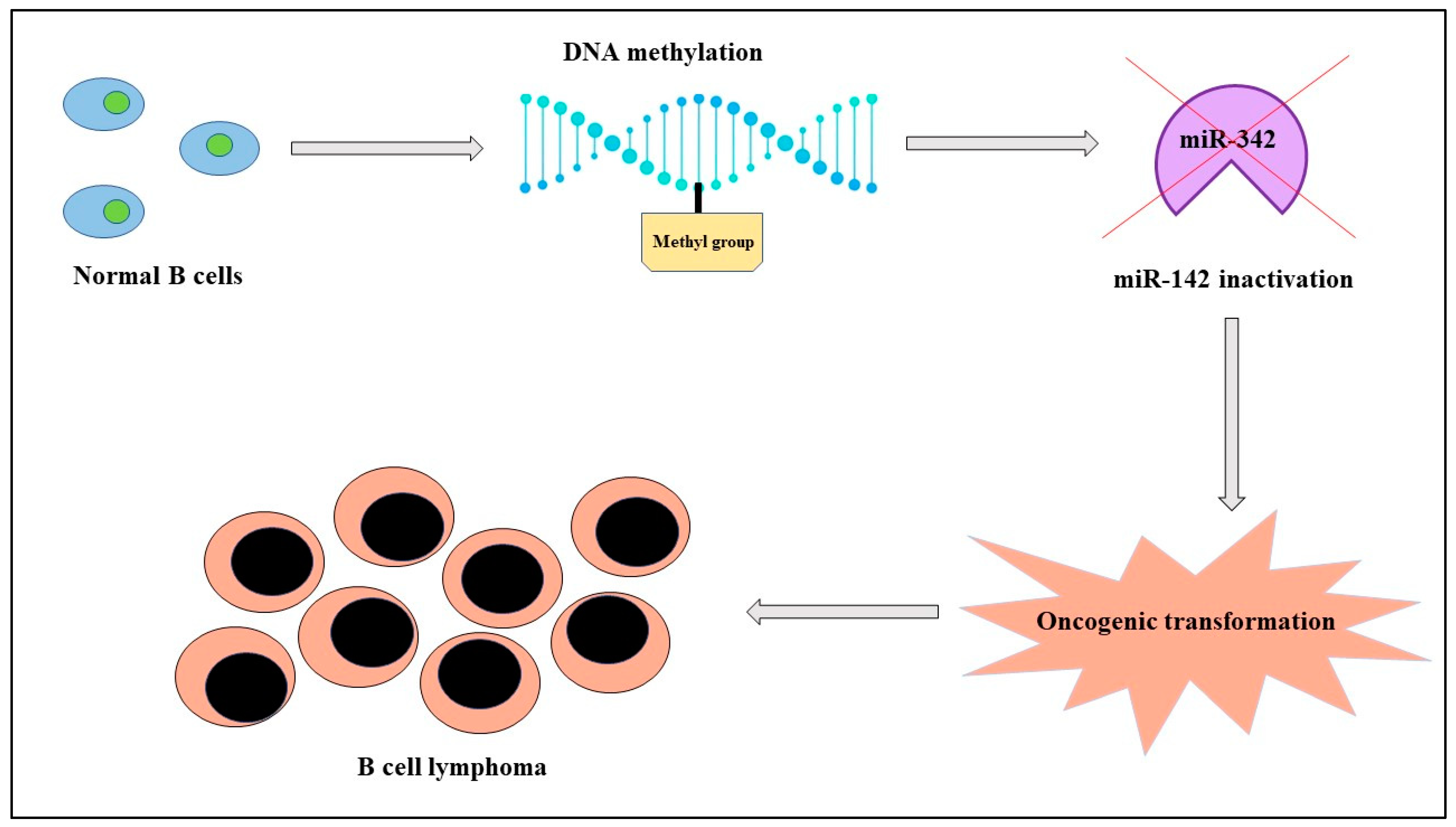
MicroRNAs are small, noncoding molecules of about twenty-two nucleotides with crucial roles in both healthy and pathological cells. Their expression depends not only on genetic factors, but also on epigenetic mechanisms like genomic imprinting and inactivation of X chromosome in females that influence in a sex-dependent manner onset, progression, and response to therapy of different diseases like cancer. There is evidence of a correlation between miRNAs, sex, and cancer both in solid tumors and in hematological malignancies; as an example, in lymphomas, with a prevalence rate higher in men than women, miR-142 is “silenced” because of its hypermethylation by DNA methyltransferase-1 and it is blocked in its normal activity of regulating the migration of the cell. This condition corresponds in clinical practice with a more aggressive tumor. In addition, cancer treatment can have advantages from the evaluation of miRNAs expression; in fact, therapy with estrogens in hepatocellular carcinoma determines an upregulation of the oncosuppressors miR-26a, miR-92, and miR-122 and, consequently, apoptosis. 

  • sex differences
  • male
  • female
  • X chromosome inactivation
  • genomic imprinting
  • epigenetics
  • miRNA
  • cancer
  • apoptosis
  • oncoMir

1. Introduction

1.1. General Considerations on miRNAs

MicroRNAs are small, noncoding molecules of about twenty-two nucleotides and have crucial roles in both healthy and pathological cells; in fact, they are involved in various intra- and intercellular mechanisms like proliferation and apoptosis, so their incorrect function is involved in the onset of different diseases, such as cancer [1][2]. In detail, a miRNA could have a lot of genes as targets and, at the same time, a gene could be regulated by different miRNAs [3]; according to the scientific literature, they control about 60% of human genes [4]. miRNAs derive from a long molecule of ribonucleic acid called pri-miRNA that is caped and polyadenylated in the nucleus from polymerase II and then processed from DiGeorge syndrome critical region 8 (DGCR8) and Drosha proteins to form the pre-miRNA, consisting of about seventy nucleotides. Next, this molecule moves to the cytoplasm through the exportin 5–RAs-related nuclear protein (RAN) complex and is cut from an endonuclease into the mature miRNA. They can be secreted into vesicles called exosomes in the extracellular microenvironment or circulate tied to lipoproteins and argonaut proteins [3] and act by tying to the 3′ untranslated regions of mRNA and, consequently, blocking its translation or promoting protein degradation [5][6]. Some miRNAs are expressed in certain tissues and in a specific sex; for example, molecules from the miR-35 family are involved in sex determination, while miR-532 and miR-660 are highly expressed but lowly methylated in females rather than males [3][6][7]. Furthermore, in the X chromosome, there are a lot of miRNAs, while in the Y chromosome, there are only a few [8]. In addition to genetic mechanisms, epigenetics regulate their expression also [3]. In fact, external factors like infections, food, traumatic experiences, chemical pollutants, cold, and heat lead to changes in miRNA expression that can be transmitted to the next generations. Epigenetic mechanisms include the X chromosome inactivation in females and genomic imprinting. It is well known that, during embryo development, the inactivation of one of the two X chromosomes occurs in order to maintain a balanced expression of X chromosomes with men having just one; an incomplete inactivation of the X chromosome leads to a biallelic expression of miRNA [8]. The genomic imprinting consists of the expression of just one allele in all the genes of a subset of maternal and paternal origin, localized in the so-called “Differentially Methylated Regions” of the human genome. A particular type of imprinting is “hormonal imprinting”, a mechanism during which the hormonal receptor ties for the first time with a hormone, and the latter becomes its life-long target; however, during endocrine system genesis, receptors do not have an absolute specificity, so synthetic molecules like drugs or other hormones similar to the target one could tie to the receptor, leading to wrong hormonal imprinting and, consequently, to life-long events transmitted to future generations [7]. Cancer cells present alterations in the expression of genes related to molecules that regulate proliferation, death, and survival, including miRNAs. In this context, tumor-suppressor micro-RNAs (oncoMirs) have the capacity to promote cancerogenesis by suppressing tumor-suppressor genes and, consequently, inhibiting cell-death pathways like apoptosis; they are upregulated in tumor cells and in the tumor microenvironment. For example, in B-cell lymphoma, miR-17-92 inhibits the “pro-apoptotic protein Bcl-2-like protein 1” (Bcl-2L1), inducing resistance to apoptosis and promoting the proliferation of immature lymphoid cells. On the other hand, antagoMirs are synthetic miRNAs, antagonists first developed as silencing molecules of miRNAs in the first years of this millennium; in detail, they are antisense oligonucleotides (ASOs) that tie to specific ribonucleic acid (RNA) sequences and, by inducing RNase H-mediated cleavage, influence the expression of specific miRNAs, leading to a reduction of cancer-related ones [9].

1.2. Sex Differences, miRNAs, and Cancer

MiRNAs have several roles in cancer also related to sex chromosomes. In fact, the X chromosome contains a high density of these small noncoding molecules involved in the regulation of immune response in humans, specifically, of immunosurveillance against the onset and progression of cancer [8]. The immune system has demonstrated roles also in cancer therapy as shown by a new class of drugs called “immune checkpoint inhibitors” [10]. We know tumor cells have the ability to escape from immune recognition as nonself-factors through the action of molecules like programmed cell death protein 1, a receptor that is tied by its proteic targets programmed death-ligand 1 (PD-L1) and programmed death-ligand 2 (PD-L2) with the result of an inhibition of T cells and the consequent block of apoptosis of cancer cells. However, PD-L1 expression is regulated by X-linked miRNAs such as miR-106b and miR-20b. The scientific literature showed their oncogenic function and their ability, especially for miR106a, in downregulating the anti-inflammatory activity of cytokine IL-10. Other miRNAs involved in potentiating inflammation with an obvious stimulus to cancerogenesis are miR-18 and -19, since they act on the nuclear factor-κB (NF-κB) [8][11][12]. Furthermore, miRNAs can also be secreted by the pituitary gland and, since the last is dependent on sex hormones, menopause in women and other hormonal changes in men conduct sex-related changes in miRNA production from the pituitary gland. In particular, an increase in proapoptotic miRNAs and a decrease in antiapoptotic ones verifies in this sex-related manner leading to an inhibition of all stimulating mechanisms, such as proliferation and neoangiogenesis, and to the induction of apoptosis, with a possible antitumoral effect [6]. In some cases, scientists succeeded in identifying the role of specific miRNAs; for example, the capacity of miR-22 to act on methylenetetrahydrofolate dehydrogenase 2 and methylenetetrahydrofolate reductase blocking and, consequently, the S-adenosylmethionine synthesis with the result of cancer cell death. Moreover, in females but not in males, an important expression of miR-27a-3p has been observed in cases of high tumor stage, while the expression of miR-17-5p and miR-20a-5p has been observed to be reduced in metastatic cancer cells [13]. The onset of cancer was recently associated with the so-called “XCI genes”, genes that escape from X chromosome inactivation with the consequent failed balance between male and female genomic balance; among them, researchers found that the connector enhancer of kinase suppressor of ras 2 (CNKSR2), lysine demethylase 6A (KDM6A), alpha-thalassemia/mental retardation, X-linked (ATRX) and Lysine-specific demethylase 5C (KDM5C) are mutated in males with cancer but not in females. On the other hand, toll-like receptor 7 (TLR7), chromosome X open reading frame 21 (CXORF21), and CD40L are double expressed in women, determining an augmented risk of autoimmune diseases [9]. The scientific literature demonstrates that miRNA expression can be influenced by natural substances such as genistein, a molecule belonging to the chemical class of isoflavones, with described activities of phytoestrogen and of an angiogenesis inhibitor; it can be found in different vegetables like fava beans, soybeans, lupin, and coffee. Experiments conducted using genistein put in evidence its capability to cause in vitro the stop of the cell cycle at the G2/M phase by interacting with proteins involved in cell proliferation and cancer growth such as kinesin family member 20 (KIF20) of the kinesins family. Western blot analysis showed which genistein is capable of inhibiting the translation of the (Cdc25C) protein, cyclin-dependent kinase 1 (CDk1), cyclin A, and cyclin B, and stimulating the expression of cyclin-dependent kinase (CDK) inhibitor and cyclin-dependent kinase inhibitor 1 (p21 CIP1/WAF1); furthermore, it modulates the RAS/RAF pathway, stabilizing activation and phosphorylation of MAPK and inhibits the Akt and JAK/STAT pathways, both crucial for cell survival. miRNA expression can be influenced by genistein; in fact, it has been shown that this natural substance is capable of downregulating miR-151, responsible for invasion and cell migration in prostate cancer, and the minichromosome maintenance (MCM) gene family, involved in DNA replication and cancerogenesis. However, there are obstacles to the daily use of genisteins because of their poor water solubility and low serum availability; thus, solid–lipid–particulate systems (SLPs) have been proposed. In fact, these carriers consent to the drug being dissolved, encapsulated, and attached to a nanoparticle matrix. For example, a combination of genistein and doxorubicin has been associated with lower adverse effects compared with doxorubicin hydrochloride alone and induces a decrease in ROS production by prostate cancer cells [14][15].

2. Hematological Malignancies

Hematopoietic stem cells derive from the mesoderm during embryogenesis and are crucial for the correct development of blood cells and the functioning of the immune system [15][16][17]; both genetic and environmental factors such as chemical substances, radiation, and infections can cause their dysfunction and lead to hematological malignancies. Hematological malignancies, like leukemia, lymphoma, and myeloma, represent 7% of new cancer cases globally every year and, despite new treatments including drugs and bone marrow transplantation, they have survival rates far from 100% [18].
Studies demonstrated that miRNAs are relevant in the determination of hematological diseases and their complications [19][20].
Hematological malignancies show larger rates of incidence and mortality in men than in women. For example, there is a 6.1% risk of acute myeloid leukemia and a 4.3% mortality rate in males vs. the 4.2% risk and mortality rate of 2.8% in females; the same sex correlation can be observed for myelodysplastic syndromes and for lymphomas (especially for mantle-cell type and less for marginal zone lymphoma). Therefore, it can be stated that sex could be considered a negative prognostic factor both in the onset and progression of hematological malignancies [21]. Studies of molecular biology demonstrated that miR-342, localized in the Enah/Vasp-Like (EVL) gene of chromosome 14, regulates cytoskeleton remodeling and, consequently, the capacity of migration of cells. In B-cell lymphomas, miR-342 is silenced because of the hypermethylation of the EVL gene in its promoter region and the treatment with decitabine leads to the re-expression of miR-342 and the translation of the EVL protein. It is well known which DNA methyltransferase-1 normally determines the hypermethylation of genes; in this case, miR-342 leads to a downregulation of this enzyme and, consequently, to the hypomethylation of cells and a reduction in B-cell lymphoma aggressiveness [22]. Furthermore, men have a worse outcome in terms of systemic effects like sarcopenia than women and this condition can be related to the fact that hormones such as estradiol in females reduce the viscosity of the mitochondrial membrane, improving the activity of the skeletal muscle; in this mechanism, miR-486 is involved and supports the synthesis of myotubes from myoblasts, so it can be considered a sex-related biomarker of cancer systemic effects [23] (Figure 1).
Figure 1. In B-cell lymphomas, the activity of miR-142, localized in the EVL gene of chromosome 14, is blocked by the hypermethylation of DNA, leading to cancerogenesis.
Table 1 summarizes  miRNAs involved in apoptosis and autophagy regulation of several types of cancer.
Table 1. MiRNAs involved in apoptosis and autophagy regulation of several types of cancer. 

Cancer

miRNAs

UP/DOWN Regulated

Mechanism of Action

Onset/Prognosis/Response to Therapy Involvement

References

B-cell lymphoma

miR-142

DOWN

Altered migration of cells

Poor prognosis

[22]

Papillary thyroid cancer

miR-21

miR-26a

miR-181a

miR-181b

miR-219

miR-221

miR-222

miR-245

UP

Modulation in protein p27Kip1 expression

Onset

[24]

Hepatocellular carcinoma

miR-371a-5p

UP

Transition from G to S phase of cell cycle

Onset

[25]

Colorectal cancer

miR-16

miR-22

miR-142-3p

miR-22 UP; miR-16 and miR-142-3p DOWN

Inhibition of autophagy (miR-16, miR-142-3p) and inhibition of estrogen activity

Onset (miR-16, miR-142-3p) and better response to therapy (miR-22)

[8]

Gastric cancer

miR-125

UP

Block of apoptosis

Onset

[26]

Lung cancer

miR-143

miR-145

miR-153-3p

UP

Apoptosis induction

Good prognosis

[22]

Melanoma

miR-23a

miR-221

miR-222

UP

Block of cell proliferation

Onset

[8]

Breast cancer

miR-17

miR-21

miR-124

UP

BRCA1 inactivation

Onset

[27][28]

Glioblastoma

hsa-miR-1909-5p

hsa-let-7c-5p

miR-206-5p

hsa-miR-1909-5p and

hsa-let-7c-5p UP; miR-206-5p DOWN

Promotion of cell migration and invasion (hsa-miR-1909-5p,

hsa-let-7c-5p) and modulation of apoptosis (miR-206-5p)

Onset

[29]

This entry is adapted from the peer-reviewed paper 10.3390/ijms241411544

References

  1. Allegra, A.; Murdaca, G.; Gammeri, L.; Ettari, R.; Gangemi, S. Alarmins and MicroRNAs, a New Axis in the Genesis of Respiratory Diseases: Possible Therapeutic Implications. Int. J. Mol. Sci. 2023, 24, 1783.
  2. Amato, G.; Vita, F.; Quattrocchi, P.; Minciullo, P.L.; Pioggia, G.; Gangemi, S. Involvement of miR-142 and miR-155 in Non-Infectious Complications of CVID. Molecules 2020, 25, 4760.
  3. Kraczkowska, W.; Stachowiak, L.; Pławski, A.; Jagodziński, P.P. Circulating miRNA as potential biomarkers for diabetes mellitus type 2: Should we focus on searching for sex differences? J. Appl. Genet. 2022, 63, 293–303.
  4. Hasakova, K.; Bezakova, J.; Vician, M.; Reis, R.; Zeman, M.; Herichova, I. Gender-Dependent Expression of Leading and Passenger Strand of miR-21 and miR-16 in Human Colorectal Cancer and Adjacent Colonic Tissues. Physiol. Res. 2017, 66, S575–S582.
  5. Umansky, S. Aging and aging-associated diseases: A microRNA-based endocrine regulation hypothesis. Aging 2018, 10, 2557–2569.
  6. Warnefors, M.; Mössinger, K.; Halbert, J.; Studer, T.; VandeBerg, J.L.; Lindgren, I.; Fallahshahroudi, A.; Jensen, P.; Kaessmann, H. Sex-biased microRNA expression in mammals and birds reveals underlying regulatory mechanisms and a role in dosage compensation. Genome Res. 2017, 27, 1961–1973.
  7. Bezler, A.; Braukmann, F.; West, S.M.; Duplan, A.; Conconi, R.; Schütz, F.; Gönczy, P.; Piano, F.; Gunsalus, K.; Miska, E.A.; et al. Tissue- and sex-specific small RNAomes reveal sex differences in response to the environment. PLOS Genet. 2019, 15, e1007905.
  8. Bellenghi, M.; Puglisi, R.; Pontecorvi, G.; De Feo, A.; Carè, A.; Mattia, G. Sex and Gender Disparities in Melanoma. Cancers 2020, 12, 1819.
  9. Otmani, K.; Rouas, R.; Lewalle, P. OncomiRs as noncoding RNAs having functions in cancer: Their role in immune suppression and clinical implications. Front. Immunol. 2022, 13, 913951.
  10. Caserta, S.; Innao, V.; Musolino, C.; Allegra, A. Immune checkpoint inhibitors in multiple myeloma: A review of the literature. Pathol. Res. Pract. 2020, 216, 153114.
  11. Carè, A.; Bellenghi, M.; Matarrese, P.; Gabriele, L.; Salvioli, S.; Malorni, W. Sex disparity in cancer: Roles of microRNAs and related functional players. Cell Death Differ. 2018, 25, 477–485.
  12. Fernández-Torrón, R.; García-Puga, M.; Emparanza, J.-I.; Maneiro, M.; Cobo, A.-M.; Poza, J.-J.; Espinal, J.-B.; Zulaica, M.; Ruiz, I.; Martorell, L.; et al. Cancer risk in DM1 is sex-related and linked to miRNA-200/141 downregulation. Neurology 2016, 87, 1250–1257.
  13. Tomeva, E.; Krammer, U.D.B.; Switzeny, O.J.; Haslberger, A.G.; Hippe, B. Sex-Specific miRNA Differences in Liquid Biopsies from Subjects with Solid Tumors and Healthy Controls. Epigenomes 2023, 7, 2.
  14. Javed, Z.; Khan, K.; Herrera-Bravo, J.; Naeem, S.; Iqbal, M.J.; Sadia, H.; Qadri, Q.R.; Raza, S.; Irshad, A.; Akbar, A.; et al. Correction: Genistein as a regulator of signaling pathways and microRNAs in different types of cancers. Cancer Cell Int. 2022, 22, 256.
  15. Shiau, J.-P.; Chuang, Y.-T.; Yen, C.-Y.; Chang, F.-R.; Yang, K.-H.; Hou, M.-F.; Tang, J.-Y.; Chang, H.-W. Modulation of AKT Pathway-Targeting miRNAs for Cancer Cell Treatment with Natural Products. Int. J. Mol. Sci. 2023, 24, 3688.
  16. Caserta, S.; Zaccuri, A.M.; Innao, V.; Musolino, C.; Allegra, A. Immune thrombocytopenia: Options and new perspectives. Blood Coagul. Fibrinolysis 2021, 32, 427–433.
  17. De Luca, F.; Allegra, A.; Di Chio, C.; Previti, S.; Zappalà, M.; Ettari, R. Monoclonal Antibodies: The Greatest Resource to Treat Multiple Myeloma. Int. J. Mol. Sci. 2023, 24, 3136.
  18. Musolino, C.; Oteri, G.; Allegra, A.; Mania, M.; D’ascola, A.; Avenoso, A.; Innao, V.; Allegra, A.G.; Campo, S. Altered microRNA expression profile in the peripheral lymphoid compartment of multiple myeloma patients with bisphosphonate-induced osteonecrosis of the jaw. Ann. Hematol. 2018, 97, 1259–1269.
  19. Allegra, A.; Tonacci, A.; Giordano, L.; Musolino, C.; Gangemi, S. Targeting Redox Regulation as a Therapeutic Opportunity against Acute Leukemia: Pro-Oxidant Strategy or Antioxidant Approach? Antioxidants 2022, 11, 1696.
  20. Taverna, S.; Tonacci, A.; Ferraro, M.; Cammarata, G.; Cuttitta, G.; Bucchieri, S.; Pace, E.; Gangemi, S. High Mobility Group Box 1: Biological Functions and Relevance in Oxidative Stress Related Chronic Diseases. Cells 2022, 11, 849.
  21. Cui, X.; Zhao, X.; Liang, Y. Sex differences in normal and malignant hematopoiesis. Blood Sci. 2022, 4, 185–191.
  22. Matarrese, P.; Mattia, G.; Pagano, M.T.; Pontecorvi, G.; Ortona, E.; Malorni, W.; Carè, A. The Sex-Related Interplay between TME and Cancer: On the Critical Role of Estrogen, MicroRNAs and Autophagy. Cancers 2021, 13, 3287.
  23. Anonymous. Correction to: Hormonally Regulated Myogenic miR-486 Influences Sex-specific Differences in Cancer-induced Skeletal Muscle Defects. Endocrinology 2022, 163, bqac120, Erratum in: Endocrinology 2021, 162, 35939403.
  24. Huang, C.; Cai, Z.; Huang, M.; Mao, C.; Zhang, Q.; Lin, Y.; Zhang, X.; Tang, B.; Chen, Y.; Wang, X.; et al. miR-219–5p Modulates Cell Growth of Papillary Thyroid Carcinoma by Targeting Estrogen Receptor α. J. Clin. Endocrinol. Metab. 2015, 100, E204–E213.
  25. Bai, P.-S.; Hou, P.; Kong, Y. Hepatitis B virus promotes proliferation and metastasis in male Chinese hepatocellular carcinoma patients through the LEF-1/miR-371a-5p/SRCIN1/pleiotrophin/Slug pathway. Exp. Cell Res. 2018, 370, 174–188.
  26. Liu, B.; Zhou, M.; Li, X.; Zhang, X.; Wang, Q.; Liu, L.; Yang, M.; Yang, D.; Guo, Y.; Zhang, Q.; et al. Interrogation of gender disparity uncovers androgen receptor as the transcriptional activator for oncogenic miR-125b in gastric cancer. Cell Death Dis. 2021, 12, 441.
  27. Li, E.W.; Bai, Y. Computational Identification of Sex-Biased Biomarker MicroRNAs and Genes Associated with Immune Infiltration in Breast Cancer. Genes 2021, 12, 570.
  28. Singh, H. Role of Molecular Targeted Therapeutic Drugs in Treatment of Glioblastoma: A Review Article. Glob. Med. Genet. 2023, 10, 42–47.
  29. Stupp, R.; Taillibert, S.; Kanner, A.A.; Kesari, S.; Steinberg, D.M.; Toms, S.A.; Taylor, L.P.; Lieberman, F.; Silvani, A.; Fink, K.L.; et al. Maintenance Therapy With Tumor-Treating Fields Plus Temozolomide vs Temozolomide Alone for Glioblastoma. JAMA 2015, 314, 2535–2543.
More
This entry is offline, you can click here to edit this entry!
Video Production Service