Endothelial Senescence on Angiogenesis in Alzheimer’s Disease: History
Please note this is an old version of this entry, which may differ significantly from the current revision.
Contributor: , , ,

Endothelial cells are constantly exposed to environmental stress factors that, above a certain threshold, trigger cellular senescence and apoptosis. The altered vascular function affects new vessel formation and endothelial fitness, contributing to the progression of age-related diseases. 

  • angiogenesis
  • cellular senescence
  • aging
  • extracellular vesicles
  • oxidative stress
  • Alzheimer’s disease

1. Introduction

Angiogenesis is a complex biological process that involves the formation of new blood vessels from preexisting ones and should not be confused with vasculogenesis, in which blood vessels emerge de novo from endothelial progenitor cells [1]. It plays a crucial role during embryonic development and later in tissue growth and repair, wound healing, and reproduction. Still, it must be carefully regulated to avoid excessive or insufficient vascularization. New vessels emerge from sprouting endothelial cells (EC), the leading players, toward an angiogenic stimulus (sprouting angiogenesis) or by insertion into existing vessels and division into new ones (splitting angiogenesis) [2]. It is led by a tip cell that elongates and explores the environment while transmitting signals to the stalk cells that follow behind to proliferate and form tubular networks. The entire process is highly complex and difficult to imitate in vitro, highlighting the need for development of reliable models to study it [3][4][5]. Angiogenesis is governed by a strict balance between pro- and antiangiogenic factors, which, if broken, leads to uncontrolled cell proliferation (cancer, atherosclerosis, rheumatoid arthritis) or mitotic inhibition (aging and neurodegenerative diseases) [6][7][8]. Excessive angiogenesis can promote inflammation and tissue damage, while insufficient angiogenesis can lead to ischemia and cell death. Most studies are focused on the involvement of angiogenesis in cancer and cardiovascular diseases (CVD). Fewer examine its contribution to neurodegeneration, although it is correlated with the impairment of angiogenesis [9], endothelial senescence [7] and the occurrence of cerebrovascular angiopathy (a process in which small blood vessels burst and cause hemorrhages) [10][11]. The altered blood circulation in the elderly contributes to the lengthy process of wound healing and inadequate recovery of ischemic tissues due to the lack of response from aged ECs. Typically, ECs’ physiological functions are suppressed in time because of accumulated stress and induction of cellular senescence and apoptosis [12], leading to alterations in the regulation of angiogenesis and insufficient or excessive vascularization [6]. Age-related vasculature dysfunction has been implicated in the pathogenesis of various neurodegenerative diseases, including Alzheimer’s disease [13], Parkinson’s disease [14], and Huntington’s disease [15]. It may contribute to their progression by modulating the delivery of nutrients and oxygen and clearing of waste products from the brain.
Alzheimer’s disease (AD) is a debilitating condition characterized by progressive cognitive decline and behavioral changes that severely impact the daily lives of suffering individuals. Similarly, to other neurodegenerative diseases, aging is an essential factor contributing to its onset. There is overwhelming research aiming to find the causes, better ways for detection, treatment and, if possible, ways to avoid it altogether (reviewed elsewhere [16][17][18][19][20]). Factors involved in angiogenesis have roles in the birth of new neurons (neurogenesis), the prevention or mitigation of neuronal injury (neuroprotection), and the pathogenesis of stroke, AD and motor neuron disease [21]. Indeed, axon and blood vessel growth and migration are braided together via chemo-repulsive and attractive signals in which the vascular endothelial growth factor (VEGF) and the Delta-Notch signaling have a direct effect on both nervous and vascular systems [22], confirming that angiogenesis is closely related to neurodegeneration. AD patients exhibit changes in the number, diameter and density of blood vessels, which lead to decreased brain perfusion and BBB disruption.

2. The Dual Nature of Cellular Senescence

Cellular senescence is a fundamental process associated with tissue homeostasis during development, first described by Hayflick and Moorhead [23]. The authors observed a terminal pause in cell division of normal human fibroblasts after several cycles of passaging. They concluded that cultured cells cease to proliferate upon a finite number of doublings and, therefore, could be used as a model for aging. Today, this is referred to as the Hayflick limit. The processes of senescence and aging are intertwined in the sense that aging progresses with time and associates with increased numbers of senescent cells. Therefore, cellular senescence is also accepted as a hallmark of aging and a risk factor for age-related neurodegenerative diseases. However, senescence occurs during the full lifespan of an individual and is not restricted to later life stages. The resulting inability to divide is a consequence of irreversible cell cycle arrest, caused by the accumulation of various stress factors such as DNA damage, inflammation, telomere shortening, chromatin perturbations, and oncogene induction [12][24][25][26]. Senescence is believed to have evolved as a protective mechanism against cancer, but it also contributes to age-related physiological decline [27]. Additionally, loss of senescence during embryonic development allows the progression of unhealthy cells in embryos [28]. In contrast, while protecting against the propagation of mutated DNA, senescence harms long-living organisms, as it inhibits tissue renewal and function. These observations gave rise to the idea that there is a “right time to senesce”, arguing that the end goal of the fight against aging is not to completely eliminate senescent cells (SCs) but to learn how to tame them [29].

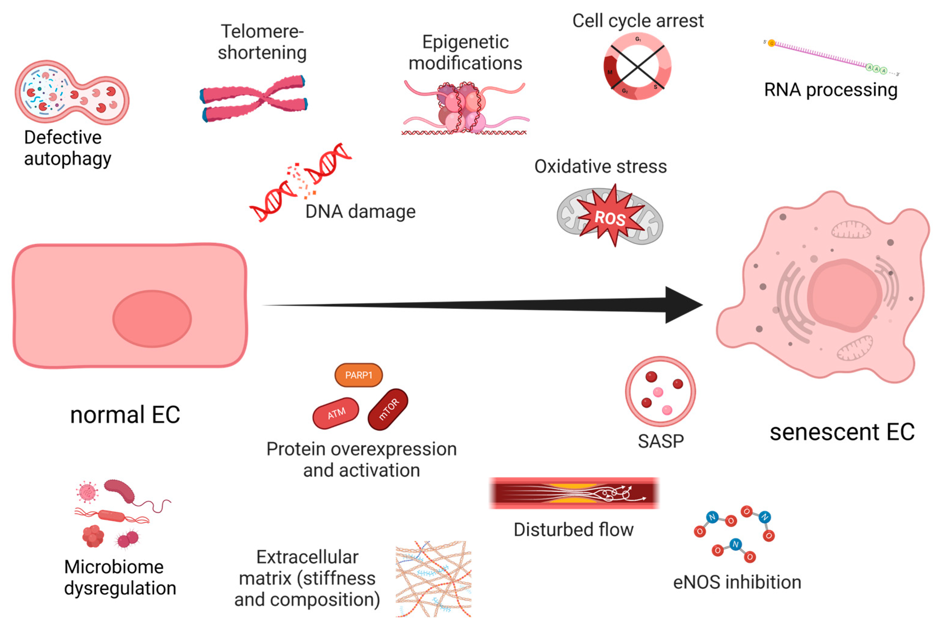
3. Endothelial Senescence

Aging and prolonged exposure to environmental factors, such as toxins, ROS, shear stress, and extracellular matrix (ECM) perturbations, induce senescence in ECs (Figure 1). Interestingly, unlike most SCs, senescent ECs (sen-ECs) remain susceptible to apoptosis [30], a mechanism most likely evolved to rearrange the microvasculature and counteract proliferation. Senescence in ECs is usually triggered by telomere shortening [26], which can be avoided by the exogenous introduction of telomerase [6]. Ionizing radiation can also geroconvert human microvascular cells in a time- and dose-dependent manner, predominantly by uncoupling Complex II of the mitochondrial respiratory chain [31], demonstrating ECs’ susceptibility to OS. In any case, the balance between senescence and angiogenesis becomes dysregulated during aging and neurodegenerative diseases, but the underlying mechanisms remain elusive. The negative consequences of vascular aging are apparent in older people in whom the regeneration of blood flow after ischemia or wounding is a slow and tedious process [32]. The accumulated stress over time reduces the proliferative capacity of ECs and modifies their interaction with the already altered ECM [33]. Furthermore, aging reduces the general expression of vascular endothelial growth factor (VEGF) [6] and promotes angiogenic incompetence in ECs, making them unable to respond to VEGF [7]. Some of the suggested reasons for the VEGF insensitivity are the age-related loss of VEGF receptor 2 (VEGFR2) [34], androgen resistance [35] and reduction in nitric oxide (NO) [6]. Furthermore, the SASP can directly inhibit angiogenesis by secreting factors that block endothelial cell proliferation and migration. At the same time, SCs can induce angiogenesis by secreting pro-inflammatory cytokines that promote neovascularization.
Figure 1. Hallmarks of endothelial senescence. Created with https://www.BioRender.com (accessed on 7 July 2023).

4. Unveiling the Interplay between Hypoxia and Oxidative Stress-Induced Endothelial Senescence

The main reason for O2’s negative manifestation is that it is responsible for the generation of reactive oxygen species (ROS), which cause DNA damage and induce senescence. It is still unclear whether there are different mechanisms of senescence activation, depending on the source of ROS and/or the place of accumulation [36]. For example, in CVD, mitochondrial dysfunction often triggers age-associated perturbations in the production of NO and VEGF [27][33], which can be mitigated by reduced mitochondrial oxidative phosphorylation in mammals [37]. On the other hand, mitochondrial ROS in the model organism Caenorhabditis elegans increases its longevity [38]. In addition, reduced mitochondrial mass and alterations in the electron transport chain (ETC) due to a decline in cytochrome C oxidase and Complex IV [39] and upregulated NADPH oxidases (NOX) increase OS and shorten telomeres [40]. The role of mitochondria in senescence was also confirmed by global transcriptomic analysis, where the expression of 38% of senescence-associated genes was reversed in mitochondrial-depleted fibroblasts [41]. A direct link between ROS, telomere shortening and senescence was evidenced by assessing the number of SA-β-Gal+ ECs after exposure to H2O2 or glutathione (GSH) peroxidase inhibitors (it should be noted that other senescence markers were not used) [40]. Since OS is a prominent contributor to endothelial senescence, it is natural to assume that low levels of O2 could prevent this process. Interestingly, low ROS delay DNA replication and cell cycle progression via a CDK2-dependent mechanism [42]. Therefore, lower ROS levels and prolonged cell division could potentially prevent replicative EC senescence due to excessive telomere shortening and reduced DNA damage.

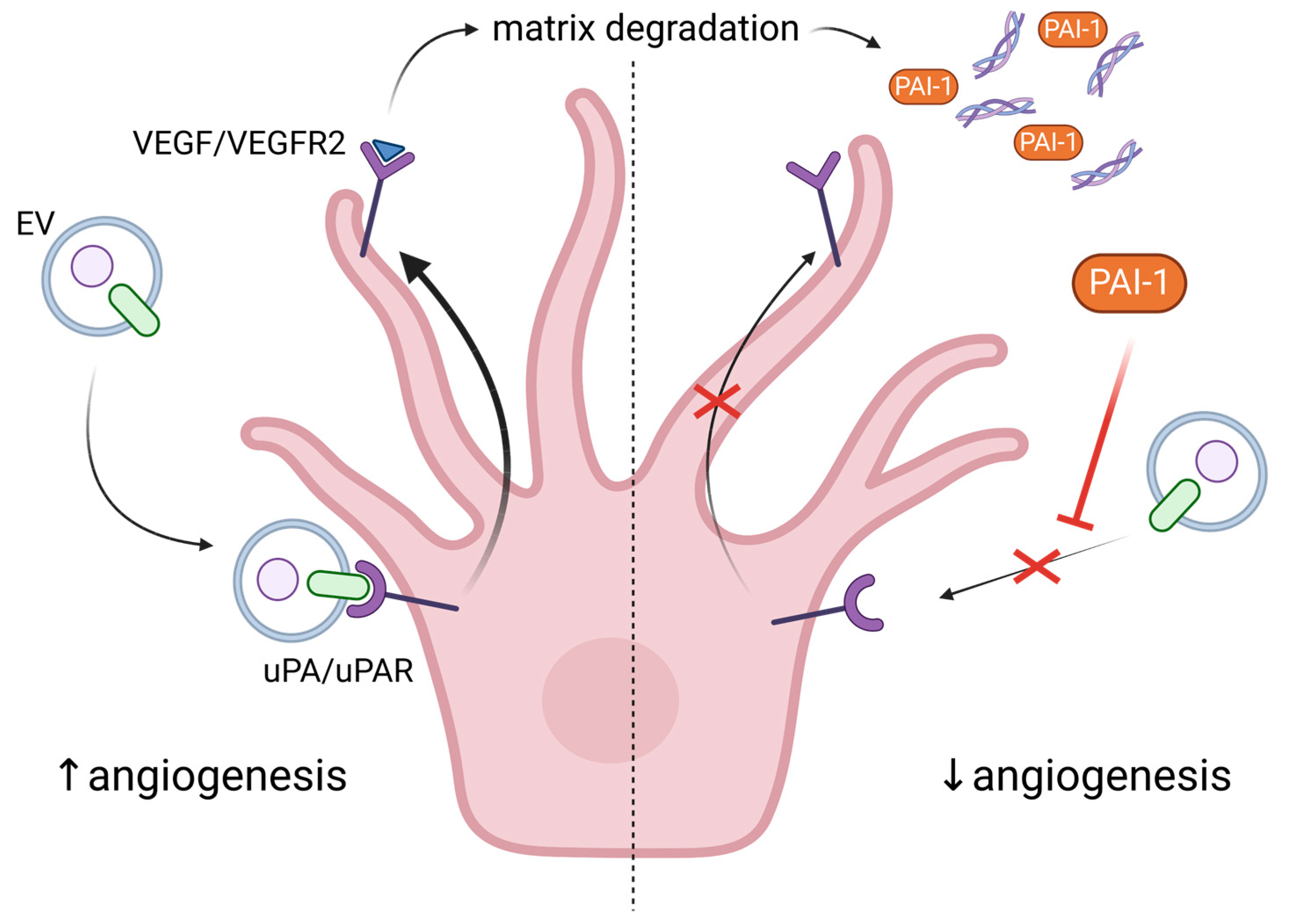
5. Exploring the Role of Extracellular Vesicles in Angiogenesis and Senescence

Legends about the infamous Hungarian Countess Elizabeth Bathory tell the story of her supposed anti-aging process of bathing in the blood of young girls. A similar idea governs the myths for vampires, which might not necessarily stay young, but become immortal by feeding on human blood. Surprisingly, there seems to be some truth in these myths, as recent studies showed that blood exchange from young to old mice rejuvenates them, but the opposite transfusion leads to senescence in the young [43][44][45]. The latter highlights the role of SASP in aging, which assists the immune response and, in the context of angiogenesis, influences new vessel formation. In addition to soluble factors, such as chemokines, inflammatory cytokines and growth factors, extracellular vesicles (EVs) are key components of SASP (reviewed in [46]). EVs are a very heterogeneous group of membranous structures, roughly categorized into three main groups based on size and origin: apoptotic bodies (ABs), microvesicles (MVs) that range from 50 to 5000 nm and are formed by outward budding and fission of the plasma membrane, and exosomes (30–100 nm) that are produced by the fusion of multivesicular endosomes with the plasma membrane, releasing intraluminal vesicles into the extracellular space. The EVs play important roles in intercellular communication, and their release is a strictly regulated process [47]. They are involved in both physiological and pathological processes and play a role in intercellular communication through the transfer of proteins, lipids, and nucleic acids [48][49]. EVs are implicated in cancer etiology due to their ability to promote cancer cell migration, transformation of non-malignant cells and pro-angiogenic activity [50]. While healthy cells release EVs as part of normal cellular homeostasis, senescent cells secrete EVs that have a significant role in angiogenesis and neurodegenerative disease progression. The presence of pro-angiogenic molecules like HIF-1α, VEGF, MMPs, and microRNAs in EVs [51] may lead to homeostasis disruption and non-productive angiogenesis. The role of EVs as key functional components of SASP is further highlighted by the observation that secretion of EVs is much higher in different types of senescent cells, including ECs, as compared to young ones [52][53]. A possible explanation for this is the observed upregulation of neutral sphingomyelinase and dysfunction of lysosomal activity in senescent cells [54]. One study even suggests that hypoxia prevents senescence by decreasing the SASP, rather than reducing the number of senescent cells [55].
The important role of EVs from ECs, as well as other blood cell types, in angiogenesis is summarized here [47]. More specifically, EVs from ECs are rich in β1 integrins and metalloproteinases (MMP-2 and MMP-9), which allow them to penetrate the ECM, to remodel it and to form tubular capillary-like structures. Stimulation with VEGF and FGF-2 facilitates the association of the active and proenzyme forms of the MMPs with EC-derived vesicles [56]. EVs can also transport urokinase plasminogen activator/uPA receptor (uPA/uPAR), which are both pro-angiogenic. It was shown that uPAR modulates VEGF-induced EC migration by balancing the proteolysis of the ECM and the cell motility through integrin-associated focal adhesion (Figure 2). Revu Ann Alexander and colleagues demonstrated that VEGF causes endocytosis of αVβI integrin and activation of uPA/uPAR, resulting in matrix degradation [57]. Another active participant in this process is the inhibitor of uPA—plasminogen activator inhibitor (PAI-1), which is released from the degraded matrix and internalized, further directing the balance toward invasive cell migration, i.e., angiogenesis (Figure 2). Inhibition or deficiency of uPAR suppressed VEGF-induced angiogenesis in tumor cells [58] or in mice [59], respectively. Moreover, uPAR stimulated angiogenesis through VEGFR2, which upon internalization activates other pro-angiogenic stimuli [59]. In confluent ECs, the expression of uPAR is down-regulated compared to sub-confluent proliferative cells, thus preventing VEGF-activated signaling and angiogenesis [60]. In addition, levels of PAI-1 are elevated in senescent and aged ECs, making it a useful marker for senescence [61]. Besides inhibiting uPAR, PAI-1 also induces p53 and p21, activity that is suppressed by SIRT1 overexpression in endothelial cells. SIRT1 is also able to induce eNOS activity, protecting ECs from endothelial dysfunction [61]. The pro-angiogenic properties of exosomes from ECs may also be attributed to EV-associated micro RNAs such as miR-214 [62]. More specifically, the latter prevents senescence through silencing ATM in recipient cells.
Figure 2. Regulation of angiogenesis by urokinase plasminogen activator and its receptor. uPA/uPAR are carried by extracellular vesicles (EVs) to ECs. Upon receptor binding, VEGF-mediated matrix degradation is stimulated via VEGFR2. The processed matrix releases the plasminogen activator inhibitor-1 (PAI-1), which inhibits uPA/uPAR recognition and subsequent VEGFR2 activation. This feedback loop prevents excessive angiogenesis. The red X depicts the obstruction of uPA/uPAR recognition under the influence of PAI-1 and the inability of uPA/uPAR to activate VEGFR2. Created with https://www.BioRender.com (accessed on 30 May 2023).
Depending on their source and the specific experimental conditions, EVs may also have anti-angiogenic properties. For example, NO production and angiogenesis are impaired by EC-derived EVs under oxidative stress, via Src kinase- and NOX-dependent mechanisms [33][63][64]. Moreover, in contrast to EVs from young cells, those derived from senescent cells exert mostly negative effects on EC functions and angiogenesis. More specifically, senescent osteoblasts secrete EVs that induce senescence and apoptosis and decrease proliferation of ECs through transfer of miR-139-5p [65]. Likewise, senescent HUVEC cells secrete exosomes that interfere with cell growth and downregulate expression of adherent junction proteins, resulting in impaired endothelial migration of young ECs and endothelial barrier dysfunction [66].
Interestingly, the effect of EVs on angiogenesis may be swayed in opposite directions depending on the dose. Namely, it was found that a low dose of EVs exhibited pro-angiogenic activity, which was suppressed below control levels upon increasing the concentration of the EC-derived EVs [67]—an effect dependent on uPA activity. The inhibitory effect of EC-derived EVs on endothelial cell tube formation was confirmed by another study in which even higher concentrations of EVs were used, and it was shown that the inhibition was dependent on NF-kB signaling and eNOS pathway suppression [68]. EVs are carriers of damaged genomic DNA molecules whose concentration increases in EVs upon induction of senescence [52] and under pathological conditions [69]. Functioning as intercellular vectors, EVs may transfer their DNA into the cytoplasm of recipient cells, leading to activation of the cGAS-STING signaling and consequently EC senescence, eNOS suppression and endothelial dysfunction [70]. Therefore, the hormetic effect of EC-derived EVs on EC tube formation, as well as the inhibitory effect of EVs from senescent ECs on angiogenesis, may possibly be due to EV DNA-induced cGAS-STING activation. Shedding more light on these processes and mechanisms would be a particularly interesting direction for further studies.

6. The Non-Productive Angiogenesis in Alzheimer’s Disease

Currently, there are two main hypotheses for the development of AD—the accumulation of amyloid plaques (Aβ) due to an error in the metabolism of the amyloid precursor protein (APP); and the hyperphosphorylation of Tau (or p-Tau), resulting in microtubule polymerization catastrophe and formation of fibrils [16]. APP is a transmembrane glycoprotein separated into an intracellular C-terminal, Aβ transmembrane and N-terminal extracellular domains. Its primary function is interneuronal communication, and once it performs it, APP is degraded by α- and γ-secretase to a soluble, non-amyloid form, or by β- and γ-secretase to insoluble Aβ1–40 and Aβ1–42 isoforms [16]. In animal models, elevated levels of Aβ1–42 and p-Tau were correlated with cerebrovascular dysfunction, chronic hypoperfusion and worsened AD symptoms [71][72]. One of the most affected brain areas in AD is the hippocampus, which is normally able to continue with adult neurogenesis. Thus, a decline in neurogenesis could be used as a marker for AD progression in animal models [73]. In fact, the researchers demonstrated worsened long-term memory and anxiety in a rat model of icvAβ1–42 concomitant with pinealectomy (AD with melatonin deficiency). These behaviors are controlled by the hippocampus and corresponded with increased OS in the structure [74].
Pro-inflammatory cytokines, such as interleukin-1β, become abundant during AD and induce the expression of VEGF, yielding new blood vessels [18]. Although angiogenesis is initiated around Aβ plaques, the process is non-productive, leading to the disassembly of Aβ plaque-associated blood vessels and the phagocytic activity of microglia [75]. However, there is conflicting evidence relating the cause of AD and whether there is an increase or decrease in blood vessel density [75][76][77][78][79][80][81][82] (Table 1).
Table 1. The role of Aβ in cerebral blood vessels.
↑—indicates an increase in the process; ↓—indicates a decrease in the process.
Joe Steinman, Hong-Shuo Sun and Zhong-Ping Feng provide a reasonable explanation for the discrepancies—“An overall measure of vessel density may indicate loss of vessels due to holes [note: from plaque deposits], without accounting for the increase in vessels surrounding holes” [83]. Although angiogenesis might not be beneficial for AD’s progression, it seems to alleviate some of the cognitive disabilities. For instance, one study showed that AD patients and AD mouse models accumulated Aβ in arterioles and experienced apoptosis of ECs [81]. When the same mouse model TgCRND8 was treated with VEGF, the growth factor was able to rescue vascular loss. And, most importantly, it significantly improved the behavior and memory of the subjects [81]. However, this observation could not be repeated in vitro on Matrigel®, where Aβ maintained low vascular density regardless of VEGF in tube formation assays, demonstrating the inability to always correlate in vivo and in vitro studies. A natural way to suppress Aβ accumulation is through melatonin. Besides its function as a radical scavenger, research shows that melatonin disrupts amyloid fibril formation [20] and exhibits anti-angiogenic properties [84][85]. Thus, by hindering Aβ plaque formation and reducing OS, melatonin deflects their role in non-productive angiogenesis and endothelial senescence. Taken together, these observations support the use of the hormone as an adjuvant therapy in AD.

7. Therapeutic Approaches to Endothelial Senescence and Dysfunction

Understanding the underlying mechanism of aging and neurodegenerative diseases will one day provide us with the means to treat them. Along with DNA damage, OS, and insufficient or disturbed blood flow, behavioral and social cues guiding unhealthy lifestyle choices also accelerate the aging process. It is urban knowledge that chronic stress with high cortisol levels, high-calorie food, lack of exercise, etc. worsens life quality and expectancy.

7.1. Exercise Improves CBF, Vascular Function and Cognitive Performance

Angiogenesis in the brain microvasculature can improve tissue oxygenation, but if done improperly, it can provoke vascular leakage and neurodegeneration. A way to ensure positive angiogenesis is exercise, which stimulates eNOS by increasing the CBF [86] and potentially reduces OS by hypoxia-mediated inhibition of oxidative phosphorylation. In addition, aerobic exercise increases energy consumption (mimicking CR), while alleviating basal membrane dysfunction [87] and age-related behavior changes [88].
In an eight-week comparative study between old sedentary and exercised male rats, moderate exercise decreased the mean arterial blood pressure in favor of the trained group [86]. It also improved CBF, VEGF, eNOS expression, capillary density and astrocyte growth [86][89][90]. Furthermore, malondialdehyde (MDA—a marker for lipid peroxidation) levels were reduced in the exercised aged group [86]. Exercise also reduced the levels of fibrin and fibrinogen in old mice, improving the activity of neurovascular units (microvascular ECs, basement membrane, pericytes and astrocytes) [87]. Increased CBF, by regular treadmill running, prevented the loss of BDNF, which usually leads to learning and memory deficiencies [88]. Aerobic running on a treadmill or cycling induces EV secretion before reaching an anaerobic state [91]. In contrast, Brahmer et al. collected EV samples of athletes before, during and after cycling to exhaustion [92]. They observed a significant increase in CD63+ EVs post exercise (at the highest lactate levels), with some also carrying CD105 and CD146 (markers for ECs). Thus, exercise itself rather than the intensity influences EV release. The EV release is very likely to be Ca2+-dependent, and since muscle activation leads to Ca2+ flux, this could be a potential cause of EV accumulation [93]. Meanwhile, Ca2+ signaling is impaired in senescent ECs and impedes the contraction of vascular smooth muscle cells in mesenteric arteries of aged (24–26 month old) mice [94]. Taken together, these findings support speculation that the increase in plasma Ca2+ due to exercise could improve the vasomotor control of the arteries. Furthermore, exercise-induced moderate hypoxia causes metabolic conversion to anaerobic glycolysis, securing NAD+ availability when the TCA cycle and the ETC are subdued. The resulting buildup of lactate provokes the expression of VEGFR2 in ECs [95] and stimulates reparative angiogenesis in ischemic tissues [96]. Furthermore, lactate secreted by skeletal muscle can travel through the blood and bind to the lactate receptor HCAR1, enriched in cells lining the brain’s blood vessels, inducing VEGF expression and cerebral angiogenesis [89]. This was positively impacted by high-intensity interval training (HIIT) or lactate injections and led to increased capillary density in the brain of WT mice and not in HCAR1-KO. The authors linked this effect with the activation of ERK1/2 and Akt, which are upstream positive regulators of VEGF [89]. In general, physical activity improves physical and cognitive function by enhancing CBF and reducing OS, neuroinflammation and vascular dysfunction, and positively impacts AD’s symptoms.

7.2. Caloric Restriction Reduces OS and Vascular Aging

Already, Ciceron has suggested that moderate eating and exercise are key factors for longevity. Therefore, caloric restriction (CR) could be beneficial for people, as it activates autophagy and triggers the cells to recycle and renew themselves [97]. Under CR, high temperatures or excessive competition, C. elegans undergoes a dramatic metamorphosis into a dauer form. In this state, the worms close their mouth apparatus, switch their metabolism from the TCA cycle to gluconeogenesis and seize their development until food becomes available. The incredible thing is that dauers live at least twice as long compared to adult worms [98]. This is one of the reasons why C. elegans is the go-to system when studying senescence. However, the restricted activity of mitochondria ultimately leads to their deterioration [38]. In a recent study, mice meeting their caloric needs but consuming less protein and branched fatty acids had lower adiposity, higher metabolic rates and lifespans [99]. The authors attributed this to lower activation of mTORC1 by amino and fatty acids, rather than CR itself. With aging, mTORC1 is upregulated, which correlates with eNOS uncoupling and O2•− generation, which are significantly reduced in senescent ECs treated with rapamycin (an mTOR inhibitor) [100] and in old mice under CR diet [101]. A detailed review by Christopher R. Martens and Douglas R. Seals describes other stress-induced cellular mechanisms inhibited in senile ECs—NO synthesis mediated by AMPK and SIRT1, autophagy (detailed review of autophagy factors promoting longevity [102]), and ECM stiffening through elastin proteolysis by MMP-9 and AGEs-induced inflammation of the arterial wall that can be ameliorated by CR [103]. Furthermore, the activity of SIRTs as histone deacetylases, hence, the epigenetic regulation of senescence and aging, is promoted by CR [104]. Although there is substantial evidence that CR can reduce and delay the deteriorating effects of aging and maintain our longevity, more controlled research is necessary to establish good CR protocols accounting for personal needs.

7.3. Role of Resveratrol in the Vascular Biology and Senescence Process

In general antioxidants such as reduced glutathione (GSH) and melatonin inhibit cell senescence by reducing reactive oxygen species (ROS) generation [105]. Resveratrol (3,5,40-trihydroxystilbene) (R), which is a non-flavonoid polyphenolic compound and derivative of stilbene, exhibits its pleotropic function also by decreasing ROS production and improving the antioxidant levels [106]. As mentioned above, EPCs are critical circulating components of the endothelium and are identified as key factors in endothelial repair. In this respect, resveratrol treatment can reverse EPC dysfunction by decreasing oxidative stress and increasing proliferation and capillary-like structure formation, and, by increasing the angiogenic factors like (NO), can reverse stress-induced senescence [107].

This entry is adapted from the peer-reviewed paper 10.3390/ijms241411344

References

  1. Patel-Hett, S.; D’Amore, P.A. Signal transduction in vasculogenesis and developmental angiogenesis. Int. J. Dev. Biol. 2011, 55, 353–363.
  2. Adair, T.H.; Montani, J.-P. Overview of Angiogenesis. In Angiogenesis; Morgan & Claypool Life Sciences: San Rafael, CA, USA, 2010.
  3. Weinstein, N.; Mendoza, L.; Gitler, I.; Klapp, J. A network model to explore the effect of the micro-environment on endothelial cell behavior during angiogenesis. Front. Physiol. 2017, 8, 960.
  4. Brassard-Jollive, N.; Monnot, C.; Muller, L.; Germain, S. In vitro 3D Systems to Model Tumor Angiogenesis and Interactions With Stromal Cells. Front. Cell Dev. Biol. 2020, 8, 594903.
  5. Stryker, Z.I.; Rajabi, M.; Davis, P.J.; Mousa, S.A. Evaluation of angiogenesis assays. Biomedicines 2019, 7, 37.
  6. Lähteenvuo, J.; Rosenzweig, A. Effects of aging on angiogenesis. Circ. Res. 2012, 110, 1252–1263.
  7. Ungvari, Z.; Tarantini, S.; Kiss, T.; Wren, J.D.; Giles, C.B.; Griffin, C.T.; Murfee, W.L.; Pacher, P.; Csiszar, A. Endothelial dysfunction and angiogenesis impairment in the ageing vasculature. Nat. Rev. Cardiol. 2018, 15, 555–565.
  8. Carmeliet, P.; Jain, R.K. Molecular mechanisms and clinical applications of angiogenesis. Nature 2011, 473, 298–307.
  9. Ambrose, C.T. Pro-Angiogenesis Therapy and Aging: A Mini-Review. Gerontology 2017, 63, 393–400.
  10. Vinters, H.V.; Gilbert, J.J. Cerebral amyloid angiopathy: Incidence and complications in the aging brain. II. The distribution of amyloid vascular changes. Stroke 1983, 14, 924–928.
  11. Beckmann, N.; Schuler, A.; Mueggler, T.; Meyer, E.P.; Wiederhold, K.H.; Staufenbiel, M.; Krucker, T. Age-Dependent Cerebrovascular Abnormalities and Blood Flow Disturbances in APP23 Mice Modeling Alzheimer’s Disease. J. Neurosci. 2003, 23, 8453–8459.
  12. Childs, B.G.; Baker, D.J.; Kirkland, J.L.; Campisi, J.; Van Deursen, J.M. Senescence and apoptosis: Dueling or complementary cell fates? EMBO Rep. 2014, 15, 1139–1153.
  13. Singh, C.; Pfeifer, C.G.; Jefferies, W.A. Pathogenic Angiogenic Mechanisms in Alzheimer’s Disease. In Physiologic and Pathologic Angiogenesis-Signaling Mechanisms and Targeted Therapy; BoD–Books on Demand: Norderstedt, Germany, 2017.
  14. Bradaric, B.D.; Patel, A.; Schneider, J.A.; Carvey, P.M.; Hendey, B. Evidence for Angiogenesis in Parkinson’s disease, Incidental Lewy Body disease, and Progressive Supranuclear Palsy. J. Neural Transm. 2012, 119, 59.
  15. Ellison, S.M.; Trabalza, A.; Tisato, V.; Pazarentzos, E.; Lee, S.; Papadaki, V.; Goniotaki, D.; Morgan, S.; Mirzaei, N.; Mazarakis, N.D. Dose-dependent Neuroprotection of VEGF165 in Huntington’s DiseaseStriatum. Mol. Ther. 2013, 21, 1862.
  16. Fontana, I.C.; Zimmer, A.R.; Rocha, A.S.; Gosmann, G.; Souza, D.O.; Lourenco, M.V.; Ferreira, S.T.; Zimmer, E.R. Amyloid-β oligomers in cellular models of Alzheimer’s disease. J. Neurochem. 2020, 155, 348–369.
  17. Butterfield, D.A.; Halliwell, B. Oxidative stress, dysfunctional glucose metabolism and Alzheimer disease. Nat. Rev. Neurosci. 2019, 20, 148–160.
  18. Jefferies, W.A.; Price, K.A.; Biron, K.E.; Fenninger, F.; Pfeifer, C.G.; Dickstein, D.L. Adjusting the compass: New insights into the role of angiogenesis in Alzheimer’s disease. Alzheimer’s Res. Ther. 2013, 5, 64–69.
  19. Tchekalarova, J.; Tzoneva, R. Oxidative Stress and Aging as Risk Factors for Alzheimer’s Disease and Parkinson’s Disease: The Role of the Antioxidant Melatonin. Int. J. Mol. Sci. 2023, 24, 3022.
  20. Tadokoro, K.; Ohta, Y.; Inufusa, H.; Loon, A.F.N.; Abe, K. Prevention of Cognitive Decline in Alzheimer’s Disease by Novel Antioxidative Supplements. Int. J. Mol. Sci. 2020, 21, 1974.
  21. Greenberg, D.A.; Jin, K. From angiogenesis to neuropathology. Nature 2005, 438, 954–959.
  22. Ribatti, D.; Guidolin, D. Morphogenesis of vascular and neuronal networks and the relationships between their remodeling processes. Brain Res. Bull. 2022, 186, 62–69.
  23. Hayflick, L.; Moorhead, P.S. The serial cultivation of human diploid cell strains. Exp. Cell Res. 1961, 25, 585–621.
  24. López-Otín, C.; Blasco, M.A.; Partridge, L.; Serrano, M.; Kroemer, G. The hallmarks of aging. Cell 2013, 153, 1194.
  25. Schmauck-Medina, T.; Molière, A.; Lautrup, S.; Zhang, J.; Chlopicki, S.; Madsen, H.B.; Cao, S.; Soendenbroe, C.; Mansell, E.; Vestergaard, M.B.; et al. New hallmarks of ageing: A 2022 Copenhagen ageing meeting summary. Aging 2022, 14, 6829–6839.
  26. Kumari, R.; Jat, P. Mechanisms of Cellular Senescence: Cell Cycle Arrest and Senescence Associated Secretory Phenotype. Front. Cell Dev. Biol. 2021, 9, 485.
  27. Han, Y.; Kim, S.Y. Endothelial senescence in vascular diseases: Current understanding and future opportunities in senotherapeutics. Exp. Mol. Med. 2023, 55, 1–12.
  28. Ramos-Ibeas, P.; Gimeno, I.; Cañón-Beltrán, K.; Gutiérrez-Adán, A.; Rizos, D.; Gómez, E. Senescence and Apoptosis During in vitro Embryo Development in a Bovine Model. Front. Cell Dev. Biol. 2020, 8, 1646.
  29. De-Carvalho, D.P.; Jacinto, A.; Saúde, L. The right time for senescence. Elife 2021, 10, e72449.
  30. Wagner, M.; Hampel, B.; Bernhard, D.; Hala, M.; Zwerschke, W.; Jansen-Dürr, P. Replicative senescence of human endothelial cells in vitro involves G1 arrest, polyploidization and senescence-associated apoptosis. Exp. Gerontol. 2001, 36, 1327–1347.
  31. Lafargue, A.; Degorre, C.; Corre, I.; Alves-Guerra, M.C.; Gaugler, M.H.; Vallette, F.; Pecqueur, C.; Paris, F. Ionizing radiation induces long-term senescence in endothelial cells through mitochondrial respiratory complex II dysfunction and superoxide generation. Free Radic. Biol. Med. 2017, 108, 750–759.
  32. Nakae, I.; Fujita, M.; Miwa, K.; Hasegawa, K.; Kihara, Y.; Nohara, R.; Miyamoto, S.; Ueda, K.; Tamaki, S.I.; Sasayama, S. Age-dependent impairment of coronary collateral development in humans. Heart Vessels 2000, 15, 176–180.
  33. Ungvari, Z.; Tarantini, S.; Donato, A.J.; Galvan, V.; Csiszar, A. Mechanisms of vascular aging. Circ. Res. 2018, 123, 849–867.
  34. Baffert, F.; Thurston, G.; Rochon-Duck, M.; Le, T.; Brekken, R.; McDonald, D.M. Age-Related Changes in Vascular Endothelial Growth Factor Dependency and Angiopoietin-1-Induced Plasticity of Adult Blood Vessels. Circ. Res. 2004, 94, 984–992.
  35. Lecce, L.; Lam, Y.T.; Lindsay, L.A.; Yuen, S.C.; Simpson, P.J.L.; Handelsman, D.J.; Ng, M.K.C. Aging Impairs VEGF-Mediated, Androgen-Dependent Regulation of Angiogenesis. Mol. Endocrinol. 2014, 28, 1487–1501.
  36. Welford, S.M.; Giaccia, A.J. Hypoxia and Senescence: The impact of oxygenation on tumor suppression. Mol. Cancer Res. 2011, 9, 538.
  37. Donato, A.J.; Eskurza, I.; Silver, A.E.; Levy, A.S.; Pierce, G.L.; Gates, P.E.; Seals, D.R. Direct evidence of endothelial oxidative stress with aging in humans: Relation to impaired endothelium-dependent dilation and upregulation of nuclear factor-kappaB. Circ. Res. 2007, 100, 1659–1666.
  38. Yang, W.; Hekimi, S. A Mitochondrial Superoxide Signal Triggers Increased Longevity in Caenorhabditis elegans. PLoS Biol. 2010, 8, 1000556.
  39. Ungvari, Z.; Tucsek, Z.; Sosnowska, D.; Toth, P.; Gautam, T.; Podlutsky, A.; Csiszar, A.; Losonczy, G.; Valcarcel-Ares, M.N.; Sonntag, W.E.; et al. Aging-induced dysregulation of dicer1-dependent microRNA expression impairs angiogenic capacity of rat cerebromicrovascular endothelial cells. J. Gerontol. A Biol. Sci. Med. Sci. 2013, 68, 877–891.
  40. Kurz, D.J.; Decary, S.; Hong, Y.; Trivier, E.; Akhmedov, A.; Erusalimsky, J.D. Chronic oxidative stress compromises telomere integrity and accelerates the onset of senescence in human endothelial cells. J. Cell Sci. 2004, 117, 2417–2426.
  41. Correia-Melo, C.; Marques, F.D.; Anderson, R.; Hewitt, G.; Hewitt, R.; Cole, J.; Carroll, B.M.; Miwa, S.; Birch, J.; Merz, A.; et al. Mitochondria are required for pro-ageing features of the senescent phenotype. EMBO J. 2016, 35, 724–742.
  42. Kirova, D.G.; Judasova, K.; Vorhauser, J.; Zerjatke, T.; Leung, J.K.; Glauche, I.; Mansfeld, J. A ROS-dependent mechanism promotes CDK2 phosphorylation to drive progression through S phase. Dev. Cell 2022, 57, 1712–1727.e9.
  43. Conboy, I.M.; Conboy, M.J.; Wagers, A.J.; Girma, E.R.; Weissman, I.L.; Rando, T.A. Conboy, 2005, Nature, Rejuvenecimento celular e nicho.pdf. Nature 2005, 433, 760–764.
  44. Villeda, S.A.; Plambeck, K.E.; Middeldorp, J.; Castellano, J.M.; Mosher, K.I.; Luo, J.; Smith, L.K.; Bieri, G.; Lin, K.; Berdnik, D.; et al. Young blood reverses age-related impairments in cognitive function and synaptic plasticity in mice. Nat. Med. 2014, 20, 659–663.
  45. Jeon, O.H.; Mehdipour, M.; Gil, T.H.; Kang, M.; Aguirre, N.W.; Robinson, Z.R.; Kato, C.; Etienne, J.; Lee, H.G.; Alimirah, F.; et al. Systemic induction of senescence in young mice after single heterochronic blood exchange. Nat. Metab. 2022, 4, 995–1006.
  46. Oh, C.; Koh, D.; Jeon, H.B.; Kim, K.M. The Role of Extracellular Vesicles in Senescence. Mol. Cells 2022, 45, 603–609.
  47. Todorova, D.; Simoncini, S.; Lacroix, R.; Sabatier, F.; Dignat-George, F. Extracellular Vesicles in Angiogenesis. Circ. Res. 2017, 120, 1658–1673.
  48. Van Niel, G.; D’Angelo, G.; Raposo, G. Shedding light on the cell biology of extracellular vesicles. Nat. Rev. Mol. Cell Biol. 2018, 19, 213–228.
  49. Raposo, G.; Stoorvogel, W. Extracellular vesicles: Exosomes, microvesicles, and friends. J. Cell Biol. 2013, 200, 373–383.
  50. Kuriyama, N.; Yoshioka, Y.; Kikuchi, S.; Azuma, N.; Ochiya, T. Extracellular Vesicles Are Key Regulators of Tumor Neovasculature. Front. Cell Dev. Biol. 2020, 8, 611039.
  51. Olejarz, W.; Kubiak-Tomaszewska, G.; Chrzanowska, A.; Lorenc, T. Exosomes in Angiogenesis and Anti-angiogenic Therapy in Cancers. Int. J. Mol. Sci. 2020, 21, 5840.
  52. Takahashi, A.; Okada, R.; Nagao, K.; Kawamata, Y.; Hanyu, A.; Yoshimoto, S.; Takasugi, M.; Watanabe, S.; Kanemaki, M.T.; Obuse, C.; et al. Exosomes maintain cellular homeostasis by excreting harmful DNA from cells. Nat. Commun. 2017, 8, 15287.
  53. Riquelme, J.A.; Takov, K.; Santiago-Fernández, C.; Rossello, X.; Lavandero, S.; Yellon, D.M.; Davidson, S.M. Increased production of functional small extracellular vesicles in senescent endothelial cells. J. Cell. Mol. Med. 2020, 24, 4871–4876.
  54. Choi, E.J.; Kil, I.S.; Cho, E.G. Extracellular Vesicles Derived from Senescent Fibroblasts Attenuate the Dermal Effect on Keratinocyte Differentiation. Int. J. Mol. Sci. 2020, 21, 1022.
  55. van Vliet, T.; Varela-Eirin, M.; Wang, B.; Borghesan, M.; Brandenburg, S.M.; Franzin, R.; Evangelou, K.; Seelen, M.; Gorgoulis, V.; Demaria, M. Physiological hypoxia restrains the senescence-associated secretory phenotype via AMPK-mediated mTOR suppression. Mol. Cell 2021, 81, 2041–2052.e6.
  56. Taraboletti, G.; D’Ascenzo, S.; Borsotti, P.; Giavazzi, R.; Pavan, A.; Dolo, V. Shedding of the matrix metalloproteinases MMP-2, MMP-9, and MT1-MMP as membrane vesicle-associated components by endothelial cells. Am. J. Pathol. 2002, 160, 673–680.
  57. Alexander, R.A.; Prager, G.W.; Mihaly-Bison, J.; Uhrin, P.; Sunzenauer, S.; Binder, B.R.; Schütz, G.J.; Freissmuth, M.; Breuss, J.M. VEGF-induced endothelial cell migration requires urokinase receptor (uPAR)-dependent integrin redistribution. Cardiovasc. Res. 2012, 94, 125–135.
  58. Breuss, J.M.; Uhrin, P. VEGF-initiated angiogenesis and the uPA/uPAR system. Cell Adh. Migr. 2012, 6, 535.
  59. Herkenne, S.; Paques, C.; Nivelles, O.; Lion, M.; Bajou, K.; Pollenus, T.; Fontaine, M.; Carmeliet, P.; Martial, J.A.; Nguyen, N.Q.N.; et al. The interaction of uPAR with VEGFR2 promotes VEGF-induced angiogenesis. Sci. Signal. 2015, 8, ra117.
  60. Brunner, P.M.; Heier, P.C.; Mihaly-Bison, J.; Priglinger, U.; Binder, B.R.; Prager, G.W. Density enhanced phosphatase-1 down-regulates urokinase receptor surface expression in confluent endothelial cells. Blood 2011, 117, 4154–4161.
  61. Vaughan, D.E.; Rai, R.; Khan, S.S.; Eren, M.; Ghosh, A.K. PAI-1 is a Marker and a Mediator of Senescence. Arterioscler. Thromb. Vasc. Biol. 2017, 37, 1446.
  62. van Balkom, B.W.M.; de Jong, O.G.; Smits, M.; Brummelman, J.; den Ouden, K.; de Bree, P.M.; van Eijndhoven, M.A.J.; Pegtel, D.M.; Stoorvogel, W.; Würdinger, T.; et al. Endothelial cells require miR-214 to secrete exosomes that suppress senescence and induce angiogenesis in human and mouse endothelial cells. Blood 2013, 121, 3997–4006.
  63. Ramakrishnan, D.P.; Hajj-Ali, R.A.; Chen, Y.; Silverstein, R.L. Extracellular vesicles activate a CD36-dependent signaling pathway to inhibit microvascular endothelial cell migration and tube formation. Arterioscler. Thromb. Vasc. Biol. 2016, 36, 534–544.
  64. Mezentsev, A.; Merks, R.M.H.; O’Riordan, E.; Chen, J.; Mendelev, N.; Goligorsky, M.S.; Brodsky, S.V. Endothelial microparticles affect angiogenesis in vitro: Role of oxidative stress. Am. J. Physiol.-Heart Circ. Physiol. 2005, 289, 1106–1114.
  65. Lu, Q.; Qin, H.; Tan, H.; Wei, C.; Yang, X.; He, J.; Liang, W.; Li, J. Senescence Osteoblast-Derived Exosome-Mediated miR-139-5p Regulates Endothelial Cell Functions. Biomed Res. Int. 2021, 2021, 5576023.
  66. Wong, P.F.; Tong, K.L.; Jamal, J.; Khor, E.S.; Lai, S.L.; Mustafa, M.R. Senescent HUVECs-secreted exosomes trigger endothelial barrier dysfunction in young endothelial cells. Excli J. 2019, 18, 764–776.
  67. Lacroix, R.; Sabatier, F.; Mialhe, A.; Basire, A.; Pannell, R.; Borghi, H.; Robert, S.; Lamy, E.; Plawinski, L.; Camoin-Jau, L.; et al. Activation of plasminogen into plasmin at the surface of endothelial microparticles: A mechanism that modulates angiogenic properties of endothelial progenitor cells in vitro. Blood 2007, 110, 2432–2439.
  68. Ou, Z.J.; Chang, F.J.; Luo, D.; Liao, X.L.; Wang, Z.P.; Zhang, X.; Xu, Y.Q.; Ou, J.S. Endothelium-derived microparticles inhibit angiogenesis in the heart and enhance the inhibitory effects of hypercholesterolemia on angiogenesis. Am. J. Physiol. Endocrinol. Metab. 2011, 300, 661–668.
  69. Li, Y.; Bax, C.; Patel, J.; Vazquez, T.; Ravishankar, A.; Bashir, M.M.; Grinnell, M.; Diaz, D.; Werth, V.P. Plasma-derived DNA containing-extracellular vesicles induce STING-mediated proinflammatory responses in dermatomyositis. Theranostics 2021, 11, 7144–7158.
  70. Yu, H.; Liao, K.; Hu, Y.; Lv, D.; Luo, M.; Liu, Q.; Huang, L.; Luo, S. Role of the cGAS-STING Pathway in Aging-related Endothelial Dysfunction. Aging Dis. 2022, 13, 1901.
  71. Park, J.H.; Hong, J.H.; Lee, S.W.; Ji, H.D.; Jung, J.A.; Yoon, K.W.; Lee, J.I.; Won, K.S.; Song, B.I.; Kim, H.W. The effect of chronic cerebral hypoperfusion on the pathology of Alzheimer’s disease: A positron emission tomography study in rats. Sci. Rep. 2019, 9, 14102.
  72. Qiu, L.; Ng, G.; Tan, E.K.; Liao, P.; Kandiah, N.; Zeng, L. Chronic cerebral hypoperfusion enhances Tau hyperphosphorylation and reduces autophagy in Alzheimer’s disease mice. Sci. Rep. 2016, 6, 23964.
  73. Babcock, K.R.; Page, J.S.; Fallon, J.R.; Webb, A.E. Adult hippocampal neurogenesis in aging and Alzheimer’s disease. Stem Cell Rep. 2021, 16, 681–693.
  74. Adamovich, Y.; Ladeuix, B.; Golik, M.; Koeners, M.P.; Asher, G. Rhythmic Oxygen Levels Reset Circadian Clocks through HIF1α. Cell Metab. 2017, 25, 93–101.
  75. Cameron, D.J.; Galvin, C.; Alkam, T.; Sidhu, H.; Ellison, J.; Luna, S.; Ethell, D.W. Alzheimer’s-Related Peptide Amyloid-b Plays a Conserved Role in Angiogenesis. PLoS ONE 2012, 7, e39598.
  76. Bennett, R.E.; Robbins, A.B.; Hu, M.; Cao, X.; Betensky, R.A.; Clark, T.; Das, S.; Hyman, B.T. Tau induces blood vessel abnormalities and angiogenesis-related gene expression in P301L transgenic mice and human Alzheimer’s disease. Proc. Natl. Acad. Sci. USA 2018, 115, E1289–E1298.
  77. Alvarez-Vergara, M.I.; Rosales-Nieves, A.E.; March-Diaz, R.; Rodriguez-Perinan, G.; Lara-Ureña, N.; Ortega-de San Luis, C.; Sanchez-Garcia, M.A.; Martin-Bornez, M.; Gómez-Gálvez, P.; Vicente-Munuera, P.; et al. Non-productive angiogenesis disassembles Aß plaque-associated blood vessels. Nat. Commun. 2021, 12, 3098.
  78. Tarkowski, E.; Issa, R.; Sjögren, M.; Wallin, A.; Blennow, K.; Tarkowski, A.; Kumar, P. Increased intrathecal levels of the angiogenic factors VEGF and TGF-β in Alzheimer’s disease and vascular dementia. Neurobiol. Aging 2002, 23, 237–243.
  79. Angom, R.S.; Wang, Y.; Wang, E.; Pal, K.; Bhattacharya, S.; Watzlawik, J.O.; Rosenberry, T.L.; Das, P.; Mukhopadhyay, D. VEGF receptor-1 modulates amyloid β 1-42 oligomer-induced senescence in brain endothelial cells. FASEB J. 2019, 33, 4626.
  80. Kouznetsova, E.; Klingner, M.; Sorger, D.; Sabri, O.; Großmann, U.; Steinbach, J.; Scheunemann, M.; Schliebs, R. Developmental and amyloid plaque-related changes in cerebral cortical capillaries in transgenic Tg2576 Alzheimer mice. Int. J. Dev. Neurosci. 2006, 24, 187–193.
  81. Religa, P.; Cao, R.; Religa, D.; Xue, Y.; Bogdanovic, N.; Westaway, D.; Marti, H.H.; Winblad, B.; Cao, Y. VEGF significantly restores impaired memory behavior in Alzheimer’s mice by improvement of vascular survival. Sci. Rep. 2013, 3, srep02053.
  82. Jullienne, A.; Quan, R.; Szu, J.I.; Trinh, M.V.; Behringer, E.J.; Obenaus, A. Progressive Vascular Abnormalities in the Aging 3xTg-AD Mouse Model of Alzheimer’s Disease. Biomedicines 2022, 10, 1967.
  83. Steinman, J.; Sun, H.S.; Feng, Z.P. Microvascular Alterations in Alzheimer’s Disease. Front. Cell. Neurosci. 2021, 14, 472.
  84. Rahbarghazi, A.; Siahkouhian, M.; Rahbarghazi, R.; Ahmadi, M.; Bolboli, L.; Keyhanmanesh, R.; Mahdipour, M.; Rajabi, H. Role of melatonin in the angiogenesis potential; highlights on the cardiovascular disease. J. Inflamm. 2021, 18, 4.
  85. Goradel, N.H.; Asghari, M.H.; Moloudizargari, M.; Negahdari, B.; Haghi-Aminjan, H.; Abdollahi, M. Melatonin as an angiogenesis inhibitor to combat cancer: Mechanistic evidence. Toxicol. Appl. Pharmacol. 2017, 335, 56–63.
  86. Viboolvorakul, S.; Patumraj, S. Exercise training could improve age-related changes in cerebral blood flow and capillary vascularity through the upregulation of VEGF and eNOS. Biomed Res. Int. 2014, 2014, 230791.
  87. Soto, I.; Graham, L.C.; Richter, H.J.; Simeone, S.N.; Radell, J.E.; Grabowska, W. APOE Stabilization by Exercise Prevents Aging Neurovascular Dysfunction and Complement Induction. PLoS Biol. 2015, 13, 1002279.
  88. Archer, T. Physical exercise alleviates debilities of normal aging and Alzheimer Õ s disease. Acta Neurol. Scand. 2011, 123, 221–238.
  89. Morland, C.; Andersson, K.A.; Haugen, Ø.P.; Hadzic, A.; Kleppa, L.; Gille, A.; Rinholm, J.E.; Palibrk, V.; Diget, E.H.; Kennedy, L.H.; et al. Exercise induces cerebral VEGF and angiogenesis via the lactate receptor HCAR1. Nat. Commun. 2017, 8, 15557.
  90. Ding, Y.; Li, J.; Zhou, Y.; Rafols, J.A.; Clark, J.C.; Ding, Y. Cerebral Angiogenesis and Expression of Angiogenic Factors in Aging Rats after Exercise. Curr. Neurovascular Res. 2006, 3, 15–23.
  91. Frühbeis, C.; Helmig, S.; Tug, S.; Simon, P.; Krämer-Albers, E.M. Physical exercise induces rapid release of small extracellular vesicles into the circulation. J. Extracell. Vesicles 2015, 4, 28239.
  92. Brahmer, A.; Neuberger, E.; Esch-Heisser, L.; Haller, N.; Jorgensen, M.M.; Baek, R.; Möbius, W.; Simon, P.; Krämer-Albers, E.M. Platelets, endothelial cells and leukocytes contribute to the exercise-triggered release of extracellular vesicles into the circulation. J. Extracell. Vesicles 2019, 8, 1615820.
  93. Nederveen, J.P.; Warnier, G.; Di Carlo, A.; Nilsson, M.I.; Tarnopolsky, M.A.; Mccarthy, J.J. Extracellular Vesicles and Exosomes: Insights From Exercise Science. Front. Physiol. 2021, 11, 604274.
  94. Boerman, E.M.; Everhart, J.E.; Segal, S.S. Advanced age decreases local calcium signaling in endothelium of mouse mesenteric arteries in vivo. Am. J. Physiol.-Heart Circ. Physiol. 2016, 310, H1091–H1096.
  95. Kumar, V.B.S.; Viji, R.I.; Kiran, M.S.; Sudhakaran, P.R. Endothelial cell response to lactate: Implication of PAR modification of VEGF. J. Cell. Physiol. 2007, 211, 477–485.
  96. Porporato, P.E.; Payen, V.L.; De Saedeleer, C.J.; Préat, V.; Thissen, J.P.; Feron, O.; Sonveaux, P. Lactate stimulates angiogenesis and accelerates the healing of superficial and ischemic wounds in mice. Angiogenesis 2012, 15, 581–592.
  97. Nencioni, A.; Caffa, I.; Cortellino, S.; Longo, V.D. Fasting and cancer: Molecular mechanisms and clinical application. Nat. Rev. Cancer 2018, 18, 707–719.
  98. Ewald, C.Y.; Castillo-Quan, J.I.; Blackwell, T.K. Untangling longevity, dauer, and healthspan in Caenorhabditis elegans insulin/IGF-1-signalling. Gerontology 2018, 64, 96.
  99. Solon-Biet, S.M.; McMahon, A.C.; Ballard, J.W.O.; Ruohonen, K.; Wu, L.E.; Cogger, V.C.; Warren, A.; Huang, X.; Pichaud, N.; Melvin, R.G.; et al. The ratio of macronutrients, not caloric intake, dictates cardiometabolic health, aging, and longevity in ad libitum-fed mice. Cell Metab. 2014, 19, 418–430.
  100. Rajapakse, A.G.; Yepuri, G.; Carvas, J.M.; Stein, S.; Matter, C.M.; Scerri, I.; Ruffieux, J.; Montani, J.P.; Ming, X.F.; Yang, Z. Hyperactive S6K1 Mediates Oxidative Stress and Endothelial Dysfunction in Aging: Inhibition by Resveratrol. PLoS ONE 2011, 6, e19237.
  101. Donato, A.J.; Walker, A.E.; Magerko, K.A.; Bramwell, R.C.; Black, A.D.; Henson, G.D.; Lawson, B.R.; Lesniewski, L.A.; Seals, D.R. Life-Long Caloric Restriction Reduces Oxidative Stress and Preserves Nitric Oxide Bioavailability and Function in Arteries of Old Mice. Aging Cell 2013, 12, 772.
  102. Aman, Y.; Schmauck-Medina, T.; Hansen, M.; Morimoto, R.I.; Simon, A.K.; Bjedov, I.; Palikaras, K.; Simonsen, A.; Johansen, T.; Tavernarakis, N.; et al. Autophagy in healthy aging and disease. Nat. Aging 2021, 1, 634–650.
  103. Martens, C.R.; Seals, D.R. Practical alternatives to chronic caloric restriction for optimizing vascular function with ageing. J. Physiol. 2016, 594, 7177–7195.
  104. Custodia, A.; Ouro, A.; Romaus-Sanjurjo, D.; Pías-Peleteiro, J.M.; de Vries, H.E.; Castillo, J.; Sobrino, T. Endothelial Progenitor Cells and Vascular Alterations in Alzheimer’s Disease. Front. Aging Neurosci. 2022, 13, 811210.
  105. Liao, N.; Shi, Y.; Zhang, C.; Zheng, Y.; Wang, Y.; Zhao, B.; Zeng, Y.; Liu, X.; Liu, J. Antioxidants inhibit cell senescence and preserve stemness of adipose tissue-derived stem cells by reducing ROS generation during long-term in vitro expansion. Stem Cell Res. Ther. 2019, 10, 306.
  106. Sovernigo, T.C.; Adona, P.R.; Monzani, P.S.; Guemra, S.; Barros, F.D.A.; Lopes, F.G.; Leal, C.L.V. Effects of supplementation of medium with different antioxidants during in vitro maturation of bovine oocytes on subsequent embryo production. Reprod. Domest. Anim. 2017, 52, 561–569.
  107. Guillot, E.; Lemay, A.; Allouche, M.; Vitorino Silva, S.; Coppola, H.; Sabatier, F.; Dignat-George, F.; Sarre, A.; Peyter, A.C.; Simoncini, S.; et al. Resveratrol Reverses Endothelial Colony-Forming Cell Dysfunction in Adulthood in a Rat Model of Intrauterine Growth Restriction. Int. J. Mol. Sci. 2023, 24, 9747.
More
This entry is offline, you can click here to edit this entry!
Video Production Service