Your browser does not fully support modern features. Please upgrade for a smoother experience.
Please note this is an old version of this entry, which may differ significantly from the current revision.
Subjects:
Food Science & Technology
Vitamins are a group of complex chemical compounds with very low content in the human body but extremely important functions. Compared with traditional analytical methods, electrochemical techniques, with the advantages of low cost, high sensitivity, flexible detection strategies, easy integration, and miniaturization, have gradually become the main tools in vitamin detection.
- vitamin
- electrochemical sensor
- nanomaterials
1. Electrochemical Sensors for Water-Soluble Vitamins
1.1. Vitamin B1 Sensors
Adsorptive chronopotentiometric stripping analysis (AdCSA) and non-specific adsorption of VB1 onto a mercury film electrode were used for VB1 determination [74]. Under an optimal experimental condition, the linear detection range of VB1 was 5–50 mg L−1 with a LOD (limit of detection) of 1.64 mg L−1. This method was applied for the analysis of VB1 in pharmaceutical products.
1.2. Vitamin B2 Sensors
Various sensitive and accurate electrochemical detection methods were developed for VB2 detection [58,64,75,76,77,78,79,80,81,82,83,84,85,86,87,88,89,90,91]. For example, Wu et al. grew a covalent-organic framework nanobelt (COFTFPB-Thi) vertically on three-dimensional porous carbon (3DKSC) to achieve double signal ratiometric electrochemical detection of RF [10]. Due to the reduction of COFTFPB-Thi and its conjugate structure, when two different response signals were used, the linear ranges were 0.13 μM–0.23 mM and 0.30 μM–0.23 mM, respectively, and the LODs were 44 nM and 90 nM respectively. Double signal-based detection can correct the measurement results and improve their reliability.
The combination of nanocomposites and the screen-printed electrode (SPE) was conducive to the construction of highly sensitive miniaturized RF sensing devices. The screen-printed carbon electrode (SPCE) was modified with Ni/NiO nanocomposites, and the sensor could achieve high detection performance after optimizing the parameters, such as the proportion of nanocomposites. In Britton Robinson (BR) buffer, the linear range was 0.5–75 μM, and the LOD was 0.15 μM [38].
Using binary transition metal oxides (ZnO-MnO) to construct core-shell nanocomposites, its unique structural morphology could improve the specific surface area and increase the number of active sites [60], thus showing a superior electron transfer rate. Based on this electrode, a wider linear range (0.05–1102 μM) could be achieved in RF detection, and the LOD also decreased to 13 nM.
1.3. Vitamin B3 Sensors
A graphite paste microelectrode (GPE) modified with 5,10,15,20-tetrakis(4-methoxyphenyl)21H,23H-porphine cobalt (II) (CoTMPP) was used to detect NA, and a wide linear detection range (1.00 × 10⁻⁷–1.00 × 10⁻⁴ M) with a LOD of 3.03 × 10⁻⁸ M could be achieved [92].
1.4. Vitamin B6 Sensors
Many methods have been used to quantify VB6 accurately. [36,37,93,94,95,96,97]. Based on the high recognition ability of MIP (3-amino benzoic acid), good selectivity and sensitivity could be obtained when VB6 is detected on MIP modified carbon fiber paper electrode (CFPE). Its linear detection range was 0.6 μM to 700 μM with a LOD of 0.010 μM [16].
The glassy carbon electrode was modified with nickel zeolite/carbon black to form a special durable layer [15]. The synergistic effect produced by the unique characteristics of zeolite material and carbon black provides excellent repeatability and short-term stability, as well as good reproducibility and advanced electrical performance. When determining VB6 with DPV, the linear range was 0.050–1.0 mg L−1 with a LOD of 15 μg L−1.
Using the unique properties of different materials will help to improve the sensitivity and specificity of VB6 detection. Vu et al. electrodeposited poly(1,8-diaminonaphthalene) onto graphene/GCE using potential cycling technology [17]. Then the electrode was immersed in copper (II) aqueous solution to adsorb copper ions onto poly(1,8-diaminonaphthalene)/graphene-modified GCE. The synthetic materials not only obtained the ability of graphene to promote electron transfer but also had the electrocatalytic capacity of copper for the oxidation of pyridoxine. In SWV detection within 0.1 M phosphate buffer solution (pH 7.4), the linear detection range of pyridoxine was 0.58–24.51 μM, and the LOD was 0.3 μM.
1.5. Vitamin B9 Sensors
In order to achieve highly sensitive and accurate electrochemical sensing detection of FA, special recognition elements are often required [98,99,100,101]. Ali et al. developed a FA sensor based on UV-Vis spectroelectrochemical technology using Ni-tipped carbon nanofibers (Ni-CNFs) anchored over a transparent ITO electrode, which could simultaneously detect the electrochemical response of FA oxidized by Ni-CNF electrocatalyst and the optical sensing signal of FA oxidation products [19]. In the presence of interfering molecules, the linear detection range of the FA sensor was 1–100 μg mL−1 with LOD 0.14 μg mL−1.
Because UV-Vis spectroelectrochemistry can simultaneously obtain complementary information from both electrochemistry and spectroscopy, as well as the unique electrical activity and optical characteristics of FA, this method has significant application value in FA quantitative analysis. Olmo et al. [20] used this method to quantitatively analyze FA concentration under various conditions and obtained ideal results (the linear detection range of the FA sensor was 5–100 μM with LOD ~1 μM). Especially, spectral analysis can more accurately capture the dynamic electrochemical response signals of FA, providing conditions for real-time analysis.
1.6. Vitamin B12 Sensors
The determination of VB12 in human plasma, urine, food and drugs is of great significance in the diagnosis and treatment of metabolic diseases [102,103,104,105]. Guo [21] et al. modified the GCE with a nanocomposite of Au and polypyrrole nanoparticles and functionalized carbon nanotubes (Au-PPy NPs@f-CNTs), and conducted electrochemical detection using DPV and amperometric techniques. Due to the high conductivity of Au NPs, PPy NPs, and f-CNTs, as well as the uniform distribution of electrochemically active sites on CNTs, the stable and strong electrochemical signals could be obtained, and the linear range, LOD, and sensitivity of electrochemical detection of VB12 were respectively 0–85 μM, 0.9 nM and 4.3597 μA μM−1.
1.7. Vitamin C Sensors
The electrochemical AA sensor is mainly divided into enzyme sensors and non-enzyme sensors [42,44,106,107,108,109,110,111,112,113,114,115,116,117,118,119,120,121,122,123,124,125,126,127,128,129]. The enzyme sensor uses the super ability of biological enzymes in recognition and catalysis, with high sensitivity and selectivity, but it still has limitations in cost and preservation. The non-enzymatic method is the focus of current research [50]. Common bare electrodes are gradually replaced by various modified electrodes due to their low electrical activity and easy passivation by electrochemical reactants, and nanomaterials have become the most commonly used modifiers [35,52,130,131,132].
Transition metal oxides have high electrochemical activity and are common modifiers for electrochemical electrodes. Sampathkumar et al. used a molybdenum disulfide (MoS2) modified GCE to determine AA electrochemically [25]. The linear range of detection was 90 to 590 nM, the sensitivity was 5.83 × 10⁻² μA μM−1, and the LOD was 41 nM. It could achieve excellent detection effects in artificial urine samples with complex components.
Tortolini et al. studied the effect of different nanostructured modified gold electrodes on the electrochemical detection of AA [23]. Using CV and EIS, the developed sensor had strong electrocatalytic activity against the oxidation of AA. Compared with the bare gold electrode (1.0 × 10⁻² μA μM−1 cm−2), the detection sensitivity of gold single-walled carbon nanotubes (Au/SWCNT) and gold modified electrode with high nanoporous gold (h-nPG) film was improved to 1.2 × 10⁻² μA μM−1 cm−2 and 2.5 × 10⁻² μA μM−1 cm−2, respectively. The h-nPG electrode was also successfully used to determine AA in human urine.
Compared with ordinary nanomaterials, nanocomposites have more advantages in porosity and synergistic effect of various electrochemical active substances. Ahmed et al. [133] developed an AA electrochemical sensor based on porous silicon-mesoporous carbon nanocomposites (PSi-MC NCs) modified GCE (Figure 1a), which could measure AA in a very wide range (0.5–2473 μM) in PBS with a sensitivity of 0.1982 μA μM−1 cm−2 and a LOD of 30.0 ± 0.1 nM. The detection of AA in human blood showed good anti-interference ability and high selectivity.
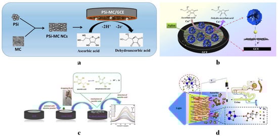
Figure 1. (a) Schematic representation of PSi-MC/GCE-based ascorbic acid electrochemical sensor. Reprinted with permission from Ref. [133]. Copyright 2022, Wiley-VCH GmbH. (b) Au-CEW-Cu3(PO4)2-GO HNFs-based electrochemical sensor for AA detection. Reprinted with permission from Ref. [57]. Copyright 2021, IOP Publishing Ltd. (c) The fabrication of Au-MoS2/NiO composite on GCE for AA detection. Reprinted with permission from Ref. [24]. Copyright 2022, the Korean Institute of Chemical Engineers. (d) Schematic diagram of Dy-SCN/FTO PEC sensor detecting AA. Reprinted with permission from Ref. [61]. Copyright 2022, MDPI.
Gold nanoparticles (Au NPs), chicken egg white (CEW), copper phosphate (Cu3(PO4)2), and graphene oxide (GO) were assembled together to form organic/inorganic hybrid nanoflowers (Au-CEW-Cu3(PO4)2-GO HNFs) (Figure 1b) [57]. When HNFs-modified GCE was used for electrochemical detection of AA, a linear range of 8–300 μM, the LOD of 2.67 μM, and a high sensitivity of 6.01 × 10⁻³ μA μM−1 cm−2 could be achieved due to many reasons, such as the high conductivity of Au NPs, the large specific surface area of GO, the strong electrocatalytic activity of HNFs, and so on.
Combined with the outstanding electrical activity of transition element oxides and the superior performance of nanostructures on the surface, Atacan et al. studied the electrochemical sensing of AA with Au-MoS2/NiO modified GCE (Figure 1c) [24]. The detection linear range and LOD were 2–50 μM and 0.13 μM, respectively, realizing the detection of AA in vitamin C tablets.
Zhao et al. designed a photoelectrochemical (PEC) sensor based on Dy-OSCN for the detection of AA (Figure 1d) [61]. Dy-OSCN single crystal has a regular microstructure, which accelerates the separation and transmission of internal carriers under light excitation, and can effectively improve the sensing performance. Under the best conditions, the linear range of AA sensing detection was from 7.94 μM to 1.113 × 10⁴ μM, and LOD was 3.35 μM. The analysis of human urine samples was realized.
1.8. Vitamin P Sensors
Rutin is an effective antioxidant polyphenol with an important protective effect [134,135]. In BR electrolyte, it was determined by differential pulse stripping voltammetry (DPSV) using GCE modified with graphene oxide nanocomposite reduced (rGO) by ferric oxide containing cerium (Ce) and chromium (Cr) [26]. The integration of Ce4+, Cr3+, and rGO has a synergistic effect on the sensor performance and excellent activity. Under the optimized construction conditions of the nanocomposite, the linear range of rutin detection was 0.075–12.00 μM, and the LOD was 52 nM.
Elancheziyan et al. developed a highly porous graphitic-activated carbon-coated iron oxide nanocomposite (Fe2O3/GAC) and then deposited it on an SPE to construct Fe2O3-GAC/SPE for electrochemical detection of rutin [27]. The synergistic effect of Fe2O3 and GAC can enhance the electrocatalytic reaction ability. A wide linear detection range (0.1–130 μM) could be obtained by DPV, and the LOD was 0.027 μM.
1.9. Simultaneous Analysis of Multiple Water-Soluble Vitamins
Since various vitamins in the body have their own unique and important functions, it is of great significance to detect and analyze them [136,137,138,139,140,141,142]. Simultaneous detection of multiple vitamins can greatly improve detection efficiency, which is also a research hotspot in recent years. Based on the requirements of the electrochemical detection environment, vitamin samples that can be detected at the same time basically have similar solubility. There are two common detection and discrimination methods for different water-soluble vitamins. One is to rely on the difference of electrochemical response signals of a specific electrode material to different vitamins, such as different detection peak positions. The other is to select or design electrode materials with selectivity for each of several target vitamins and then combine them in a certain means to detect and discriminate their mixtures.
In B vitamins, VB2 and VB6 play an essential role in the normal function of the body and are interrelated, so Buleandra tried to detect and analyze them simultaneously by electrochemical method [51]. Preliminary cyclic voltammetry analysis showed that VB2 underwent a quasi-reversible electron transfer reaction in BR buffer with a pH of 5.0, the oxidation of VB6 was irreversible, and their electrochemical properties were of obvious differences. Therefore, PGE and SWV could be used to determine both of them simultaneously. For VB2, the two linear concentration ranges of 1.00 × 10⁻⁷–5.00 × 10⁻⁵ M and 5.00 × 10⁻⁵–7.50 × 10⁻⁴ M were achieved, and for VB6, the linear range was 2.50 × 10⁻⁵–2.50 × 10⁻³ M. The detection limits of VB2 and VB6 were 7.38 × 10⁻⁸ M and 1.10 × 10⁻⁵ M, respectively. The method was further realized in the simultaneous determination of VB2 and VB6 in tablets. Using magnetite nanoparticles to improve the detection sensitivity of disposable electrochemical paper-based analysis equipment, Pereira et al. also realized simultaneous quantitative analysis of VB2 and VB6 [49]. The calibration plot for VB2 was in the range of 2.0 to 20.0 μM with a detection limit of 0.25 μM, and the calibration plot for VB6 was in the range of 0.2 to 2.0 mM with a detection limit of 29.5 μM.
Porada et al. used a nickel zeolite/carbon black modified GCE (NiZCBGCE) to determine VB2, VB9, VB12, and VB3 simultaneously [9]. Based on the enhancement of zeolite and carbon black on the vitamin reduction signal, the linear response range of vitamin detection was 0.008–0.24 mg L−1, 0.004–0.22 mg L−1, 0.003–0.1 mg L−1, and 0.15–10 mg L−1, and LOD was 2.3 μg L−1, 1.3 μg L−1, 1.092 μg L−1 and 45 μg L−1 for VB2, VB9, VB12, and VB3 respectively. Chitosan, in which nitrogen and sulfur co-doped graphene quantum dots are immobilized, was used to modify GCE for monitoring a variety of B vitamins [63]. Using the optimized parameters, vitamins B2, B6 and B12 were detected by SWV with detection limits of 0.30, 30.1, and 0.32 nM, respectively, and vitamins in energy drinks could be quantified.
2. Electrochemical Sensors for Fat-Soluble Vitamins
Like water-soluble vitamins, fat-soluble vitamins are essential nutrients in the human body and play an important role in human growth and development. It is also important to effectively detect their concentrations in the human body and supplementary nutrients. However, compared with water-soluble vitamins, there are relatively few electrochemical studies on fat-soluble vitamins. The use of various organic solvents requires higher chemical inertness of various devices, and the selection of organic materials is often limited by the physical and chemical properties of vitamins to be tested. Due to the non-polar characteristics of organic solutions, the charge movement is far inferior to the polar water environment. Therefore, the detection and exploration of various fat-soluble vitamins are still of great concern.
2.1. Vitamin D Sensors
Serum 25 hydroxyvitamin D (25-OHD) has been considered a new biomarker of vitamin D deficiency, and its concentration analysis is the focus of vitamin D-related electrochemical detection [143,144,145]. Immunological methods are often used for this purpose. Anusha et al. proposed a label-free impedimetric immunosensor, using a carbodiimide chemical method to covalently immobilize Ab-25-OHD antibody on GCN-b-CD@Au/GCE for specific recognition of 25-OHD in the serum [41]. The combination of antigen and antibody was detected by EIS, and the linear concentration range was 0.1–500 ng mL−1 with a LOD of 0.01 ng mL−1. Zirconia nanoparticles modified with L-cysteine functionalized gold (Cys-Au@ZrO2 NPs) were electrodeposited onto ITO substrate to enhance the electrochemical behavior, stability, and availability of covalent binding of functional groups and biomolecules [31]. Immunosensing analysis of 25-OHD3 could be realized by further modifying the electrode with its antibody (Ab-25-OHD3). Its sensitivity was increased to 2.01 μA ng−1 mL cm−2, and the linear detection range and LOD were 1–50 ng mL−1 and 3.54 ng mL−1, respectively. Polli et al. modified the graphite SPE with cysteamine-functionalized core-shell magnetic nanoparticles and then fixed the 25-OHD3 antibody through glutaraldehyde cross-linking [29]. Utilizing DPV, the linear detection range was between 7.4 and 70 ng mL−1, and the LOD was 2.4 ng mL−1.
Aptamers are also widely used in the highly sensitive and selective electrochemical analysis. Yin et al. fixed the DNA tetrahedron on the gold surface and then assembled the hairpin DNA probe-25-OHD3 complex [40]. Its electrochemical detection showed a wide linear range of 0.1–1000 nM, and the LOD was 0.026 nM. Park et al. prepared heterogeneous nanostructures composed of molybdenum disulfide and an electrochemically reduced graphene oxide composite to modify the electrode for amplifying the electrochemical signal [43]. Then, 1,2,4-triazol-3,5-dione (MB-TAD) labeled with methylene blue was modified on the electrode, where MB was used as the redox mediator to generate electrons, and TAD could combine with 25-OHD3 to form the aptamer/25-OHD3/MB-TAD complex. The binding of the aptamer to 25-OHD3 was detected by DPV. At the same time, 1,6-hexanedithiol was modified to avoid nonspecific adsorption and reduce the steric hindrance between aptamers. The linear range of 0.1–150 ng mL−1 could be obtained by electrochemical detection, and the LOD was 0.02 ng mL−1.
Similarly, as an important specificity analysis technology, molecular imprinting is also used in vitamin D determination. GCE was modified with CuCo2O4/nitrogen-doped carbon nanotubes and phosphorus-doped oxygraphene nanocomposites and then coated by electropolymerization of 25-OHD3 imprinted polypyrrole to construct a specific recognition electrochemical sensor [30]. Using ferricyanide as the signal mediator, the signal was significantly reduced after recombining 25-OHD3 on the electrode, and the linear sensing detection range and LOD were 0.002–10 μM and 0.38 nM, respectively.
Vitamin D (such as VD3) can be directly detected by electrochemical analysis in biological samples [146,147,148]. For example, Bora et al. used nitrogen doped carbon nanotubes to improve the hydrophilicity and specific surface area of the working electrode, thereby improving the electrochemical detection performance [55]. The VD3 sensor based on it could provide high performance in the concentration range of 0–10 nM, with a LOD of 16 pM, and the sensitivity value 0.000495 mA cm−2 nM−1 was achieved.
2.2. Vitamin E Sensors
Jashari et al. used the SWV method to simultaneously detect three natural isomers of tocopherol (α, γ, and δ) [32]. Under optimized conditions, there were similar linear detection ranges (3.0 × 10⁻⁶–1.0 × 10⁻⁵ M) for these isomers. For α, γ, and δ-tocopherol, quantification limits were 11.28, 2.70, and 3.67 × 10⁻⁶ M, and LODs were 3.72, 0.89, and 1.21 × 10⁻⁶ M, respectively. This method achieved the same detection effect as the popular chromatographic methods.
2.3. Vitamin K Sensors
Rostami-Javanroudi et al. modified PGE by electrodepositing silver nanoparticles and 2-amino-5chloro benzophenone [149]. Utilizing their electro-catalytic performance for the reduction of VK1, the sensor could achieve a linear detection range of 50–700 nM with an LOD of 16.58 nM, and it was applied in the quantifying of VK1 in human blood serums.
2.4. Simultaneous Analysis of Multiple Fat-Soluble Vitamins
Similar to water-soluble vitamins, it has outstanding application value in detecting multiple fat-soluble vitamins simultaneously [150,151]. Avan et al. modified GCE by using a β-cyclodextrin/multi-wall carbon nanotube and constructed an electrochemical sensor for the detection of various fat-soluble vitamins (VA, VD3, VE, and VK1) in an aqueous media of micellar solutions [65]. In the BR buffer at pH 5.0, the linear calibration curves of VA, VD3, VE, and VK1 could be obtained as 8–100, 0.8–60, 0.5–60, and 0.1–20 µM, respectively. The detection results have high reproducibility and selectivity.
3. Simultaneous Detection of Vitamins and Non-Vitamin Substances
In actual biomedical samples (such as serum), besides vitamins, there are often many other substances related to health. Simultaneous quantitative analysis of biological components concerned is an important goal of inspection science, and it is also the goal of many new electrochemical sensors [152,153,154,155,156,157,158,159,160].
Based on Pt-Pd nanoparticles/chitosan/nitrogen-doped graphene (N-Gra) nanocomposite, Luo et al. established an electroanalytical method for the simultaneous detection of AA, sulfite, and oxalic acid (OA) [46]. The nanocomposites have remarkable electrochemical activity for the electrooxidation of sulfite and OA. At the same time, the full separation of multiple oxidation peaks ensured high electrochemical resolution, allowing simultaneous quantification of three substances of interest. The linear ranges of AA, sulfite, and OA measured by DPV were 2–400 μM, 8–600 μM, and 1.5–500 μM, respectively. Accordingly, the LODs were 0.97 μM, 5.5 μM, and 0.84 μM, respectively.
GCE modified by Zn-Al layered double hydroxide (Zn-Al LDH) and methyl red (PMR) polymer film [47] has a high electrocatalytic activity for the oxidation of AA and aspirin (ASA). There were well-spaced anodic peaks in the CV sensing detection spectrum, which could simultaneously identify trace AA and ASA. The anode peak current and the concentration of AA and ASA were of good linear range in 0.10–53.17 μM, and LODs were 1.26 and 1.27 μM, respectively. Meanwhile, the quantification limits of AA and ASA were 4.21 and 4.25 μM, respectively. On the other hand, according to the DPV method, the LODs of AA and ASA oxidation were 0.47 and 0.21 μM, respectively.
The GCE modified with sunset yellow (SY) food dye could be used to simultaneously detect AA, dopamine (DA), and uric acid (UA) [39]. Under the optimal conditions, the linear ranges of AA, DA, and UA electrochemical detection were 7–320, 0.2–45, and 0.2–50 μM, respectively, the LODs were low (4.78, 0.12, 0.12 μM, respectively), and the stability was high.
The electrochemical sensor constructed by molecular imprinted polymer (MIP) could realize the simultaneous determination of paracetamol, AA, and uric acid mixture in pharmaceutical samples [73]. Good linearity at μM level, LOD (1–24 μM), and repeatability could be obtained.
A summary of various types of electrochemical vitamin sensors is shown in Table 1.
Table 1. A summary of electrochemical vitamin sensors.
| Vitamin | Electrode | Technique | Medium | Linear Range | LOD | Application | Ref. |
|---|---|---|---|---|---|---|---|
| VB2 | ZnO NPS-CPE | CV, SWV | PBS (pH 6) | 0.005–10 μM | 0.7 ± 0.01 nM | Beverage, milk | [12] |
| Bi2WO6 (PVP + NaOH)/GCE |
DPV | 0.05 M PBS (pH 7.0) | 0.03–457 μM | 3.65 nM | Almond milk, soymilk | [11] | |
| Ru/S-GCN-SPCE | CV, DPV | 0.1 M PBS (pH 7.0) | 0.003–75 μM 95–260 μM |
54.3 pM | Oral solution, syrup, tablets | [81] | |
| VB6 | IONCPE | DPV | 0.1 M PBS (pH 6.0) | 8.88–1000.0 μM | 9.06 μM | Urine, pharmaceuticals | [37] |
| Pt/β-CD-GR/PGE | DPV | 0.1 M PBS (pH 7.0) | 5–205 nM | 1.2 nM | Juice | [36] | |
| P-doped/PGE | DPV | 0.1 M PBS (pH 3.0) | 0.5–300 μM | 0.219 μM | Beverage | [90] | |
| VB9 | ZnFe2O4MNPs/SPE | DPV | 0.1 M PBS (pH 7.0) | 1.0–100.0 μM | 0.3 μM | Tablets, urine | [99] |
| GCE/f-MWCNT-Ni(OH)2-Si4Pic+Cl− | DPV | PBS (pH 7.0) | 0.5–26 μM | 0.095 μM | Dietary supplement, fortifier compound, wheat flour | [100] | |
| VB12 | Copper oxide- GUITAR |
LSV | Neutral (pH = 7) solutions |
0.15–7378 nM | 0.59 nM | Bacterial strains | [103] |
| Mn-CPE | SWV | Acetate buffer (pH 4.6) | 13.86–1500 ng L−1 | 4.34 ng L−1 | Tablets, dietary supplements | [48] | |
| VC | Pyrolytic graphite sheet | CV, SWV | KNO3 (pH 7.0) | 1.0–400 μM | 0.4 μM | Extract of cultivated arugula |
[50] |
| Gr/NiHCF | CV, Amp * | 0.01 M PBS (pH 7.4) | 1–16,280 μM | 0.25 μM | Supplements, fruit juices | [130] | |
| PBNPs/GCE | CV, Amp | PBS (pH 5) | 1–1100 μM | 0.47 μM | Domestic water, medicine samples | [131] | |
| VP | Co-BPDC-MOF/GCE | DPV, Amp | 0.1 M PBS (pH 7) | 0.5–1000 μM | 0.03 μM | BROMEZER tablet |
[134] |
| Co/ZIF-C/GCE | DPV | BR buffer (pH 2.0) | 0.1–30 μM | 22 nM | Tablets | [135] | |
| VD (25-OHD3) | Ab-25(OH)D3-Cys/Au/MoS2/FTO | DPV | 5 mM [Fe(CN)6]3−/4− + 0.9% KCl(pH 7.4) | 1 pg mL−1–100 ng mL−1 | 0.38 pg mL−1 | - | [143] |
| Glut/Au-Pt/APTES/FTO | DPV | 50 mM PBS (pH 7.4, 0.9% NaCl) + [Fe(CN)6]3−/4− | 0.1 pg mL−1–1 μg mL−1 | 0.49 pg mL−1 | Serum | [145] | |
| VD3 | Co-Ag/PANI-PPY/IL/GCE | CV, SWV, Amp |
PBS (pH 7) | 0.0125–22.5 μM | 0.0073 μM | Serum, urine | [146] |
| Paper-sensor | CV, SWV, Amp |
PBS (pH 7) | 0.025–0.125 μM | 0.015 μM | Serum, urine | [146] | |
| GQD-Au | EIS | 0.1 M PBS (pH 7.0) | 1–500 nM | 0.70 nM | Serum | [147] | |
| CuNPs-NiNPs@reduced-fullerene- C60/GCE |
CV, SWV |
PBS (pH 7.0) | 1.25–475 μM | 0.0025 μM | Serum, urine | [148] |
* Amp: Amperometry.
This entry is adapted from the peer-reviewed paper 10.3390/chemosensors10110494
This entry is offline, you can click here to edit this entry!