Environmental Pollution and the Risk of Metabolic Disorders: History
Please note this is an old version of this entry, which may differ significantly from the current revision.
Contributor: , , , ,

Metabolic disorders are a spectrum of diseases that affect normal metabolic functioning and regulation. More than 500 metabolic disorders exist, two of the most common being diabetes mellitus and obesity. Obesity is a multifactorial disorder involving the interaction of genetics, lifestyle and environment. It results in excessive adipose tissue deposition and is defined by a body mass index (BMI) greater than 30 kg/m2. In addition, obesity is the leading cause of metabolic syndrome and insulin resistance. Type 2 diabetes is characterized by an elevated blood glucose level, a chronic hyperglycemic state caused by a combination of pancreatic β-cell loss through apoptosis and insulin resistance in peripheral tissues, such as skeletal muscle. By contrast, type 1 diabetes is caused by autoimmune attack upon pancreatic β-cells, causing an almost complete loss of insulin production and secretion. Whilst a genetic predisposition can underlie the onset of type 1 diabetes, with particular loci of interest having been identified, environmental factors may also contribute. Well-established risk factors for type 2 diabetes and obesity are a sedentary lifestyle, poor nutrition, insulin resistance, environmental factors and genetics.

  • persistent organic pollutants
  • atmospheric pollution
  • obesity
  • type 1 diabetes
  • type 2 diabetes
  • gestational diabetes
  • metabolic disorders
  • heavy metals
  • adipogenesis

1. The Impact of Pollution on Obesity

1.1. The Development of Obesity

Obesity is an ongoing pandemic affecting many nations across the globe, with an exponential growth in prevalence over recent years. Obesity has many risk factors associated with its development, including genetic predisposition and environmental impacts. Three main mechanisms have been proposed for the link between pollution and obesity: physical inactivity, oxidative stress caused by pollutants and epigenetic modifications [1].
Rodent studies indicate that ambient air pollution behaves as an obesogenic factor, disrupting typical adipose function [1]. These ambient air pollutants alter specific metabolic functions, such as storage and breakdown of fats, leading to an increase in weight [2]. Although not conclusive, especially in humans, there is a growing field of research surrounding the impact of pollution due to the growth of cities, the increased density of urban areas and the overall global industrial shift over the years likely contributing to the alarming prevalence of metabolic disorders such as obesity and diabetes (Figure 1).
Figure 1. Effect of environmental pollution in developing metabolic disorders: rapid industrialization, toxic chemicals and a polluted environment generate a massive number of endocrine disruptor compounds (EDCs), for example, advanced glycation end products (AGEs), persistent organic pollutants (POPs) and metal pollutants, that affect the normal physiological functions in the human body. Disruption of cellular and physiological activities in response to EDCs increases the risk for development of obesity and diabetes.

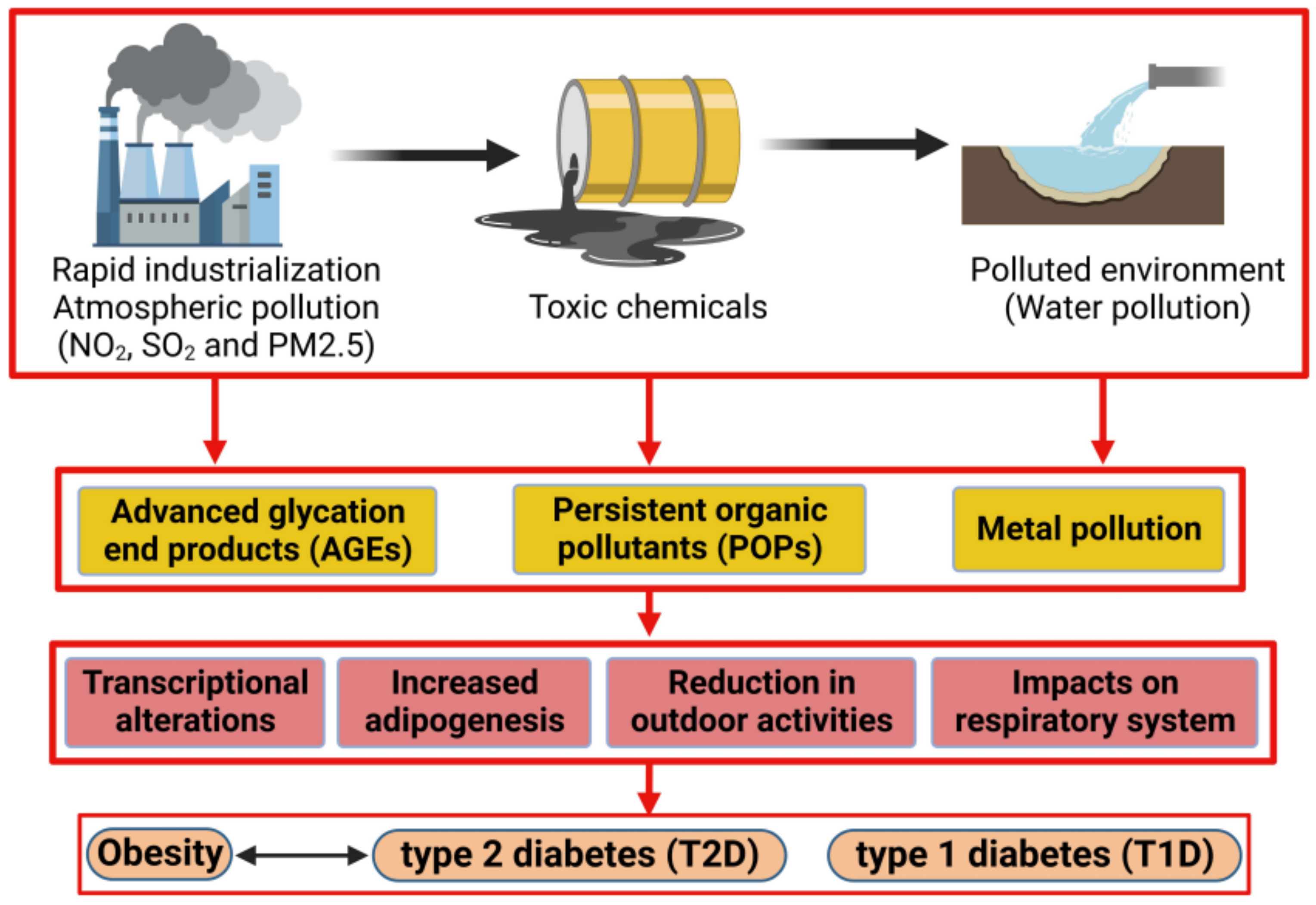
1.2. Advanced Glycation End Product (AGE) Impact on Obesity Prevalence

As many nations continue to industrialize, fast food and processed foods have become increasingly prevalent. This has drastically altered human dietary intake. Through the processing of foods, advanced glycation end products (AGEs) have become an important aspect of food consumption [3][4]. AGEs are produced when proteins, lipids or nucleic acids are glycated through the unnatural processing of foods that involves various chemicals used for increasing product storage life [3][4]. Common AGEs are pentosidine and N-[carboxymethyl]-lysine (CML), caused by the increased presence of glucose [5]. AGEs function by forming a link between the membrane of molecules and specific receptors known as receptors for advanced glycation end products (RAGEs) [6]. The ability of AGEs to bind to the basement membrane of the extracellular matrix alters cell function and structure [6]. The recruitment of RAGEs further changes the cell’s ability to function [6] (Figure 2). Through their binding, AGEs can alter collagen, laminin and vitronectin [6]. These three proteins are integral parts of vascular homeostasis; thus, AGEs have an impact microscopically and macroscopically [6]. The increase in processed foods has paralleled the timeline and growth of the obesity pandemic, and research has investigated whether there is a correlation between these AGEs and obesity. A study of 4245 participants found that individuals who consumed higher amounts of AGEs had increased abdominal obesity [3]. Rather than the quantity of food or the caloric intake, it was the consumption of AGEs that caused the increased adiposity [3].
Figure 2. Mechanism of persistent organic pollutant (POP)-induced obesity: (upper panel) illustration showing interaction of advanced glycation end products (AGEs) and receptor of AGE (RAGE) in cells (including adipocytes). AGE binds to RAGE expressed in cells as well as in the extracellular matrix (ECM), altering the structure of ECM and causing cellular damage. (Lower panel) diagram illustrating the effect of phthalate, DDT and PBDE in adipogenesis. Phthalate and DDT induce stem cells to differentiate to adipocytes. PBDE binds to the peroxisome proliferator-activated receptor-gamma (PPAR-γ) and enhances lipid accumulation. An upward red arrow indicates an increase and a downward red arrow indicates a decrease in the rate of the specified metabolic pathway.

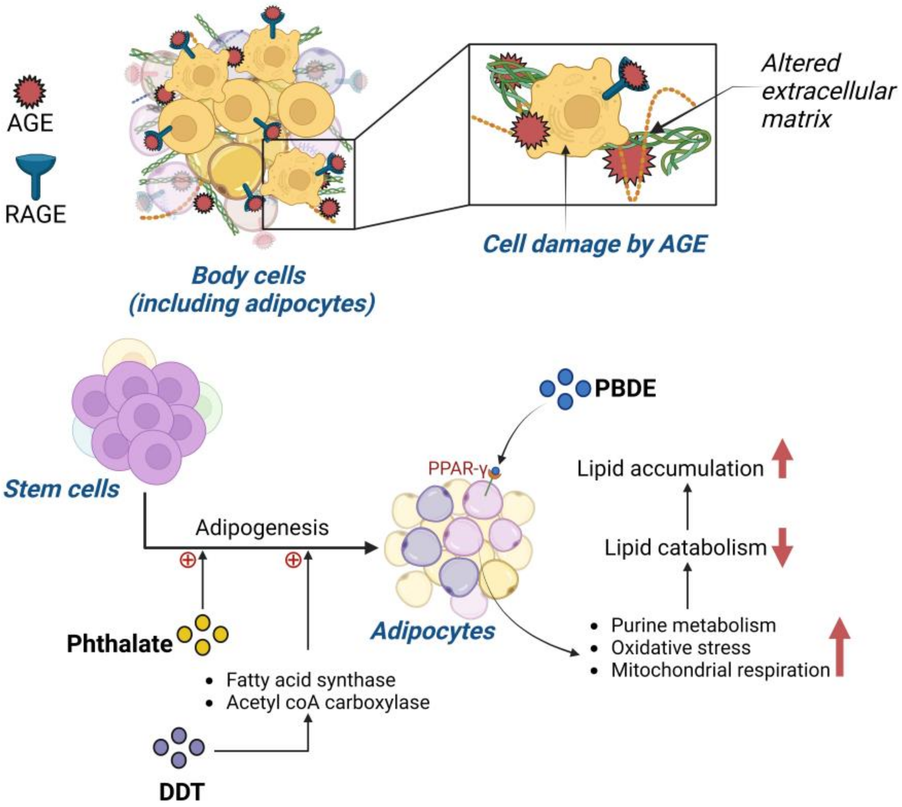
1.3. Impact of Persistent Organic Pollutants on Obesity

Bisphenol A has a link to obesity and body mass index (BMI), mainly through the alteration of adipogenesis regulators [7][8]. BPA leads to the increase in mRNA for 11b-hydroxysteroid dehydrogenase type 1 (HSD1), an enzyme affecting adipogenesis regulation [9]. In addition, bisphenol A binds to glucocorticoid receptors activating 11b-HSD1, accelerating the process of adipogenesis [9].
Due to the negative impact of BPA, other components used in plastics were evaluated for their effects on the human body. One of those components was phthalate and another was POP. Urinary content analysis found that increased phthalate metabolites were positively correlated to three factors: obesity, triglycerides and increased blood pressure [7][10]. A remarkable yet detrimental feature of phthalate function is the ability to promote adipogenesis from stem cells rather than osteoblast genesis, causing increased fat build-up rather than required bone quality improvement [7][11] (Figure 3). Phthalates, specifically mono-2ethylhexyl-phthalate, bind to PPAR-γ, a receptor that influences adipocyte differentiation [12]. PPAR-γ is a key regulatory receptor that influences adipogenesis through a transcriptional cascade [12]. Many genes associated with adipogenesis are activated and upregulated once PPAR-γ is activated. This receptor also contributes to adipocyte insulin sensitization, disrupting blood glucose homeostasis [13]. POPs found in plastic similarly impact human physiology and are worrisome as POP-infused plastics are widely used for storing water, food and other commonly used objects.
Another widely used POP is DDT. A study conducted on 298 individuals in Spain found that lipid DDT levels positively correlated with BMI. A more recent study on 429 adults found that DDT and its metabolites alter the fasted glucose state, further contributing to obesity development [7][14][15]. Laboratory investigations confirmed the potency of DDT and its adipogenicity effects; DDT was found to promote adipogenesis by increasing the presence of fatty acid synthase, acetyl-CoA carboxylase and lipid accumulation [7][14][16]. The main mechanism for these increases relates to DDT and its subsequent upregulation of PPAR-γ, leading to the transcriptional cascade influencing adipogenesis [14][17]. DDT has also been seen to have an influence on insulin resistance of adipocytes [14][17], thus leading to further dysregulation of lipid and carbohydrate metabolism, adipose tissue accumulation and metabolic disruption [14][17] (Figure 3).
A major concern is that the accumulation of PBDEs in adipose tissue closely correlates with insulin resistance and therefore the development of diabetes [7][14][18][19]. Laboratory studies have shown that obesogenic effects are correlated to the presence of PBDEs, specifically in the promotion of adipogenesis through lipid level alteration [7][14][20]. PBDE accumulation was found to have links to certain obesity biomarkers such as leptin and adiponectin, as well as increased PPAR-γ expression [7]. However, another hypothesized obesogenic mechanism for PBDE is through the activation of purine metabolism, with an increase in oxidative stress and increased mitochondrial respiration [7]. These three factors were seen to decrease lipid catabolism, allowing lipids to accumulate within adipose tissue [7].
Perfluorinated compounds, another form of POP, have been shown to be inducers of adipogenesis [7][14]. These compounds, like PBDEs, also play a role in the insulin signaling pathway, which raises concerns about their promotion of other metabolic disorders [7][14][21]. Perfluorinated compounds influence insulin-stimulated glucose uptake by an increase in both glucose transporter type 4 (GLUT4) transportation and insulin receptor substrate-1 (IRS1) [7]. PCBs have already been banned in many places due to their ability to imitate key hormones of the endocrine system [7][14][21]. A linear relationship between PCBs and waist circumference has been documented [7][14][22]. Lipid metabolism is another target of these chemicals, due to their accumulation in adipose tissue [7][14][22]. PCBs were found to accumulate specifically within lipid droplets. They have a role in regulating PPAR-γ as well as CCAAT/enhancer-binding protein-alpha, two receptors that are adipogenic factors [7]. Perfluorinated compounds influence insulin-stimulated glucose uptake by an increase in both GLUT4 transportation and IRS1 [7].
Another group of POPs in common use are the polycyclic aromatic hydrocarbons (PAHs). Investigation of 3189 urine samples from those exposed to PAHs found a positive association between body mass index (BMI), obesity and PAH concentration [7][14][23]. PAHs impair adipose tissue lipolysis, as well as affect PPAR-γ [23]. Reducing exposure to POPs, particularly in infants and children, is important in efforts to reduce the prevalence of obesity and other metabolic disorders [7][14][23].

1.4. Pollution and the Impact on Childhood Obesity

Several disorders have roots in the pre-natal environment as, when the pre-natal environment is disrupted, a higher prevalence of certain disorders occurs in later life. In studies researching the impact of pollutants on the pre-natal environment, pre-natal exposure to various types of pollutants can promote childhood obesity [24]. In three separate studies using Project Viva (a Boston-area pre-birth cohort including 1649 children), mothers living in high traffic density areas had children with a higher likelihood of developing obesity between the ages of six months and 10 years of age [24][25][26]. Smoking was another factor studied to determine the impact on the pre-natal environment and the potential risks it could impose on the future child. In two studies, one with 21,063 mother–child pairs and one with 13,188 singleton children, it was demonstrated that smoking or high exposure to secondhand smoking (tobacco) led to overweight children through evaluation of BMI at the ages of three and seven years [27][28]. A positive association between pollutants in the pre-natal environment and childhood obesity was found with polycyclic aromatic hydrocarbons (PAHs). Children with increased pre-natal exposure to PAH were seen to have an increased prevalence of obesity from 20.6% at age 5 to 33% at age 11 in comparison to those with no or minimal PAH exposure [29][30]. Post-natal early exposure to pollution was also shown to be detrimental to long-term metabolic health. When evaluating children at an average age of 6.6 years old, it was found that those living in high-density traffic areas had a 2.6 kg/m2 increase in their BMI compared to those in less dense areas at three-year follow up [31]. Whether due to pollution from vehicle exhausts or a pollution-induced reduction in physical activity, these children were put at metabolic risk from living near dense traffic [31]. A study in mice demonstrated that early life exposure to particulate matter could lead to many risk factors associated with obesity such as insulin resistance and accumulation of macrophages within adipose tissue [2]. A reduction in early life pollution exposure is important for reducing the prevalence of metabolic disorders occurring in later life.
Obesity’s prevalence continues its dramatic increase worldwide and it is critical to find a way to curb this epidemic. Lifestyle changes are key to reversing obesity, but these changes are challenging for patients to maintain. Nutritionally healthy meals and increased physical activity are the cornerstones of lifestyle modification. Exercise can “flush out” POPs, due to the progressive loss of adipose tissue, the major storage site of POPs [32]. Exercise is not only beneficial for weight loss and improving overall health, but also for reducing the impact of pollutants on the body through the mechanism noted above [32]. Obesity itself is a risk factor for the development of type 2 diabetes (T2D) and the continuing increase in obesity is reflected in the increasing prevalence of T2D.

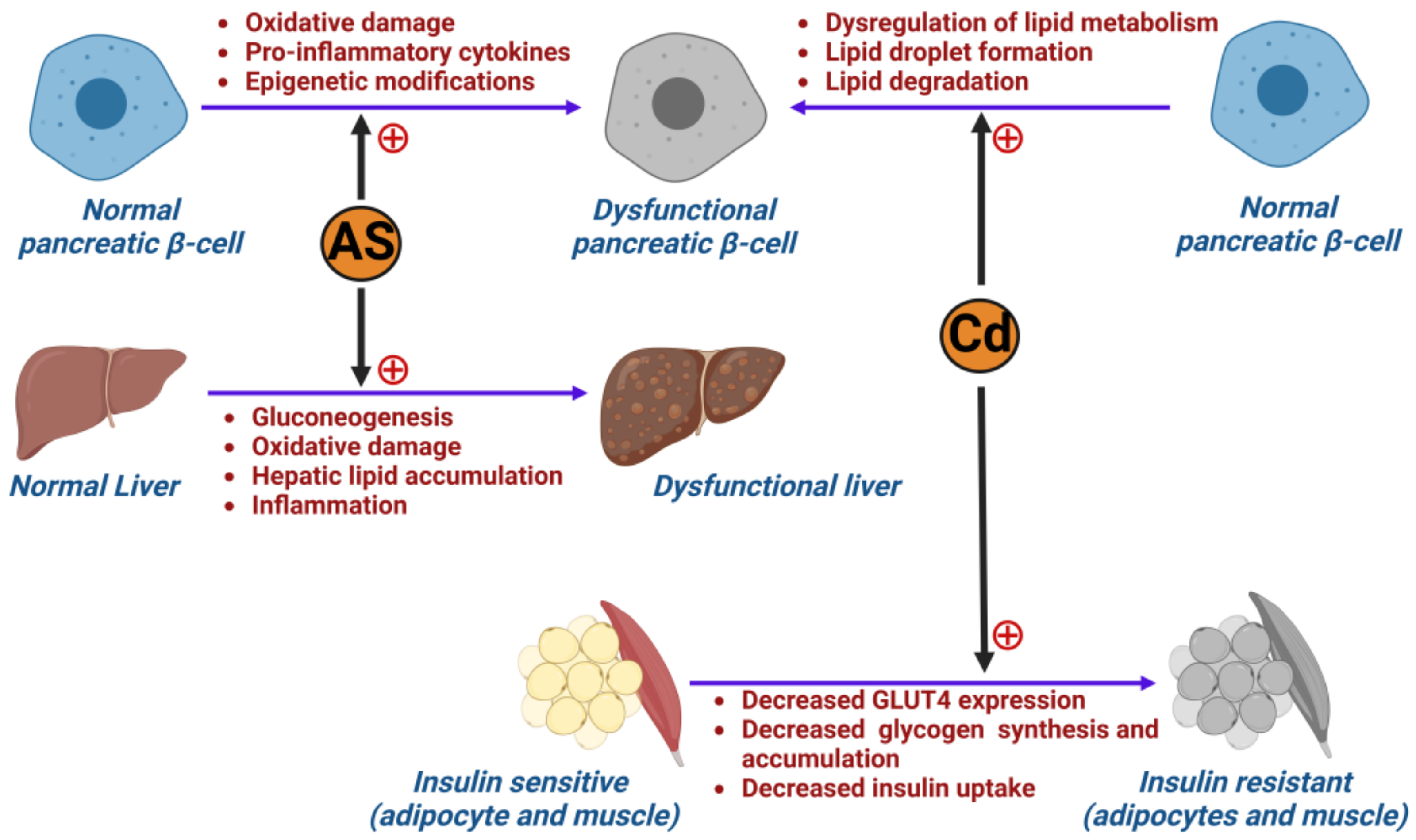
2. Impact of Pollution on Type 2 Diabetes Mellitus (T2D)

2.1. The Development of Type 2 Diabetes Mellitus

Characteristics of T2D are hyperglycemia, increased peripheral insulin resistance and increased lipid oxidation. Chronic hyperglycemia is due to an inability of the body to consistently break down the glucose required to keep up with energy requirements because of peripheral insulin resistance. Peripheral insulin resistance results in a consequent reduction in the uptake of glucose by insulin-dependent glucose transporters (GLUT4), resulting in an increase in circulating glucose, and subsequent lipid oxidation due to impaired glucose intake [33].
While a variety of research indicates a direct link between pollutants and the onset of diabetes in human populations, several studies have deduced the association of pollutants with risk factors such as hyperglycemia, peripheral insulin resistance, hepatotoxicity and metabolic disruption, that are frequent forerunners of T2D.

2.2. The Role of Metals in Inducing Type 2 Diabetes Mellitus

The heavy metal arsenic (As) has been posited to have negative pancreatic and hepatic effects (Figure 3). A review identified that arsenic plays a role in inducing diabetes by altering tumor necrosis factor-α (TNF-α), GLUT4 and mitogen-activated protein kinase (MAPK) [33]. In a rodent study conducted by Liu et al., the control group of mice was shown to be affected by inorganic arsenic through pancreatic β-cell dysfunction with increased gluconeogenesis and oxidative damage in the liver [34]. The group of diabetic mice within the same study were shown to have even worse glucose tolerance. Ongoing arsenic exposure was also found to impact liver function as it induces hepatic lipid accumulation [35] and triggers expression of genes involved in the onset of inflammation within the liver [36]. Hyperglycemia induced by impaired liver function instigates oxidative damage and decreases glycogenesis. As such, arsenic exposure can play a role in the development of ROS through the inhibition of mitochondrial function, onset of lipid peroxidation and further mitochondrial damage [37]. Oxidative stress on the pancreas is also triggered by accumulation of arsenic within the body [38][39], resulting in diminished insulin sensitivity and β-cell apoptosis [39]. Arsenic exposure in a rodent study conducted by Khan et al. also resulted in alterations in the methylation pattern of the glucose transporter 2 gene (GLUT2), and changes to the insulin gene (INS, coding for insulin) and pancreatic and duodenal homeobox-1 (Pdx1, that regulates pancreatic cell proliferation) [35]. By altering the methylation pattern of GLUT2, a subsequent increase in GLUT2 expression was induced [38]. However, after exposure to sodium arsenite, INS and Pdx1 expression was decreased. It should also be noted that arsenic has implications in affecting fetuses through transplacental exposure. Therefore, not only were mothers seen to be negatively impacted by exposure, but their offspring as well, with future metabolic dysfunction after birth as arsenic accumulates in the liver of neonates [40]. Thus, these compounding factors would induce insulin resistance and further risk for development of T2D [36][38].
Figure 3. Effect of metal pollution in developing diabetes: illustration showing how arsenic (As) and cadmium (Cd) cause dysfunction in pancreatic β-cells, liver and insulin-sensitive tissues (adipocytes and muscle), leading to the development of diabetes.
As mentioned above, arsenic is not the sole metal found to have toxic effects over long-term exposure. The persistence of human activities, such as agriculture and industrialization practices, lifestyle and diet, has caused exposure to cadmium (Cd) [39][41] (Figure 3). Levels of exposure vary widely from country to country, with countries in Eastern Asia experiencing significantly higher exposure versus Western countries (USA and European Union) as reported by the WHO [39][42]. The exact mechanism of diabetogenesis resulting from cadmium exposure has not yet been fully elucidated. However, several studies suggest that it plays a prominent role in affecting the pancreas and the production of insulin. In a mouse study conducted by Hong et al., Cd application to MIN6 cells (a mouse insulinoma-derived cell line that serves as a model of pancreatic β-cell function) led to dysfunction in lipid metabolism and was linked to pancreatic β-cell dysfunction and death [42]. The study concluded that exposure could lead to accumulated lipids in pancreatic β-cells and cause dyslipidemia in vivo. This was demonstrated by a distinct increase in lipid droplet formation in pancreatic β-cells, with decreased lipid degradation. As Cd accumulates in the β-cells, it decreases their ability to release insulin and therefore results in increased circulating blood glucose levels [43]. Cd exposure has also reportedly caused pancreatic β-cell dysfunction via inducing hyperglycemia and affecting the lipid oxidation of pancreatic cells [41]. Some studies have also indicated that the risk of developing T2D in Cd-exposed patients is increased in those under the age of 50 via increasing the likelihood for peripheral insulin resistance [44]. This was demonstrated in a rodent study [45] where, following administration of CdCl2, expression of GLUT4 in skeletal muscle and adipocytes was decreased, resulting in further decreased glycogen synthesis and insulin-dependent uptake of glucose [45][46]. In a meta-analysis comparing high to low amounts of exposure, higher exposure translated into higher risk for developing T2D (odds ratio 1.38, 95% confidence interval 1.12–1.71) [47]. Whilst Tinkov et al. conducted a meta-analysis that found strong associations between Cd exposure and the development of pre-diabetes (p < 0.01) [47], other studies have shown no association between Cd and diabetes (p = 0.347) [48]. These conflicting findings may be due to the differing exposure markers (urine, nails, hair, HbA1c testing) used in the studies to determine Cd exposure. However, laboratory studies included in this meta-analysis concluded that Cd exposure heightened insulin resistance and, therefore, augments T2D incidence [49][50]. To support the limited studies available, more research is needed to determine whether there is a definitive link between Cd exposure and T2D.

2.3. Air Pollution and Its Impact on Developing Type 2 Diabetes (T2D)

The pathogenesis of T2D is directly associated with insulin resistance, which can be triggered by systemic inflammation and oxidative stress and has been postulated to be triggered by exposure to air pollution. In a mouse study conducted by Sun et al., 24 weeks of exposure to PM2.5 induced insulin resistance, increased visceral adipose tissue and caused immune response dysregulation [51]. The Study on the Influence of Air Pollution on Lung Function, Inflammation and Aging (SALIA) was a cohort study of women aged 54–55 years old who lived in some of Germany’s most densely polluted districts [52][53]. Consecutive cross-sectional surveys were conducted between 1985 to 1994 to identify the effects of industrial and traffic-related pollution exposure [52][53]. A follow up investigation conducted by Krämer et al. on the SALIA cohort included 1775 participants from the prior study to whom they administered self-reported questionnaires. In doing so, they were able to identify an association between NO2 exposure from traffic pollution and T2D with an incidence of 10.5% onset T2D cases [54]. Additionally, a cross-sectional study of 374 participants aged 10–18 years old conducted in Iran indicated that, with ongoing increased exposure to traffic-based and industrial air pollution (resulting in increased PM10 exposure), homoeostasis model assessment model-insulin resistance (HOMA-IR) results were elevated and concluded there to be a direct association of air pollution with insulin resistance [54][55]. While many studies have identified a positive association with pollution risk factors and development of T2D, there is a paucity of mechanistic information as to how air pollution causes T2D.

3. The Impact of Pollution on Type 1 Diabetes Mellitus (T1D)

Type 1 diabetes mellitus (T1D) is characterized by autoimmune destruction of the pancreatic β-islet cells, the cells responsible for the production of insulin. The underlying cause of this autoimmunity is still not fully understood although certain genetic predispositions to T1D development are known. Certain factors that work in concert with the underlying genetic predisposition to promote disease onset include epigenetic modifications, pancreatic β-cell destruction and inflammation [52][53]. Epigenetic alterations, transcriptional alterations and the impact of gene expression are all areas where environmental pollutants can impact upon a genetic predisposition [52][53]. Research is actively being conducted to evaluate how pollution impacts metabolic disorders, but more research directed at pollution and T1D specifically is needed.

3.1. Impact of Pollution on Pancreatic β-Islet Cells

Pollutants seem to have an affinity for pancreatic β-islet cells both during early development and once the organ is fully formed [56]. In a study investigating the impact of dioxin on pancreatic β-cells, it was reported that β-cells were unable to secrete insulin when subjected to dioxin [56]. The cause was determined to be an alteration in expression of certain genes required for the maintenance of insulin levels and β-cell function that included glucokinase (Gck), B-cell lymphoma-extra-large (Bcl-xL), Pdx1, forkhead box O1 (FoxO1), inositol-requiring enzyme 1 (IRE1), GLUT2 and musculoaponeurotic fibrosarcoma oncogene family A (MafA) [56].
Research into the impact of cadmium, a metal pollutant, on the pancreas has been minimal to date [42]. When testing the impact of cadmium on pancreatic β-cells of mice, a multifactorial effect was seen [42]. Cadmium caused increased lipid accumulation in β-cells due to upregulation of cellular lipogenesis [42]. Cadmium also provoked a proinflammatory response leading to increased production of proinflammatory cytokines, a risk factor for diabetes development [42]. These key aspects lead to β-islet cell dysfunction or death whether in vitro or in vivo, further accelerating the potential for diabetes development.

3.2. Impact of Pollution on Childhood Type 1 Diabetes Mellitus

Although the onset of type 1 diabetes (T1D) can occur throughout life, it frequently occurs in children. Pollution studies have primarily investigated the impact of toxic airborne particles on T1D in children. In a study analyzing data from 19 European countries, there was a positive association between pollution exposure and the onset of T1D for individuals in 18 of those countries [57]. The target demographic for this research was individuals diagnosed with T1D from the age of 0–15 years [57]. Emission information was gathered from the European Environmental Agency to evaluate several toxic emissions because it is recognized that individuals experience a plethora of toxins within the atmosphere concurrently rather than in isolation [57]. Findings from this article are important as it highlights the relationship between pollution and T1D and the potential that pollution reduction could lower the prevalence of this disorder.
To further emphasize the early impact of pollution on children and T1D, a study compared ozone exposure in T1D and healthy children. Children with T1D had a higher pre-diagnosis exposure to ozone than healthy children [58]. However, the key concept highlighted by this research was that certain pollutants increase the likelihood of T1D onset before the age of 5 years [58]. Sulfur dioxide (SO2) exposure was associated with a later onset of diabetes versus those with early-onset disease [58]. Therefore, ozone has been hypothesized to be a pollutant linked to the increased earlier incidence of diabetes amongst children [58]. This is of great concern, as ozone levels and other atmospheric pollutants have risen in recent years, despite the current ongoing efforts to curb atmospheric pollution levels.
Atmospheric pollution is not the only form of pollution affecting younger populations. The presence of persistent organic pollutants (POPs) is another area of concern for T1D development. When studying 442 youths for the impact of POPs, with a focus on their impact on β-cells, a clear relationship was found [59]. Initially, the study investigated the odds ratio of T1D development in the presence of POPs and found that those subjected to dichlorodiphenyldichloroethylene (DDE), a metabolite of the POP DDT, had an increased chance of T1D development [59]. When POPs were introduced to isolated β-cells, after just two days the cells were non-viable [59]. Additionally, the presence of POPs shut down insulin production due to alterations in Ins1 and Ins2 mRNA expression [59].
When examining phthalate exposure in mothers, the expression of Pdx1, a gene required for β-cell production, was downregulated [60][61]. Pdx1 not only has a role in β-cell production, but also in mitochondrial function [60][61]. During pancreas development, large quantities of energy are required for β-cell development and, with reduced functionality of mitochondria, increased production of ROS occurs [60][61]. Oxidative stress damages mitochondrial DNA and, later in life, is associated with adult-onset diabetes [60][61]. The presence of this POP found in plastic not only causes a reduction in β-cell production but also has a detrimental impact on mitochondrial function. Pre-natal and childhood periods are crucial for the growth and development of a healthy pancreas [62][63]; environmental impacts before and after birth can be detrimental in both the short and long term.
The work presented here shows that there are risk factors other than genetics associated with the onset and progression of type 1 diabetes. More work needs to be carried out to evaluate how to reduce the effect of these pollutants and reduce their impact on future generations. Pollutants have a clear and susceptible target in pancreatic β-cells by targeting critical genes. Despite T2D being more conventionally recognized as having environmental risk factors, more work must be conducted to understand how pollution causes an increase in T1D incidence as well. Though limited, the available literature has highlighted the relationship between POPs and ambient air pollution and T1D.

4. Impact of Pollution on Gestational Diabetes (GDM)

4.1. The Development of Gestational Diabetes

During pregnancy, the placenta secretes hormones to sustain the fetus, impacting insulin resistance around the 20th to 24th week of gestation. In normal conditions, the pancreas counteracts this phenomenon by increasing the production of insulin through β-cell expansion [64][65][66]. However, when underlying β-cell dysfunction and impaired insulin secretion are present, the mother cannot maintain glucose homeostasis [64][67] and gestational diabetes mellitus (GDM) results. GDM has significant short- and long-term consequences on the mother’s and offspring’s health [67]. The fetus’ primary energy source is glucose, thus making the placenta very sensitive to the prevailing hyperglycemia of GDM [68]. This intrauterine exposure increases the probability of macrosomia, congenital disabilities, stillbirth, developmental delays, pre-diabetes and obesity [64][69][70][71]. When uncontrolled, GDM causes high blood pressure, increased cardiometabolic diseases and a greater incidence of T2D development later in life despite the glucose intolerance normally regularizing in the mother after delivery [64][72]. Indeed, according to the CDC, around 50% of people with GDM develop T2D, thus contributing to the worldwide epidemic. GDM is now one of the most common metabolic disorders seen during pregnancy [73], with an incidence rate of up to 28% [64]. In 2017, 204 million people worldwide were diagnosed with GDM, with projections to reach 308 million by 2045 [64]. In the USA, the prevalence increased from 4.6% in 2006 to 8.2% ten years later [74]. Mainland China’s incidence of GDM was 14.8% between 2010 and 2017 [75]; in the Gulf Cooperation Council (GCC) countries, the prevalence increased by 4% during the last 20 years to reach 15.9% today. In those countries, the women most at risk were those who were overweight or obese, over 30 years old and those delivering their baby through C-section [76]. Pregnant women in Europe experience have a significant incidence of GDM (11%), with pregnant women in Eastern European countries experiencing the highest prevalence (31.5%) [77].

4.2. Association between Particulate Matter (PM) and Gestational Diabetes (GDM)

GDM’s well-defined risk factors cannot fully explain its rapid increase worldwide in recent decades. This has led to a growing interest in experimental and epidemiology studies to gain insight into the correlation between pollutant exposure and GDM [78]. Unfortunately, the results are inconclusive, and no direct correlation has been established. Ambient air pollution exposure has been hypothesized to increase insulin resistance inciting metabolic dysfunction, and different pathways have been proposed to explain the association.
Firstly, exposure to PM2.5 and NO could accelerate β-cell dysfunction in already susceptible pregnant women [72]. Secondly, PM could lead to altered leptin and adiponectin concentrations, regulators of neurohormonal metabolic control [72]. The reduction of adiponectin and leptin has been demonstrated in a study involving long-term exposure to PM2.5 in C57BL/6 mice [2]. Lavigne et al. demonstrated that exposure during pregnancy to PM2.5 resulted in an 11% increase in adiponectin levels, and NO2 exposure resulted in a 13% increase in adiponectin levels [79]. The proposed mechanism causing the imbalance of these adipokines involves an inflammatory response in adipose tissues following PM exposure. Maternal exposure to air pollution has also been linked to increased local or systemic inflammatory responses, causing complications during pregnancy [72].
Furthermore, metals, sulfur and organic components comprising PM have also been associated with stimulating oxidative damage through ROS production in the mitochondria, formation of AGEs, activation of protein kinase C, reduction of GLUT4 and translocation of nuclear factor kappa B (NF-κB) subunit 1 into the nucleus, thereby generating an inflammatory response that impacts insulin signaling [72]. Air pollution could also increase gut permeability by altering the gut microbiota, thereby inducing the transport of inflammatory mediators from the gut into the bloodstream [72]. Additionally, a positive association has been found between PM2.5 exposure up to 12 weeks prior to conception and fasting glucose levels, thus increasing the risk for GDM in 11,639 women in China from 2016 to 2017 [69].
Furthermore, it has been postulated that pesticides increase diabetes by increasing the accumulation of acetylcholine which causes an increase in glucose [80].
A positive association was found between the total serum concentrations of nine PCB congeners and, more significantly, with eight PBDE congeners and GDM in a study involving seventy pregnant Iranian women with no family history of diabetes between the ages of 16 and 40 years in their third trimester [81]. Another study assessed the effect of exposure to PCBs and pesticides (hexachlorobenzene, HCB, and a DDT metabolite (dichlorodiphenyldichloroethane, DDE)) in the first trimester in 939 women in Greece [82]. The study found a strong positive association between PCB serum concentration and the probability of GDM. No statistically significant results were found for DDE and HCB. A prospective cohort study involving 2292 pregnant women in their 8th to 11th week of gestation without significant comorbidities demonstrated an association between serum levels of heavily chlorinated PCBs and PBDEs (47 and 154) and GDM [83].
Conversely, a study measured serum concentrations of certain POPs in the first trimester of 1274 Canadian pregnancies with no pre-existing diabetes and found no evidence of an association between PCBs and GDM [80]. An inverse relationship between urine concentrations of pesticides (dimethylphosphate (DMP) and dimethylthiophosphate (DMTP)) and GDM was observed, a result as perplexing as it is unique, which has not been reproduced in other studies to date.
The wide disparity of results is the most significant limitation of these studies on GDM. Gestational diabetes has multifactorial risk factors, and it is hard to isolate all confounding factors to determine the direct effects of exposure of different concentrations of the POPs. Adding further complication to identifying environmental pollutants as causative, a mixture of the many comorbidities, family history of T2D, genetic predisposition, lifestyle, physical activity status, overweight/obesity status and smoking habits all affect the incidence of GDM.

This entry is adapted from the peer-reviewed paper 10.3390/ijms24108870

References

  1. Seo, M.Y.; Kim, S.-H.; Park, M.J. Air pollution and childhood obesity. Clin. Exp. Pediatr. 2020, 63, 382–388.
  2. Xu, X.; Yavar, Z.; Verdin, M.; Ying, Z.; Mihai, G.; Kampfrath, T.; Wang, A.; Zhong, M.; Lippmann, M.; Chen, L.-C.; et al. Effect of Early Particulate Air Pollution Exposure on Obesity in Mice. Arterioscler. Thromb. Vasc. Biol. 2010, 30, 2518–2527.
  3. Mirmiran, P.; Hadavi, H.; Mottaghi, A.; Azizi, F. Advanced glycation end products and risk of general and abdominal obesity in Iranian adults: Tehran lipid and glucose study. Med. J. Islam. Repub. Iran 2019, 33, 21.
  4. Uribarri, J.; Woodruff, S.; Goodman, S.; Cai, W.; Chen, X.; Pyzik, R.; Yong, A.; Striker, G.E.; Vlassara, H. Advanced glycation end products in foods and a practical guide to their reduction in the diet. J. Am. Diet. Assoc. 2010, 110, 911–916.e12.
  5. Singh, R.; Barden, A.; Mori, T.; Beilin, L. Advanced glycation end-products: A review. Diabetologia 2001, 44, 129–146.
  6. Goldin, A.; Beckman, J.A.; Schmidt, A.M.; Creager, M.A. Advanced Glycation End Products. Circulation 2006, 114, 597–605.
  7. Aaseth, J.; Javorac, D.; Djordjevic, A.; Bulat, Z.; Skalny, A.; Zaitseva, I.; Aschner, M.; Tinkov, A. The Role of Persistent Organic Pollutants in Obesity: A Review of Laboratory and Epidemiological Studies. Toxics 2022, 10, 65.
  8. Hong, S.-H.; Sung, Y.-A.; Hong, Y.S.; Ha, E.; Jeong, K.; Chung, H.; Lee, H. Urinary bisphenol A is associated with insulin resistance and obesity in reproductive-aged women. Clin. Endocrinol. 2017, 86, 506–512.
  9. Wang, J.; Sun, B.; Hou, M.; Pan, X.; Li, X. The environmental obesogen bisphenol A promotes adipogenesis by increasing the amount of 11β-hydroxysteroid dehydrogenase type 1 in the adipose tissue of children. Int. J. Obes. 2013, 37, 999–1005.
  10. Amin, M.M.; Ebrahimpour, K.; Parastar, S.; Shoshtari-Yeganeh, B.; Hashemi, M.; Mansourian, M.; Poursafa, P.; Fallah, Z.; Rafiei, N.; Kelishadi, R. Association of urinary concentrations of phthalate metabolites with cardiometabolic risk factors and obesity in children and adolescents. Chemosphere 2018, 211, 547–556.
  11. Chiu, C.-Y.; Sun, S.-C.; Chiang, C.-K.; Wang, C.-C.; Chan, D.-C.; Chen, H.-J.; Liu, S.-H.; Yang, R.-S. Plasticizer di(2-ethylhexyl)phthalate interferes with osteoblastogenesis and adipogenesis in a mouse model. J. Orthop. Res. Off. Publ. Orthop. Res. Soc. 2018, 36, 1124–1134.
  12. Bimonte, V.M.; Besharat, Z.M.; Antonioni, A.; Cella, V.; Lenzi, A.; Ferretti, E.; Migliaccio, S. The endocrine disruptor cadmium: A new player in the pathophysiology of metabolic diseases. J. Endocrinol. Investig. 2021, 44, 1363–1377.
  13. Hurst, C.H.; Waxman, D.J. Activation of PPARalpha and PPARgamma by environmental phthalate monoesters. Toxicol. Sci. Off. J. Soc. Toxicol. 2003, 74, 297–308.
  14. Yang, C.; Kong, A.P.S.; Cai, Z.; Chung, A.C.K. Persistent Organic Pollutants as Risk Factors for Obesity and Diabetes. Curr. Diab. Rep. 2017, 17, 132.
  15. Henríquez-Hernández, L.A.; Luzardo, O.P.; Valerón, P.F.; Zumbado, M.; Serra-Majem, L.; Camacho, M.; González-Antuña, A.; Boada, L.D. Persistent organic pollutants and risk of diabetes and obesity on healthy adults: Results from a cross-sectional study in Spain. Sci. Total Environ. 2017, 607–608, 1096–1102.
  16. Pesta, M.; Cedikova, M.; Dvorak, P.; Dvorakova, J.; Kulda, V.; Srbecka, K.; Muller, L.; Bouchalova, V.; Kralickova, M.; Babuska, V.; et al. Trends in gene expression changes during adipogenesis in human adipose derived mesenchymal stem cells under dichlorodiphenyldichloroethylene exposure. Mol. Cell. Toxicol. 2018, 14, 369–379.
  17. Endocrine-Disrupting Chemicals—UpToDate. Available online: https://www.uptodate.com/contents/endocrine-disrupting-chemicals?search=EDCs%20and%20reproduction&source=search_result&selectedTitle=1~150&usage_type=default&display_rank=1 (accessed on 1 February 2023).
  18. Lim, J.-S.; Lee, D.-H.; Jacobs, D.R. Association of brominated flame retardants with diabetes and metabolic syndrome in the U.S. population, 2003–2004. Diabetes Care 2008, 31, 1802–1807.
  19. Helaleh, M.; Diboun, I.; Al-Tamimi, N.; Al-Sulaiti, H.; Al-Emadi, M.; Madani, A.; Mazloum, N.A.; Latiff, A.; Elrayess, M.A. Association of polybrominated diphenyl ethers in two fat compartments with increased risk of insulin resistance in obese individuals. Chemosphere 2018, 209, 268–276.
  20. Yang, C.; Wong, C.-M.; Wei, J.; Chung, A.C.K.; Cai, Z. The brominated flame retardant BDE 47 upregulates purine metabolism and mitochondrial respiration to promote adipocyte differentiation. Sci. Total Environ. 2018, 644, 1312–1322.
  21. Inoue, K.; Goto, A.; Sugiyama, T.; Ramlau-Hansen, C.H.; Liew, Z. The Confounder-Mediator Dilemma: Should We Control for Obesity to Estimate the Effect of Perfluoroalkyl Substances on Health Outcomes? Toxics 2020, 8, 125.
  22. Lee, D.-H.; Lee, I.-K.; Porta, M.; Steffes, M.; Jacobs, D.R. Relationship between serum concentrations of persistent organic pollutants and the prevalence of metabolic syndrome among non-diabetic adults: Results from the National Health and Nutrition Examination Survey 1999–2002. Diabetologia 2007, 50, 1841–1851.
  23. Scinicariello, F.; Buser, M.C. Urinary polycyclic aromatic hydrocarbons and childhood obesity: NHANES (2001–2006). Environ. Health Perspect. 2014, 122, 299–303.
  24. Fleisch, A.F.; Luttmann-Gibson, H.; Perng, W.; Rifas-Shiman, S.L.; Coull, B.A.; Kloog, I.; Koutrakis, P.; Schwartz, J.D.; Zanobetti, A.; Mantzoros, C.S.; et al. Prenatal and early life exposure to traffic pollution and cardiometabolic health in childhood. Pediatr. Obes. 2017, 12, 48–57.
  25. Fleisch, A.F.; Rifas-Shiman, S.L.; Koutrakis, P.; Schwartz, J.D.; Kloog, I.; Melly, S.; Coull, B.A.; Zanobetti, A.; Gillman, M.W.; Gold, D.R.; et al. Prenatal exposure to traffic pollution: Associations with reduced fetal growth and rapid infant weight gain. Epidemiol. Camb. Mass 2015, 26, 43–50.
  26. Fleisch, A.F.; Aris, I.M.; Rifas-Shiman, S.L.; Coull, B.A.; Luttmann-Gibson, H.; Koutrakis, P.; Schwartz, J.D.; Kloog, I.; Gold, D.R.; Oken, E. Prenatal Exposure to Traffic Pollution and Childhood Body Mass Index Trajectory. Front. Endocrinol. 2018, 9, 771.
  27. Wen, X.; Shenassa, E.D.; Paradis, A.D. Maternal smoking, breastfeeding, and risk of childhood overweight: Findings from a national cohort. Matern. Child Health J. 2013, 17, 746–755.
  28. Hawkins, S.S.; Cole, T.J.; Law, C. Millennium Cohort Study Child Health Group An ecological systems approach to examining risk factors for early childhood overweight: Findings from the UK Millennium Cohort Study. J. Epidemiol. Community Health 2009, 63, 147–155.
  29. Rundle, A.G.; Gallagher, D.; Herbstman, J.B.; Goldsmith, J.; Holmes, D.; Hassoun, A.; Oberfield, S.; Miller, R.L.; Andrews, H.; Widen, E.M.; et al. Prenatal exposure to airborne polycyclic aromatic hydrocarbons and childhood growth trajectories from age 5-14 years. Environ. Res. 2019, 177, 108595.
  30. Rundle, A.; Hoepner, L.; Hassoun, A.; Oberfield, S.; Freyer, G.; Holmes, D.; Reyes, M.; Quinn, J.; Camann, D.; Perera, F.; et al. Association of childhood obesity with maternal exposure to ambient air polycyclic aromatic hydrocarbons during pregnancy. Am. J. Epidemiol. 2012, 175, 1163–1172.
  31. Jerrett, M.; McConnell, R.; Wolch, J.; Chang, R.; Lam, C.; Dunton, G.; Gilliland, F.; Lurmann, F.; Islam, T.; Berhane, K. Traffic-related air pollution and obesity formation in children: A longitudinal, multilevel analysis. Environ. Health 2014, 13, 49.
  32. Lee, Y.-M.; Jacobs, D.R., Jr.; Lee, D.-H. Persistent Organic Pollutants and Type 2 Diabetes: A Critical Review of Review Articles. Front. Endocrinol. 2018, 9, 712.
  33. El-Sikaily, A.; Helal, M. Environmental pollution and diabetes mellitus. World J. Meta-Anal. 2021, 9, 220–326.
  34. Liu, S.; Guo, X.; Wu, B.; Yu, H.; Zhang, X.; Li, M. Arsenic induces diabetic effects through beta-cell dysfunction and increased gluconeogenesis in mice. Sci. Rep. 2014, 4, 6894.
  35. Rajak, S.; Raza, S.; Tewari, A.; Sinha, R.A. Environmental Toxicants and NAFLD: A Neglected yet Significant Relationship. Dig. Dis. Sci. 2022, 67, 3497–3507.
  36. Kalo, M.B.; Rezaei, M. In vitro toxic interaction of arsenic and hyperglycemia in mitochondria: An important implication of increased vulnerability in pre-diabetics. Environ. Sci. Pollut. Res. Vol. 2022, 29, 28375–28385.
  37. Hill, D.J. Impact of the exposome on the development and function of pancreatic β-cells. Mol. Aspects Med. 2022, 87, 100965.
  38. Khan, F.; Hodjat, M.; Rahimifard, M.; Nigjeh, M.N.; Azizi, M.; Baeeri, M.; Bayrami, Z.; Gholami, M.; Hassani, S.; Abdollahi, M. Assessment of arsenic-induced modifications in the DNA methylation of insulin-related genes in rat pancreatic islets. Ecotoxicol. Environ. Saf. 2020, 201, 110802.
  39. WHO. Preventing Disease Through Healthy Environments. Exposure to Cadmium: A Major Public Health Concern; WHO: Geneva, Switzerland, 2019; Available online: https://apps.who.int/iris/bitstream/handle/10665/329480/WHO-CED-PHE-EPE-19.4.3-eng.pdf (accessed on 20 September 2022).
  40. Xie, Y.; Liu, J.; Benbrahim-Tallaa, L.; Ward, J.M.; Logsdon, D.; Diwan, B.A.; Waalkes, M.P. Aberrant DNA methylation and gene expression in livers of newborn mice transplacentally exposed to a hepatocarcinogenic dose of inorganic arsenic. Toxicology 2007, 236, 7–15.
  41. Jacquet, A.; Arnaud, J.; Hininger-Favier, I.; Hazane-Puch, F.; Couturier, K.; Lénon, M.; Lamarche, F.; Ounnas, F.; Fontaine, E.; Moulis, J.-M.; et al. Impact of chronic and low cadmium exposure of rats: Sex specific disruption of glucose metabolism. Chemosphere 2018, 207, 764–773.
  42. Hong, H.; Xu, Y.; Xu, J.; Zhang, J.; Xi, Y.; Pi, H.; Yang, L.; Yu, Z.; Wu, Q.; Meng, Z.; et al. Cadmium exposure impairs pancreatic β-cell function and exaggerates diabetes by disrupting lipid metabolism. Environ. Int. 2021, 149, 106406.
  43. Buha, A.; Đukić-Ćosić, D.; Ćurčić, M.; Bulat, Z.; Antonijević, B.; Moulis, J.-M.; Goumenou, M.; Wallace, D. Emerging Links between Cadmium Exposure and Insulin Resistance: Human, Animal, and Cell Study Data. Toxics 2020, 8, 63.
  44. Little, B.B.; Reilly, R.; Walsh, B.; Vu, G.T. Cadmium Is Associated with Type 2 Diabetes in a Superfund Site Lead Smelter Community in Dallas, Texas. Int. J. Environ. Res. Public. Health 2020, 17, E4558.
  45. Han, J.C.; Park, S.Y.; Hah, B.G.; Choi, G.H.; Kim, Y.K.; Kwon, T.H.; Kim, E.K.; Lachaal, M.; Jung, C.Y.; Lee, W. Cadmium induces impaired glucose tolerance in rat by down-regulating GLUT4 expression in adipocytes. Arch. Biochem. Biophys. 2003, 413, 213–220.
  46. Chen, Y.W.; Yang, C.Y.; Huang, C.F.; Hung, D.Z.; Leung, Y.M.; Liu, S.H. Heavy metals, islet function and diabetes development. Islets 2009, 1, 169–176.
  47. Tinkov, A.A.; Filippini, T.; Ajsuvakova, O.P.; Aaseth, J.; Gluhcheva, Y.G.; Ivanova, J.M.; Bjørklund, G.; Skalnaya, M.G.; Gatiatulina, E.R.; Popova, E.V.; et al. The role of cadmium in obesity and diabetes. Sci. Total Environ. 2017, 601–602, 741–755.
  48. Nie, X.; Wang, N.; Chen, Y.; Chen, C.; Han, B.; Zhu, C.; Chen, Y.; Xia, F.; Cang, Z.; Lu, M.; et al. Blood cadmium in Chinese adults and its relationships with diabetes and obesity | SpringerLink. Available online: https://link.springer.com/article/10.1007/s11356-016-7078-2 (accessed on 20 September 2022).
  49. Zhang, S.; Jin, Y.; Zeng, Z.; Liu, Z.; Fu, Z. Subchronic Exposure of Mice to Cadmium Perturbs Their Hepatic Energy Metabolism and Gut Microbiome. Chem. Res. Toxicol. 2015, 28, 2000–2009.
  50. Treviño, S.; Waalkes, M.P.; Flores Hernández, J.A.; León-Chavez, B.A.; Aguilar-Alonso, P.; Brambila, E. Chronic cadmium exposure in rats produces pancreatic impairment and insulin resistance in multiple peripheral tissues. Arch. Biochem. Biophys. 2015, 583, 27–35.
  51. Sun, Q.; Yue, P.; Deiuliis, J.A.; Lumeng, C.N.; Kampfrath, T.; Mikolaj, M.B.; Cai, Y.; Ostrowski, M.C.; Lu, B.; Parthasarathy, S.; et al. Ambient Air Pollution Exaggerates Adipose Inflammation and Insulin Resistance in a Mouse Model of Diet-Induced Obesity. Circulation 2009, 119, 538–546.
  52. Acharjee, S.; Ghosh, B.; Al-Dhubiab, B.E.; Nair, A.B. Understanding Type 1 Diabetes: Etiology and Models. Can. J. Diabetes 2013, 37, 269–276.
  53. Hasham, A.; Tomer, Y. The recent rise in the frequency of Type 1 Diabetes: Who pulled the trigger? J. Autoimmun. 2011, 37, 1–2.
  54. Krämer, U.; Herder, C.; Sugiri, D.; Strassburger, K.; Schikowski, T.; Ranft, U.; Rathmann, W. Traffic-related air pollution and incident type 2 diabetes: Results from the SALIA cohort study. Environ. Health Perspect. 2010, 118, 1273–1279.
  55. Kelishadi, R.; Mirghaffari, N.; Poursafa, P.; Gidding, S.S. Lifestyle and environmental factors associated with inflammation, oxidative stress and insulin resistance in children. Atherosclerosis 2009, 203, 311–319.
  56. Novelli, M.; Beffy, P.; Masini, M.; Vantaggiato, C.; Martino, L.; Marselli, L.; Marchetti, P.; De Tata, V. Selective beta-cell toxicity of 2,3,7,8-tetrachlorodibenzo-p-dioxin on isolated pancreatic islets. Chemosphere 2021, 265, 129103.
  57. Di Ciaula, A.; Portincasa, P. Relationships between emissions of toxic airborne molecules and type 1 diabetes incidence in children: An ecologic study. World J. Diabetes 2021, 12, 673–684.
  58. Hathout, E.H.; Beeson, W.L.; Nahab, F.; Rabadi, A.; Thomas, W.; Mace, J.W. Role of exposure to air pollutants in the development of type 1 diabetes before and after 5 yr of age. Pediatr. Diabetes 2002, 3, 184–188.
  59. Bresson, S.E.; Isom, S.; Jensen, E.T.; Huber, S.; Oulhote, Y.; Rigdon, J.; Lovato, J.; Liese, A.D.; Pihoker, C.; Dabelea, D.; et al. Associations between persistent organic pollutants and type 1 diabetes in youth. Environ. Int. 2022, 163, 107175.
  60. Fabricio, G.; Malta, A.; Chango, A.; De Freitas Mathias, P.C. Environmental Contaminants and Pancreatic Beta-Cells. J. Clin. Res. Pediatr. Endocrinol. 2016, 8, 257–263.
  61. Lin, Y.; Wei, J.; Li, Y.; Chen, J.; Zhou, Z.; Song, L.; Wei, Z.; Lv, Z.; Chen, X.; Xia, W.; et al. Developmental exposure to di(2-ethylhexyl) phthalate impairs endocrine pancreas and leads to long-term adverse effects on glucose homeostasis in the rat. Am. J. Physiol. Endocrinol. Metab. 2011, 301, E527–E538.
  62. Meier, J.J.; Köhler, C.U.; Alkhatib, B.; Sergi, C.; Junker, T.; Klein, H.H.; Schmidt, W.E.; Fritsch, H. Beta-cell development and turnover during prenatal life in humans. Eur. J. Endocrinol. 2010, 162, 559–568.
  63. Meier, J.J.; Butler, A.E.; Saisho, Y.; Monchamp, T.; Galasso, R.; Bhushan, A.; Rizza, R.A.; Butler, P.C. Beta-cell replication is the primary mechanism subserving the postnatal expansion of beta-cell mass in humans. Diabetes 2008, 57, 1584–1594.
  64. Yahaya, T.O.; Salisu, T.; Abdulrahman, Y.B.; Umar, A.K. Update on the genetic and epigenetic etiology of gestational diabetes mellitus: A review. Egypt. J. Med. Hum. Genet. 2020, 21, 13.
  65. Gestational Diabetes Mellitus (GDM). Available online: https://www.hopkinsmedicine.org/health/conditions-and-diseases/diabetes/gestational-diabetes (accessed on 25 August 2022).
  66. Butler, A.E.; Cao-Minh, L.; Galasso, R.; Rizza, R.A.; Corradin, A.; Cobelli, C.; Butler, P.C. Adaptive changes in pancreatic beta cell fractional area and beta cell turnover in human pregnancy. Diabetologia 2010, 53, 2167–2176.
  67. Robledo, C.A.; Mendola, P.; Yeung, E.; Männistö, T.; Sundaram, R.; Liu, D.; Ying, Q.; Sherman, S.; Grantz, K.L. Preconception and early pregnancy air pollution exposures and risk of gestational diabetes Mellitus. Environ. Res. 2015, 137, 316–322.
  68. Plows, J.F.; Stanley, J.L.; Baker, P.N.; Reynolds, C.M.; Vickers, M.H. The Pathophysiology of Gestational Diabetes Mellitus. Int. J. Mol. Sci. 2018, 19, 3342.
  69. Zhang, M.; Wang, X.; Yang, X.; Dong, T.; Hu, W.; Guan, Q.; Tun, H.M.; Chen, Y.; Chen, R.; Sun, Z.; et al. Increased risk of gestational diabetes mellitus in women with higher prepregnancy ambient PM2.5 exposure. Sci. Total Environ. 2020, 730, 138982.
  70. Ramanjaneya, M.; Butler, A.E.; Alkasem, M.; Bashir, M.; Jerobin, J.; Godwin, A.; Moin, A.S.M.; Ahmed, L.; Elrayess, M.A.; Hunt, S.C.; et al. Association of Complement-Related Proteins in Subjects With and Without Second Trimester Gestational Diabetes. Front. Endocrinol. 2021, 12, 641361. Available online: https://www.frontiersin.org/articles/10.3389/fendo.2021.641361 (accessed on 29 September 2022).
  71. Ramanjaneya, M.; Butler, A.E.; Bashir, M.; Bettahi, I.; Moin, A.S.M.; Ahmed, L.; Elrayess, M.A.; Hunt, S.C.; Atkin, S.L.; Abou-Samra, A.B. apoA2 correlates to gestational age with decreased apolipoproteins A2, C1, C3 and E in gestational diabetes. BMJ Open Diabetes Res. Care 2021, 9, e001925.
  72. Zhang, H.; Wang, Q.; He, S.; Wu, K.; Ren, M.; Dong, H.; Di, J.; Yu, Z.; Huang, C. Ambient air pollution and gestational diabetes mellitus: A review of evidence from biological mechanisms to population epidemiology. Sci. Total Environ. 2020, 719, 137349.
  73. Lawrence, R.L.; Wall, C.R.; Bloomfield, F.H. Prevalence of gestational diabetes according to commonly used data sources: An observational study. BMC Pregnancy Childbirth 2019, 19, 349.
  74. Zhou, T.; Sun, D.; Li, X.; Heianza, Y.; Nisa, H.; Hu, G.; Pei, X.; Shang, X.; Qi, L. Prevalence and Trends in Gestational Diabetes Mellitus among Women in the United States, 2006–2016. Diabetes 2018, 67, 121-OR.
  75. Gao, C.; Sun, X.; Lu, L.; Liu, F.; Yuan, J. Prevalence of gestational diabetes mellitus in mainland China: A systematic review and meta-analysis. J. Diabetes Investig. 2019, 10, 154–162.
  76. Al-Rifai, R.H.; Abdo, N.M.; Paulo, M.S.; Saha, S.; Ahmed, L.A. Prevalence of Gestational Diabetes Mellitus in the Middle East and North Africa, 2000–2019: A Systematic Review, Meta-Analysis, and Meta-Regression. Front. Endocrinol. 2021, 12, 668447. Available online: https://www.frontiersin.org/articles/10.3389/fendo.2021.668447 (accessed on 16 September 2022).
  77. Paulo, M.S.; Abdo, N.M.; Bettencourt-Silva, R.; Al-Rifai, R.H. Gestational Diabetes Mellitus in Europe: A Systematic Review and Meta-Analysis of Prevalence Studies. Front. Endocrinol. 2021, 12, 691033.
  78. Zhu, Y.; Zhang, C. Prevalence of Gestational Diabetes and Risk of Progression to Type 2 Diabetes: A Global Perspective. Curr. Diab. Rep. 2016, 16, 7.
  79. Lavigne, E.; Ashley-Martin, J.; Dodds, L.; Arbuckle, T.E.; Hystad, P.; Johnson, M.; Crouse, D.L.; Ettinger, A.S.; Shapiro, G.D.; Fisher, M.; et al. Air Pollution Exposure During Pregnancy and Fetal Markers of Metabolic function. Am. J. Epidemiol. 2016, 183, 842–851.
  80. Shapiro, G.D.; Dodds, L.; Arbuckle, T.E.; Ashley-Martin, J.; Ettinger, A.S.; Fisher, M.; Taback, S.; Bouchard, M.F.; Monnier, P.; Dallaire, R.; et al. Exposure to organophosphorus and organochlorine pesticides, perfluoroalkyl substances, and polychlorinated biphenyls in pregnancy and the association with impaired glucose tolerance and gestational diabetes mellitus: The MIREC Study. Environ. Res. 2016, 147, 71–81.
  81. Eslami, B.; Naddafi, K.; Rastkari, N.; Rashidi, B.H.; Djazayeri, A.; Malekafzali, H. Association between serum concentrations of persistent organic pollutants and gestational diabetes mellitus in primiparous women. Environ. Res. 2016, 151, 706–712.
  82. Vafeiadi, M.; Roumeliotaki, T.; Chalkiadaki, G.; Rantakokko, P.; Kiviranta, H.; Fthenou, E.; Kyrtopoulos, S.A.; Kogevinas, M.; Chatzi, L. Persistent organic pollutants in early pregnancy and risk of gestational diabetes mellitus. Environ. Int. 2017, 98, 89–95.
  83. Rahman, M.L.; Zhang, C.; Smarr, M.M.; Lee, S.; Honda, M.; Kannan, K.; Tekola-Ayele, F.; Buck Louis, G.M. Persistent organic pollutants and gestational diabetes: A multi-center prospective cohort study of healthy US women. Environ. Int. 2019, 124, 249–258.
More
This entry is offline, you can click here to edit this entry!
Video Production Service