Magnetic Resonance Imaging Approach in Renal Tumors: History
Please note this is an old version of this entry, which may differ significantly from the current revision.
Contributor: ,

Ultrasound and computed tomography (CT) have been the mainstay of renal mass screening and diagnosis but advances in magnetic resonance (MR) technology have made this the optimal choice when diagnosing and staging renal tumors. 

  • MRI
  • image-guided biopsy
  • solid renal mass

1. Introduction

The incidence of solid renal tumors has been increasing steadily in recent decades, with the rate at 7.1 to 10.8 cases per 100,000 between 1983 and 2002 [1][2]. Mortality from this same time period presented a paradoxical finding; the increased rate of diagnosis did not improve mortality, which increased from 1.5 to 6.5 deaths per 100 [3]. However, a more recent analysis of renal tumor staging and outcomes presents a more optimistic picture. Patel et al. showed that the rising incidence of renal cell carcinoma (RCC) has been accompanied by a steady migration of initial tumor staging toward early-stage (stage I) cancer (44% in 2004 to 48% in 2015), a decrease in diagnosis of stages III and IV (stage III: 10% in 2004 to 7% in 2015; stage IV: 13% in 2004 to 11% in 2015), and an increase in five-year overall survival from 67.9% in 2004 to 72.3% in 2010 [4]. Interestingly, the advances in survival were seen exclusively in patients with advanced RCC (stage III and IV), while survival rates for lower stages have essentially leveled off at the time of publishing in 2015. This last point suggests that additional gains in survival are largely based on improvements in treatment options as new systemic therapies were approved for advanced kidney cancer [5][6][7], while treatment of primary tumors with nephrectomy is still utilized as the mainstay therapy in low-stage kidney cancer [8]. Thus, as survivability and treatment options for the localized disease have plateaued, advancements in imaging to better predict histological subtypes and grades may further contribute to improved survivability in the coming years.
Historically, renal masses were discovered in the exam room in patients presenting with the classic triad of hematuria, pain, and a palpable mass. Furthermore, the pre-operative diagnosis of these was generally obtained by percutaneous biopsy [9]. Today, most renal masses are detected as incidental findings on cross-sectional imaging studies performed for unrelated complaints, and are often initially described as small (≤4 cm) with T1a staging [10]. A vast majority of these tumors either grow slowly or demonstrate no detectable growth over time [11]. When a renal mass is detected, the first step is to determine whether it is a benign cyst or a higher-risk solid tumor, since up to 90% of resected solid renal tumors are malignant [12]. The Bosniak classification system divides cystic renal lesions into five types based on their contrast-enhanced computed tomography (CT) imaging findings [13]. However, solid renal tumor subtypes are varied and can be placed on a large spectrum in terms of aggressiveness, ranging from benign angiomyolipomas (AML) to high-grade RCCs. Thus, significant efforts have been made over the years to establish specific diagnostic criteria and management options for solid renal tumors [14].
Advancements in high-resolution cross-sectional imaging with CT, magnetic resonance (MR) imaging, and contrast-enhanced ultrasound (CEUS) have allowed imagers to better characterize renal masses and determine appropriate management. Of these, MR imaging has been particularly effective and has provided a toolset that allows the reader to differentiate renal tumor subtypes, establish tumor grading, provide pre-operative planning, and even predict histologic subtypes in some cases.

2. Imaging Modalities

The widespread use of cross-sectional imaging has led to an increased incidence of solid renal masses and an increase in documented subtypes of renal tumors [15]. Additionally, accurate characterization of these masses is critical for determining post-diagnosis screening and the optimal therapeutic approach to reduce patient morbidity and mortality. Advancements in imaging have improved the radiologist’s ability to differentiate between benign and potentially malignant masses. However, the wide array of imaging features, as well as overlapping features of benign and malignant tumors continue to pose a clinical challenge.

2.1. Ultrasound

Ultrasound (US) is often the first-line imaging option for detecting and characterizing renal masses. The primary advantages of this modality include the lack of ionizing radiation and the need for nephrotoxic computed tomography (CT) contrast agents. US is widely available and relatively low-cost compared to CT and magnetic resonance (MR) imaging. US is currently indicated in the workup of upper urinary tract symptoms and indeterminate renal masses, with an American College of Radiology (ACR) Appropriateness Criteria rating of 8 [16]. However, US is not indicated for the staging of renal cancer (ACR Appropriateness Criteria rating 3) [16].
Sonographic evidence of solid renal masses consists of a distortion of the normal tissue architecture with variations in echogenicity, size, and location. For the most part, solid renal masses are characterized as either completely solid or partially cystic, generally as a result of necrosis. Up to 70% of renal cell carcinomas (RCCs) ≤ 30 mm are seen as hyperechoic (relatively “bright”) to the normal cortex [17]. Size is a critical factor in determining the sensitivity of US in detecting tumors; 18% of tumors ≤ 20 mm and 21% of tumors 20–25 mm are not visible [18]. Color Doppler can help aid US in diagnosing and differentiating different types of solid renal masses. Jinzaki et al. demonstrated that with the use of color Doppler, the rate of correct diagnoses increased significantly with the use of color Doppler versus using grey scale alone. However, the paper demonstrated the limitations of US in that angiomyolipomas and particularly oncocytomas cannot be reliably differentiated from RCCs, which indicates the need for cross-sectional imaging for accurate characterization of solid renal masses [19].

2.2. Computed Tomography

CT is the most commonly used modality to evaluate and stage renal cancer. Due to its increased resolution over US and wider availability and decreased scan times compared to MR imaging, CT is the first choice of imaging in most cases. The limitations of CT are the use of ionizing radiation and the need for iodinated nephrotoxic contrast agents to adequately visualize and characterize renal masses. In general, a two-phase CT scan is optimal for renal mass characterization, consisting of a non-enhanced phase and a nephrogenic phase obtained approximately 90–120 s post-intravenous contrast administration [20]. However, when characterizing a renal mass for pre-procedure planning, an additional arterial phase is added to better depict vascular supply [20]. The nephrogenic phase yields homogenous enhancement of the normal renal parenchyma allowing for the detection of small renal lesions which may have been missed during the typical corticomedullary phase of a CT scan. The nephrogenic phase is significantly more sensitive in detecting renal masses, particularly those which are small (<11 mm) and medullary-based, compared to the corticomedullary phase. In fact, studies demonstrate a 50–60% increase in the detection rate of small renal masses in the nephrogenic vs. corticomedullary phases [21]. A third, excretory phase may prove useful in further characterizing renal cell carcinoma subtypes and assessing collecting system involvement by the tumor.
The use of intravenous iodinated contrast allows the radiologist to distinguish simple from complex cysts and solid renal tumors. The accepted threshold for “enhancing” is >20 Hounsfield units (HU) difference in attenuation between non-contrast and contrast-enhanced images [22]. Previously, the “pseudoenhancement” of cystic lesions has led to a misdiagnosis of solid renal mass; however, advancements in dual-energy CT have effectively eliminated this pitfall [23]. Not all renal tumors demonstrate a >20 HU difference on contrast-enhanced imaging and instead have equivocal enhancement of 10–20 HU. More recently, MR has supplanted CT as the primary modality for characterizing renal masses found incidentally on US or CT [24].

2.3. Magnetic Resonance

MR imaging has been gaining popularity in recent years as a problem-solving tool in the evaluation of renal masses described as indeterminate using US and CT. For this indication, the ACR Appropriateness criteria rates MR an 8 which is comparable to CT for the evaluation of renal masses and staging [16]. MR is particularly useful for patients who are unable to receive ionizing radiation or contrast agents. Limitations of MR include decreased availability of scanners and long acquisition times, incompatibility with certain metallic implants, and historic concerns related to the use of gadolinium-based contrast agents (GBCA). Most institutions use Group II gadolinium-based contrast agents such as gadobutrol (Gadavist) or gadobenate dimeglumine (MultiHance) which are considered safe agents with few, if any, unconfounded cases of nephrogenic systemic fibrosis and can be used in patients with chronic kidney disease stages of 4 or 5 [25]. MR imaging has significant soft tissue delineation compared to CT, thereby offering a more comprehensive evaluation of renal masses. In particular, MR allows the reader to better discriminate solid from cystic lesions when enhancement is equivocal on CT imaging (10–20 HU) [26]. Further discussion of renal, mass-specific MR protocols and MR-specific imaging features of renal tumor subtypes will follow in subsequent sections within this entry.

3. Magnetic Resonance Imaging Approach

3.1. Magnetic Resonance Imaging Protocol
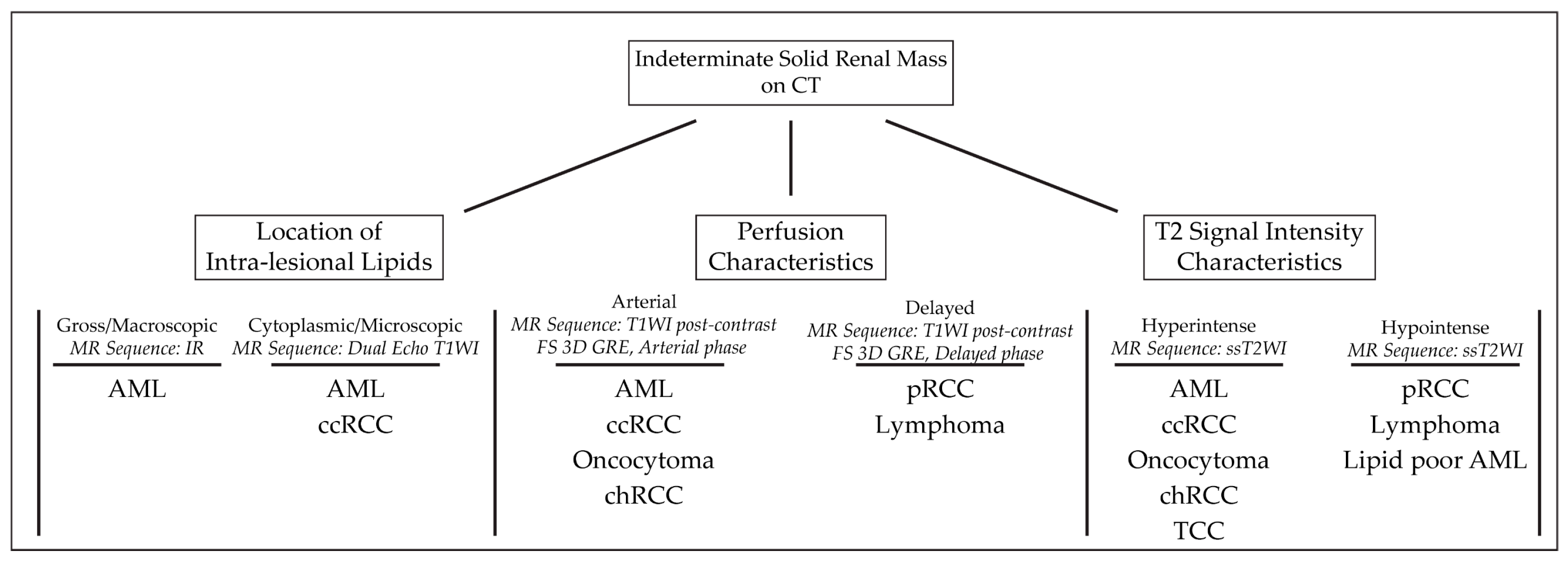
Current MR protocols for imaging solid renal masses include multiple sequences allowing for comprehensive and systematic analysis of tumor phenotype. The Society of Abdominal Radiology Disease Focused Panel recently published a MR protocol as recommended by the 13 abdominal radiologists and 10 academic institutions they represent. The proposed protocol applies to a wide range of renal mass pathology including an indeterminate renal mass on US or CT, active surveillance of a known renal mass, post-ablation surveillance, and post-nephrectomy surveillance. The protocol recommends using gadolinium-based contrast material at a volume of 0.1 mL/kg body weight followed by a 10–20 mL saline flush. The Society of Abdominal Radiology lists four sequences as its core sequences: (1) two-dimensional (2D) T2-weighted single-shot fast spin echo; (2) 2D T1-weighted echo in- and out-of-phase; (3) T1-weighted pre-gadolinium enhanced fat-suppressed three-dimensional gradient-echo (FS 3D GRE); (4) T1-weighted dynamic post-gadolinium enhanced FS 3D GRE in arterial, venous, and delayed contrast phases. Generally, T2-weighted sequences allow for the detection and characterization of cystic lesions [27]. Additionally, T2-weighted images can be helpful in suggesting certain histology, which will be discussed further in subsequent sections. T1-weighted GRE in- and out-of-phase should be included for the thorough characterization of microscopic fat and detection of hemosiderin. The dynamic timing consists of the arterial phase (30 s), venous/corticomedullary phase (90–100 s), and the delayed/nephrogenic phase (180–210 s). This multi-phase enhancement protocol is useful in determining renal cell carcinoma (RCC) subtypes. A fourth, delayed phase may be obtained at 5–7 min post contrast to capture the excretory phase and image the ureters and urinary bladder. Additionally, diffusion-weighted images (DWI) may be obtained to further detect metastatic or nodal disease.
The researchers present a systematic algorithm for the diagnosis and subtyping of small renal tumors: “virtual MR biopsy”; summarized in Figure 1.
Figure 1. Algorithmic approach to solid benign and malignant renal tumors.
An algorithmic approach to MR findings can be used to diagnose renal mass subtypes. The approach demonstrated above is based on a renal mass’s location of intralesional lipids, perfusion characteristics, and single-shot T2 signal intensity. Abbreviations: AML: angiomyolipoma; ccRCC: clear cell renal cell carcinoma; chRCC: chromophobe renal cell carcinoma; pRCC: papillary renal cell carcinoma; TCC: transitional cell carcinoma; MR: magnetic resonance; IR: inversion recovery; T1WI: T1 weighted image; ssT2WI: single-shot T2 weighted image; FS 3D GRE: fat-suppressed three-dimensional gradient-echo.
A summary of the imaging features of the discussed solid renal masses is shown in Table 1.
Table 1. Characteristic MR findings for benign and malignant renal tumors. Abbreviations: MR: magnetic resonance; GRE: gradient echo; T1W: T1-weighted; RCC: renal cell carcinoma; CC: cell carcinoma; IVC: inferior vena cava.

Type of Renal Masses


Benign Masses


Angiomyolipoma

High T2-intensity signal due to fat content.

Low T2 on fat-suppressed images.

Microscopic, intracytoplasmic fat made apparent with

in- and out-of-phase GRE

Lipid-poor Angiomyolipoma

T2-hypointense

Macroscopic fat and/or absence of fat

High arterial enhancement with subsequent washout

Oncocytoma

T2-iso-to-hyperintense relative to normal parenchyma

Central/eccentric T2-hyperintense scar

Delayed enhancement of a central scar

Segmental enhancement inversion pattern

Renal Cell Carcinomas


Clear Cell RCC

Heterogenous, high T2-intensity

Avid enhancement in corticomedullary and nephrogenic phases

Microscopic fat as see on dual echo T1W in- and out-of-phase

Invasion into surrounding vessels (esp. renal vein or IVC)

Presence of necrosis or intralesional calcification

Type 1 Papillary RCC

T2-hypointense

Uniform progressive delayed enhancement

Well-circumscribed, homogenous, peripherally-located

Type 2 Papillary RCC

Heterogenous T2 signal intensity

Heterogenous enhancement

Larger with more indistinct margin vs versus Type 1 pRCC

Chromophobe RCC

Low to intermediate T2-intensity

Intermediate, delayed enhancement

Central, stellate scar with “spoke-wheel” enhancement pattern

Peripheral, homogenous, well-circumscribed

Mimics oncocytoma on imaging

Rare Renal Masses


Renal Lymphoma

Low to intermediate T1 and T2 signal intensity

Mild, delayed, homogenous enhancement

Multiple 1–3 cm solitary masses

Metastasis

Varied presentation, usually identical to primary tumor

Multiple, atypical renal masses

History of advanced, non-renal malignancy

Transitional Cell Carcinoma

Intermediate T1 and T2 signal intensity

Delayed, heterogenous enhancement

Filling defects and soft masses when urine is present as contrast medium

This entry is adapted from the peer-reviewed paper 10.3390/cancers15102799

References

  1. Laguna, B.; Westphalen, A.C.; Guimaraes, C.T.; Whang, Z.; Simko, J.; Zagoria, R. Uncommon malignant renal tumors and atypical presentation of common ones: A guide for radiologists. Abdom. Radiol. 2019, 44, 1430–1452.
  2. Turner, R.M., 2nd; Morgan, T.M.; Jacobs, B.L. Epidemiology of the Small Renal Mass and the Treatment Disconnect Phenomenon. Urol. Clin. N. Am. 2017, 44, 147–154.
  3. Hollingsworth, J.M.; Miller, D.C.; Daignault, S.; Hollenbeck, B.K. Rising incidence of small renal masses: A need to reassess treatment effect. J. Natl. Cancer Inst. 2006, 98, 1331–1334.
  4. Patel, H.D.; Gupta, M.; Joice, G.A.; Srivastava, A.; Alam, R.; Allaf, M.E.; Pierorazio, P.M. Clinical Stage Migration and Survival for Renal Cell Carcinoma in the United States. Eur. Urol. Oncol. 2019, 2, 343–348.
  5. Escudier, B.; Eisen, T.; Stadler, W.M.; Szczylik, C.; Oudard, S.; Siebels, M.; Negrier, S.; Chevreau, C.; Solska, E.; Desai, A.A.; et al. Sorafenib in advanced clear-cell renal-cell carcinoma. N. Engl. J. Med. 2007, 356, 125–134.
  6. Hudes, G.; Carducci, M.; Tomczak, P.; Dutcher, J.; Figlin, R.; Kapoor, A.; Staroslawska, E.; Sosman, J.; McDermott, D.; Bodrogi, I.; et al. Temsirolimus, interferon alfa, or both for advanced renal-cell carcinoma. N. Engl. J. Med. 2007, 356, 2271–2281.
  7. Motzer, R.J.; Hutson, T.E.; Tomczak, P.; Michaelson, M.D.; Bukowski, R.M.; Rixe, O.; Oudard, S.; Negrier, S.; Szczylik, C.; Kim, S.T.; et al. Sunitinib versus interferon alfa in metastatic renal-cell carcinoma. N. Engl. J. Med. 2007, 356, 115–124.
  8. Ball, M.W. Surgical management of metastatic renal cell carcinoma. Discov. Med. 2017, 23, 379–387.
  9. Cornelis, F.; Grenier, N. Multiparametric Magnetic Resonance Imaging of Solid Renal Tumors: A Practical Algorithm. Semin. Ultrasound CT MR 2017, 38, 47–58.
  10. Kang, S.K.; Huang, W.C.; Pandharipande, P.V.; Chandarana, H. Solid renal masses: What the numbers tell us. AJR Am. J. Roentgenol. 2014, 202, 1196–1206.
  11. Uzosike, A.C.; Patel, H.D.; Alam, R.; Schwen, Z.R.; Gupta, M.; Gorin, M.A.; Johnson, M.H.; Gausepohl, H.; Riffon, M.F.; Trock, B.J.; et al. Growth Kinetics of Small Renal Masses on Active Surveillance: Variability and Results from the DISSRM Registry. J. Urol. 2018, 199, 641–648.
  12. Li, G.; Cuilleron, M.; Gentil-Perret, A.; Tostain, J. Characteristics of image-detected solid renal masses: Implication for optimal treatment. Int. J. Urol. 2004, 11, 63–67.
  13. Silverman, S.G.; Pedrosa, I.; Ellis, J.H.; Hindman, N.M.; Schieda, N.; Smith, A.D.; Remer, E.M.; Shinagare, A.B.; Curci, N.E.; Raman, S.S.; et al. Bosniak Classification of Cystic Renal Masses, Version 2019: An Update Proposal and Needs Assessment. Radiology 2019, 292, 475–488.
  14. Chenam, A.; Lau, C. Management of Small Renal Masses. Cancer Treat Res. 2018, 175, 105–126.
  15. Warren, A.Y.; Harrison, D. WHO/ISUP classification, grading and pathological staging of renal cell carcinoma: Standards and controversies. World J. Urol. 2018, 36, 1913–1926.
  16. Heilbrun, M.E.; Remer, E.M.; Casalino, D.D.; Beland, M.D.; Bishoff, J.T.; Blaufox, M.D.; Coursey, C.A.; Goldfarb, S.; Harvin, H.J.; Nikolaidis, P.; et al. ACR Appropriateness Criteria indeterminate renal mass. J. Am. Coll. Radiol. 2015, 12, 333–341.
  17. van Oostenbrugge, T.J.; Futterer, J.J.; Mulders, P.F.A. Diagnostic Imaging for Solid Renal Tumors: A Pictorial Review. Kidney Cancer 2018, 2, 79–93.
  18. Jamis-Dow, C.A.; Choyke, P.L.; Jennings, S.B.; Linehan, W.M.; Thakore, K.N.; Walther, M.M. Small (< or = 3-cm) renal masses: Detection with CT versus US and pathologic correlation. Radiology 1996, 198, 785–788.
  19. Jinzaki, M.; Ohkuma, K.; Tanimoto, A.; Mukai, M.; Hiramatsu, K.; Murai, M.; Hata, J. Small solid renal lesions: Usefulness of power Doppler US. Radiology 1998, 209, 543–550.
  20. Abou Elkassem, A.M.; Lo, S.S.; Gunn, A.J.; Shuch, B.M.; Dewitt-Foy, M.E.; Abouassaly, R.; Vaidya, S.S.; Clark, J.I.; Louie, A.V.; Siva, S.; et al. Role of Imaging in Renal Cell Carcinoma: A Multidisciplinary Perspective. Radiographics 2021, 41, 1387–1407.
  21. Yuh, B.I.; Cohan, R.H. Different phases of renal enhancement: Role in detecting and characterizing renal masses during helical CT. AJR Am. J. Roentgenol. 1999, 173, 747–755.
  22. Krishna, S.; Murray, C.A.; McInnes, M.D.; Chatelain, R.; Siddaiah, M.; Al-Dandan, O.; Narayanasamy, S.; Schieda, N. CT imaging of solid renal masses: Pitfalls and solutions. Clin. Radiol. 2017, 72, 708–721.
  23. Mileto, A.; Nelson, R.C.; Paulson, E.K.; Marin, D. Dual-Energy MDCT for Imaging the Renal Mass. AJR Am. J. Roentgenol. 2015, 204, W640–W647.
  24. Canvasser, N.E.; Kay, F.U.; Xi, Y.; Pinho, D.F.; Costa, D.; de Leon, A.D.; Khatri, G.; Leyendecker, J.R.; Yokoo, T.; Lay, A.; et al. Diagnostic Accuracy of Multiparametric Magnetic Resonance Imaging to Identify Clear Cell Renal Cell Carcinoma in cT1a Renal Masses. J. Urol. 2017, 198, 780–786.
  25. Weinreb, J.C.; Rodby, R.A.; Yee, J.; Wang, C.L.; Fine, D.; McDonald, R.J.; Perazella, M.A.; Dillman, J.R.; Davenport, M.S. Use of Intravenous Gadolinium-based Contrast Media in Patients with Kidney Disease: Consensus Statements from the American College of Radiology and the National Kidney Foundation. Radiology 2021, 298, 28–35.
  26. Kay, F.U.; Pedrosa, I. Imaging of Solid Renal Masses. Urol. Clin. N. Am. 2018, 45, 311–330.
  27. Schieda, N.; Lim, R.S.; McInnes, M.D.F.; Thomassin, I.; Renard-Penna, R.; Tavolaro, S.; Cornelis, F.H. Characterization of small (<4 cm) solid renal masses by computed tomography and magnetic resonance imaging: Current evidence and further development. Diagn. Interv. Imaging 2018, 99, 443–455.
More
This entry is offline, you can click here to edit this entry!
ScholarVision Creations