Your browser does not fully support modern features. Please upgrade for a smoother experience.
Please note this is an old version of this entry, which may differ significantly from the current revision.
Subjects:
Materials Science, Paper & Wood
Nanocellulosic materials have attracted special attention because of their performance in different advanced applications, biodegradability, availability, and biocompatibility. Nanocellulosic materials can assume three distinct morphologies, including cellulose nanocrystals (CNC), cellulose nanofibers (CNF), and bacterial cellulose (BC).
- cellulose nanocrystals
- cellulose nanofibers
- bacterial cellulose
1. Introduction
Cellulose was discovered by Payen Anselme, a French chemist, in 1838 [1] It is a renewable biopolymer, the most abundant in nature, with estimated natural production of about 1.5 × 10¹² tons per year [2]. Besides being abundant, cellulose is nontoxic, biocompatible, and biodegradable. The main sources of cellulose are plants, algae, bacteria, and tunicates (marine invertebrates) [3,4]. Cellulose is a natural macromolecule formed by 300–15,000 units of β-d-glucopyranose, bound by β-1,4-glycoglycodic linkages, and possessing an amorphous–crystalline supramolecular structure [5] Cellulose’s crystalline counterpart is rod-shaped and is found in the structure of elementary fibrils (EFs), where crystalline and amorphous regions alternate [5]. EFs are assembled in microfibrils and macrofibrils, which in turn make up the cell skeleton in plants (Figure 1). Cellulose crystallites can be isolated from native celluloses in different morphologic forms, and these so-called nanocelluloses are nanomaterials. By definition, nanocelluloses have at least one dimension in the nanometric range; i.e., they have a dimension of less than 100 nm [6,7], according to ISO/TS 20477:2017 [8,9]. Due to their unique properties, nanocelluloses can be used in numerous applications in different advanced materials, one of the most exciting being electronic devices and triboelectric nanogenerators (TENGs).
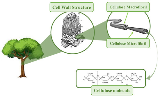
Figure 1. The simplified structural hierarchy of cellulose from a molecule to wood tissue.
This review focuses on the methods for obtaining nanocellulose and its nanotechnological applications, and is composed of two main parts. In the first part of the review, a brief characterization of the three cellulose nanomorphologies is performed, namely cellulose nanocrystals (CNCs), cellulose nanofibers (CNFs), and bacterial cellulose (BC), along with their preparation methods. It should be noted that the terms cellulose nanocrystals, cellulose nanofibers, and bacterial cellulose are strictly established in ISO/TS 20477: 2017 [8] The methods of nanocellulose preparation include such approaches as “bottom-up” or “top-down” [7,10] The mechanical, chemical, and enzymatic methods belong to a “top-down” approach for the production of cellulose nanocrystals (CNCs) and cellulose nanofibers (CNFs), while the “bottom-up” approach is commonly used for BC production. In relation to mechanical methods, methods for preparing CNFs through the disaggregation of cellulose fibers via defibrillation are reviewed. Within the category of mechanical methods, the following approaches are considered: refining, high-pressure homogenization, microfluidization, grinding, ball-milling, cryogenic crushing, steam explosion, ultrasound, extrusion, aqueous counter collision, and electrospinning [11,12]. For these mechanical methods, their functioning is presented, along with some illustrative schemes. In the scope of mechanical methods, there was also the aim to gather a set of investigations whose objective was to evaluate how operational parameters influenced the properties of CNFs. Thus, for each mechanical method and its operational parameters that interfere with the properties of CNF, a set of investigations was gathered in this study whose objective was to evaluate the relationship between the operational parameters and the properties of CNFs.
The chemical pre-treatment methods commonly applied before mechanical treatments were also reviewed. These are mainly intended for the production of CNFs. Among chemical pretreatments, acid- and alkali-catalyzed organosolvation, TEMPO-mediated oxidation, oxidation with APS and with SPS, ozone pre-treatment, extraction with ionic liquids [12,13,14,15], and two innovative methods for obtaining CNFs were described. One of these new methods consists of simultaneous bleaching and oxidative modification with TEMPO [16] and the other employs sodium persulfate (SPS) with ultraviolet light [17]. This review also presents the chemical treatments for the production of CNC, which are essentially variations of acid hydrolysis, as it degrades the amorphous part of the cellulose, leaving the crystalline part in the form of CNC [12,15]. Regarding chemical methods of CNC production, it should be noted that the TEMPO system, which has been used for the preparation of CNFs (the so-called TEMPO-CNF), is also suitable for CNC production [18]. This review also covers enzymatic hydrolysis pretreatment, considered by some researchers as the “most ecological route” for CNF production [13,15]..Enzymatic hydrolysis is carried out by cellulases, which more rapidly degrade the disordered cellulose counterpart of fibers. The three types of cellulases (endocellulases, exocellulases, and β-glucosidases), as well as their degradation modes, are specified. The first part of the review also contains a set of approaches in which chemical and enzymatic treatments were successfully combined in the production of nanocelluloses (CNFs and CNCs), and the properties of these nanocelluloses (diameter, length, and crystallinity index) are discussed.
2. Nanocellulose Nanomorphologies
Cellulosic materials can be converted into cellulose nanofibers (CNFs) and nanocrystals (CNCs), depending on the mechanical and chemical treatment applied (Table 1). In addition to these two nanomorphologies, bacterial nanocellulose, BC, also exists. These three types of nanocellulose (CNF, CNC, and BC) have different average sizes (Table 1). However, it should be noted that, for each type of nanocellulose, the values of the average sizes found in the literature present slight differences. These differences are mainly due to the different sources used and the various methods of preparation [12,19].
Table 1. Medium size, sources, and preparation of the various cellulose nanomorphologies (adapted from [1,4,11]).
| Approach | Nanomorphologies | Sources | Medium Size | Preparation |
|---|---|---|---|---|
| “Top-down” | Cellulose nanocrystals (CNCs) | Wood, cotton, hemp, linen, straw, tubers, tunicates, algae, bacteria, etc. | Diameter—5–70 nm Length—100 nm to several micrometers | Chemical treatment in the form of acid hydrolysis of cellulose (or enzyme-assisted hydrolysis) |
| “Top-down” | Cellulose nanofibers (CNFs) | Wood, cotton, hemp, beetroot, etc. | Diameter—5–60 nm Length—several micrometers | Defibrillation of wood pulp by mechanical treatment before and/or after chemical (and/or enzymatic) processes |
| “Bottom-up” | Bacterial nanocellulose (BC) | Low-molecular-weight sugars and alcohols | Diameter—20–100 nm Various kinds of nanofiber nets | Synthesized by bacteria |
It should be noted that, in the literature, the terminology used for the three types of nanocellulose is not concordant, so there are several designations for the same type of nanocellulose. For cellulose nanocrystals (CNCs), the following terms are used as synonyms: nanocrystalline cellulose, rod-type cellulose microcrystals, nanocellulose whiskers, and nanowhiskers [4,20,21]. Regarding cellulose nanofibers (CNF), the synonyms used are cellulose nanofibrils, nanofibrillated cellulose, or nanocellulose [1,4,20,22]. Additionally, bacterial cellulose (BC) is also called microbial cellulose [1,13,20,22].
Cellulose nanofibers (CNFs) are isolated from cellulose through simple mechanical disaggregation before and/or after chemical (or enzymatic) treatments [1,22]. CNFs were first produced in 1983 [12,22], and the first method of manufacture of CNF was patented in 1985 [12]. CNFs are composed of long, flexible, and scrambled “spaghetti-type” wires, with diameters below 100 nm and lengths of several micrometers [22]. In these preparations, CNFs contain amorphous and crystalline regions [23], as designated in Figure 2.
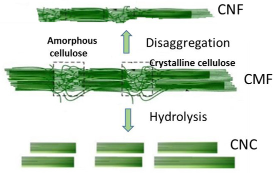
Figure 2. Schematic representation of cellulose nanofibrils (CNFs) production by disaggregation of cellulose microfibrils (CMFs) in the cell wall and the production of cellulose nanocrystals (CNCs) by the hydrolytic degradation of the amorphous part of CMF.
Cellulose nanocrystals (CNCs) appear “rice-like”, and are shorter and less flexible than CNFs (Figure 2). The reason for their lower flexibility is due to the preparation method, because they are obtained by acid hydrolysis (or enzymatic), which causes the removal of amorphous regions of cellulose; thus, they are less flexible than CNF [4,22]. The first CNCs obtained by acid hydrolysis were produced by Nickerson and Habrle in 1947 [12,15]. It should be noted that cellulose nanofibers (CNFs) and nanocrystals (CNCs) are obtained by “top-down” approaches.
Bacterial nanocellulose, BC, was first reported in 1886 by A.J. Brown [9]. BC is produced from glucose by bacterial families through a “bottom-up” approach. These bacteria, such as Gluconacetobacter xylinus, are grown in aqueous media and BC is produced by synthesizing cellulose and nanofibers in a process that takes a few days [22]. There are several families of bacteria that produce BC, namely Achromobacter, Alcaligenes, Aerobacter, Agrobacterium, Azotobacter, Komagataeibacter, Pseudomonas, Rhizobium, Sarcina, Dickeya, and Rhodobacter [24]. The most studied bacteria for this purpose are Komagataeibacter, (formerly Gluconacetobacter) [24]. BC does not contain lignin or hemicellulose, and it is said to have no impurities [19], having a high degree of purity when compared with nanocrystals and cellulose nanofibers [22]. BC is synthesized extracellularly, so hydrogen bonds between fibrils are more intense when compared with those in plant cellulose [1]. For these reasons, BC has properties that make it very attractive for various applications. These properties are its high degree of crystallinity, water retention capacity. and tensile strength [2]. The dimensions of bacterial cellulose vary depending on the family of bacteria, the conditions of cultivation, and the type of bioreactor [14].
ISO/TS 20477: 2017 [8] defines CNFs as network structures with dimensions from 3 to 100 nm, lengths that can reach 100 μm, and an aspect ratio of >10 (aspect ratio is the quotient between length and diameter). For CNCs, this ISO standard establishes a diameter of 3 to 50 nm and length from 100 nm to several microns (μm), with the aspect ratio usually in the range of 5 to 50.
This entry is adapted from the peer-reviewed paper 10.3390/ma16083104
This entry is offline, you can click here to edit this entry!