TP53 Alterations in MDS and AML: Comparison
Please note this is a comparison between Version 1 by Ramy Rahmé and Version 2 by Rita Xu.

TP53 mutations are less frequent in myelodysplastic syndromes (MDS) and acute myeloid leukemia (AML) than in solid tumors, except in secondary and therapy-related MDS/AMLs, and in cases with complex monosomal karyotype. As in solid tumors, missense mutations predominate, with the same hotspot mutated codons (particularly codons 175, 248, 273). As TP53-mutated MDS/AMLs are generally associated with complex chromosomal abnormalities, it is not always clear when TP53 mutations occur in the pathophysiological process.

  • mutant p53
  • cancer
  • myelodysplasia
  • acute myeloid leukemia

1. Introduction

The transcription factor p53, encoded by the TP53 gene in humans, is one of the most studied tumor suppressor proteins. In normal cells, p53 activity is low, but in response to DNA damage and numerous other stress signals, p53 levels rise dramatically and result in the activation and transcription of genes with important roles in cell cycle arrest, senescence, apoptosis, metabolism, and differentiation [1]. The sum of these activities is to ensure that an abnormal cell fails to arise and proliferate. During normal hematopoiesis, p53 activities preserve genome integrity and regulate several cellular processes that maintain the normal stem cell pool and serve as a barrier to tumorigenesis [2][3][2,3].
Perturbations in p53 activity or p53-dependent pathways are required for the development of most cancers [4], and there is evidence in many situations to suggest that the restoration or reactivation of physiological p53 function may be therapeutic [5][6][7][8][5,6,7,8]. Various mechanisms are responsible for the disruption of p53 activity in cancer, mainly deletion or mutation of the TP53 gene, and overexpression of the p53 negative regulators Mdm2 and Mdm4 [9][10][11][9,10,11]. Several isoforms are encoded by the TP53 gene and appear to play different roles in tumorigenesis/cancer progression [12] or response to treatment, for example in acute myeloid leukemia (AML) [13]. Regardless of the mechanism behind p53 dysfunction, the downstream consequences are profound due to the very large spectrum of biological activities in which p53 is normally implicated. The clinical correlation between p53 mutational status in cancer cells and resistance to treatment has been studied since the 1990s. In breast cancer, presence of a TP53 mutation is associated with resistance to doxorubicin [14][15][14,15]. Similarly, ovarian cancer patients harboring a TP53 mutation are less sensitive to treatment with cisplatin [16][17][16,17]. Moreover, the association of TP53 mutation with chemoresistance and poor prognosis has also been observed in lung [18], gastric, and colorectal cancers [19], as well as in hematological malignancies [20][21][20,21]. Apart from triggering chemoresistance, p53 mutants are also able to attenuate cancer response to radiotherapy [14][19][22][14,19,22].
Myelodysplastic syndromes (MDS) are a clonal hematopoietic stem cell disorders characterized by cell dysplasia, ineffective hematopoiesis that leads to cytopenias (mainly anemia) and a variable risk of progression to AML [23]. Furthermore, AML is characterized by clonal expansion of undifferentiated myeloid precursors, resulting in impaired hematopoiesis and life-threatening cytopenias. Among the prognostic factors identified in both malignancies, presence of a mutation in the TP53 gene indicates a particularly dismal prognosis irrespective of the treatment administered [24].

2. Characteristics of TP53 Mutations in MDS/AML

TP53 gene mutations are reported in about 50% of solid tumors, but only 5–10% of de novo myelodysplastic syndromes (MDS) and acute myeloid leukemia (AML). However, while these mutations are rare in MDS/AML with a normal karyotype (1%), they are seen in 40–50% of secondary and therapy-related cases [20][25][26][27][20,25,26,27], and in 80% of complex monosomal karyotypes (CK) that include 17p and/or 5q deletion [28][29][28,29]. Other inactivating mechanisms of wild-type p53 function include overexpression of MDM2 and MDM4, negative regulators of p53, in 20–30% and 40–50% of AML cases, respectively [30][31][32][33][34][30,31,32,33,34]. Unfortunately, treatment with Mdm2 inhibitors has not so far demonstrated a substantial effect in these cases [35]. Inactivation of p14-ARF, a positive regulator of p53, has been more rarely reported in AML [32][36][37][32,36,37]. As in solid tumors, most TP53 mutations in MDS/AML cluster in exons 4 to 8 that encode the DNA binding domain [38][39][38,39]. Nearly 75% of the mutants are missense: recurrent hotspot variants are also observed, as in solid tumors, and at the same frequent codons such as the contact mutants at codons 248 and 273, and the structural mutants at codons 175 and 245 [40]. The number of cooperating driver mutations is usually very low in TP53-mutated MDS/AML, and even absent in 85% of the cases [41]. TP53 alteration in MDS/AML can be monoallelic or biallelic [42]. Monoallelic mutations, with point mutation of only one TP53 allele, represent 25 to 30% of the cases. They are particularly seen in MDS with isolated 5q deletion [43][44][45][46][43,44,45,46], but can also be discovered in various MDS and AML with non-complex karyotype by systematic NGS analysis, generally at low variant allelic frequency (VAF) [42]. In 70 to 75% of the cases, TP53 alterations are biallelic, generally resulting from point mutation on one TP53 allele (usually missense) and loss of the other allele, through 17p deletion or monosomy 17. The great majority of MDS/AMLs with biallelic alteration have indeed complex monosomal karyotype resulting in 17p deletion, very often with 5q deletion [47]. On the other hand, monoallelic TP53 mutations can lead to genomic instability, chromosomal loss and chromothripsis (also known as “chromosome shattering” that leads to large structural rearrangements in chromosomes [48]) including loss of heterozygosity (LOH) through 17p deletion [49], and therefore resulting in a biallelic hit. TP53 mutations, especially in the biallelic state, are associated with resistance to most treatments, including chemotherapy, hypomethylating agents, and even allogeneic stem cell transplantation, as the risk of relapse post-transplant is very high, at least in case of biallelic alterations [50][51][52][53][50,51,52,53]. Monoallelic mutations are also, to a lesser extent, associated with a certain resistance to treatment, for example to lenalidomide–an immuno-modulatory drug used in MDS with del(5q) [43][54][43,54], but have generally a more limited impact on survival.

3. When Do TP53 Mutations Arise in MDS and AML Cells?

Whether TP53 mutations occur early or late in the evolution of MDS and AML remains uncertain in most situations. In well-performed studies in therapy-related MDS/AML, it was found that cells harboring the leukemia-specific TP53 mutation preexisted to the first cancer and were positively selected by treatment for this cancer (chemotherapy or radiation) to which they were resistant [55][56][55,56]. Progressive evolution was seen through genomic instability and chromothripsis with inactivation of the second TP53 allele, as seen above [49] (Figure 1). TP53 mutations are also among the mutations found in healthy individuals with clonal hematopoiesis of indeterminate potential (CHIP), who have an increased risk of developing various blood cancers [57][58][57,58]. However, DNMT3A, TET2, and ASXL1 mutations largely predominate in CHIPs seen in patients with no prior history of treated cancer, and CHIPs with TP53 mutation are mainly seen in patients having received chemotherapy or radiotherapy for a prior cancer, where they increase the risk of therapy-related MDS/AML, potentially by the selection mechanism just described above [55][59][60][61][62][63][64][55,59,60,61,62,63,64].
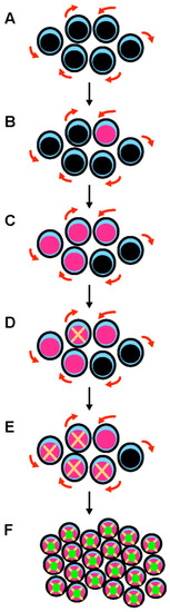
Figure 1. A multistep process leads to the development of TP53-mutated myelodysplastic syndrome and acute myeloid leukemia. (A) Hematopoietic stem cells (HSCs) are the only cells within the hematopoietic system that possess the potential for both multipotency (i.e., the potential to differentiate into all of the mature blood cell type) and self-renewal (i.e., the potential to make more stem cells, thus perpetuating the stem cell pool throughout life). Self-renewal is illustrated with red arrows. (B) A mutation in the TP53 gene can emerge in a hematopoietic stem cell (pink nucleus). This mutation can appear spontaneously and is selected in response to various stresses, including exposure to cytotoxic treatments. This mutation can be induced by these cytotoxic treatments (such as chemotherapy or radiation therapy) or by workplace exposure to toxic chemicals and carcinogenic substances such as benzene. (C) Mutant p53 confers a competitive advantage in the stem cell compartment. At this early stage, cells express both wild type and mutant p53 proteins (potential dominant-negative effect). (D) TP53-mutated HSCs acquire chromosomal changes and gene mutations that either result from mutant p53-related genomic instability and chromothripsis, or are induced by cytotoxic chemotherapy/radiation. (E) These acquired genetic changes further enhance the fitness of TP53-mutated HSCs. (F) Further chromosomal changes lead to the loss of the wild type TP53 allele by 17p deletion or monosomy 17. Loss of wild type TP53 functions and potential gain-of-function activities of mutant p53 are responsible for the development of overt acute myeloid leukemia.
It is still unclear in other MDS/AML situations when TP53 mutations arise. In MDS with isolated 5q deletion, detectable monoallelic TP53 mutations, often at very low VAF and sometimes multiple, are found in 20% of the patients at diagnosis [43][44][45][46][43,44,45,46]. Whether they occur during disease evolution or very early in minor undetectable clones (by conventional methods) that could undergo positive selection, including by treatment with lenalidomide, is unclear. In those MDS with isolated del(5q) and monoallelic TP53 mutation, as in the therapy-related model described above, progression to AML is generally preceded by acquisition of a complex karyotype, including del(17p) and biallelic TP53 inactivation [65][66][65,66] (Figure 2).
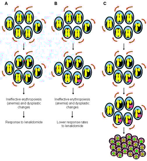
Figure 2. The special case of myelodysplastic syndrome with 5q deletion. (A) Deletion of 5q chromosome is somatically acquired and heterozygous. This chromosomal abnormality is present in the hematopoietic stem cell compartment and can be found in all lineages. The clinical phenotype of 5q- syndrome (i.e., ineffective erythropoiesis and dysplastic changes) is related to haploinsufficient gene expression of several genes such as RPS14, APC, and EGR1. Self-renewal is illustrated with red arrows. (B) Monoallelic TP53 mutations are seen at diagnosis in almost 20% of patients with 5q- syndrome, generally at low variant allelic frequency. These mutations are associated with resistance to lenalidomide, but have generally a limited impact on survival. At this stage, karyotype is non-complex. (C) Monoallelic TP53 mutations are associated with genomic instability and chromothripsis in myelodysplastic syndrome with isolated 5q deletion. Progression to higher risk myelodysplastic syndrome and acute myeloid leukemia is preceded by acquisition of a complex karyotype, including 17p deletion and biallelic TP53 inactivation.
In myeloproliferative neoplasms, TP53 mutations are seen in about 20% of the cases with progression to MDS/AML. They appear to be late events, although once again small TP53 mutant clones, undetectable by conventional techniques, may have occurred earlier in the disease course [67][68][69][70][71][72][73][67,68,69,70,71,72,73]. When TP53-mutated clones appear during the disease course would be important to determine, as their early detection in myeloid malignancies could allow to target them pharmacologically, potentially preventing progression to full-blown biallelic TP53 MDS/AML. TP53 mutations could represent early leukemogenic events in many situations, and they have been indeed reported in pre-leukemic hematopoietic stem cells (HSCs) of AML patients [74][75][74,75]. Major roles of TP53 are related to cell-cycle control, DNA repair and apoptosis. Dysregulation of these pivotal functions might be an alternative mechanism to epigenetic modifications in establishing a proleukemogenic state in HSCs [76]. By transforming HSCs into pre-leukemic stem cells (pre-LSCs), TP53 mutations substantially contribute to the development of AML and its resistance to conventional treatments [55][77][78][55,77,78]. Moreover, TP53-mutated pre-LSCs retain their ability to differentiate into mature blood cells both in patient-derived mouse xenografts and patients with AML [78]. Clonogenic assays revealed that patient-specific TP53 mutations are present in the vast majority of HSC-derived colonies (median, 97%; range, 45 to 100%), with only a paucity of cooperating mutations in known cancer genes, as seen above. Copy-number alterations, as already described, appear to be secondary events after the onset of a first TP53 mutation [79][80][79,80].
Video Production Service