Gastric motility abnormalities are common in patients with disorders of gut-brain interaction such as functional dyspepsia and gastroparesis. Accurate assessment of the gastric motility in these common disorders can help understand the underlying pathophysiology and guide effective treatment. A variety of clinically applicable diagnostic methods have been developed to objectively evaluate for presence of gastric dysmotility, including tests of gastric accommodation, antroduodenal motility, gastric emptying, and gastric myoelectrical activity. The advances in clinically available diagnostic methods for evaluation of gastric motility are discussed and the advantages and disadvantages of each test are described.
- functional dyspepsia
- gastroparesis
- gastric motility
- antroduodenal contraction
- gastric emptying
- gastric slow waves
- electrogastrography
1. Assessment of Gastric Accommodation
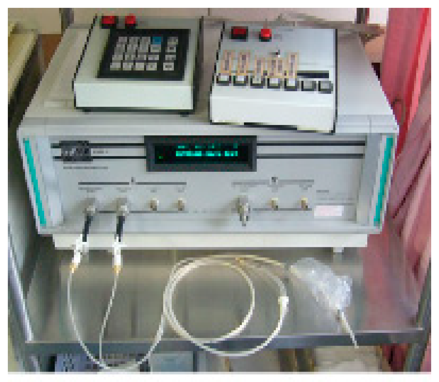
The gastric barostat is a device that currently represents a gold standard for assessment of gastric accommodation. It consists of a polyethylene balloon with a volume of up to 1.0-1.2 L, connected to the barostat device through a double-lumen polyvinyl tube (Figure 1) [1]. The principle of barostat is that it provides a constant pressure in the balloon via a build-in computerized pneumatic pump. The finely folded adherent balloon is perorally introduced into the stomach via the esophagus, attached with adhesive tape on the participant’s chin, and positioned to record volume change at a constant pressure in the proximal stomach. By maintaining a constant pressure inside the balloon and observing changes in balloon volume that correlate to changes in gastric volume, the gastric volumes can be measured. The balloon pressure can be modified in a progressive or random manner which allows for evaluation of the gastric sensation.
Satiation drinking test is a noninvasive tool developed to induce gastric distension and indirectly measure the postprandial sensorimotor functions, including gastric accommodation [2]. In this test, individuals drink a liquid of known composition and/or caloric content at a certain rate over pre-determined time-period. When maximum satiety is reached, the volume of ingested liquid is recorded to estimate the gastric accommodation and sensation [3]. A variety of satiation drinking test protocols have been reported, utilizing either water or specific nutrient solutions [3]. Drinking test protocols also differ in rapidity of the liquid intake and tested individual’s awareness of the quantity of ingestion.
Single photon emission computed tomography (SPECT) is a non-invasive approach based on the gastric mucosa property to selectively uptake and excrete 99mTc- pertechnetate. Radiolabeled 99mTc is administered intravenously and accumulates in the gastric mucosa, allowing visualization of the entire stomach wall with a SPECT gamma camera [4]. Three dimensional stomach images are produced by computerized data processing and can be used to calculate stomach volume. The gastric accommodation response after a meal can be estimated by measuring the fundic and total stomach volumes during both fasting and postprandial periods. It has been demonstrated that postprandial volume changes measured by SPECT correspond with barostat volume measurements [5].
Transabdominal ultrasonography is another non-invasive method with demonstrated utility and validity for the investigation of gastric accommodation, gastric emptying and gastroduodenal flow [6]. A conventional ultrasound probe is used to visualize the stomach and can indirectly estimate gastric volumes by measuring the gastric diameter and area [7]. Serial measurements of variations in the area of antral cross-section can serve as an indicator to estimate gastric emptying. Duplex sonography can be used to visualize luminal contents movement [8].
2. Assessment of Antroduodenal Motility
Antroduodenal manometry (ADM) is a method allowing simultaneous assessment of gastric and duodenal motility by measuring the frequency and characteristics of contraction patterns [9]. In this method, a water-perfused or solid-state (high-resolution) manometric catheter is typically positioned transnasally through the pylorus into duodenum. Consequently, evaluation of the number and amplitude of gastric contractions in both fasting and postprandial states is possible [10]. However, given its invasive nature, ADM is typically applied in selected patients with marked symptoms and presumed severely impaired gastrointestinal function, in whom understanding the pathogenesis of the motility disorder is necessary to guide better treatment [11].
Dynamic magnetic resonance imaging (MRI) is a noninvasive technique that uses body-surface mapping and assessment of contractions to simultaneously analyze gastric motility, anatomy and emptying with high spatial and temporal resolution [12]. Gastric MRI requires consumption of a test meal containing paramagnetic ions, such as manganese and gadolinium, to allow adequate reconstruction of stomach images [13]. Using both liquid and solid meals, gastric MRI has been validated to accurately investigate the gastric volume content and emptying rate [14]. An MRI acquisition protocol and image processing pipeline were proposed in a recent study to assess gastric wall motion and emptying in healthy adults [15]. This methodology enables direct visualization of peristaltic waves along the gastric wall and can provide personalized profiles of gastric contractions and emptying.
3. Assessment of Gastric Emptying
Gastric emptying scintigraphy is a conventional method to measure the rate of gastric emptying that is considered gold standard. A standardized meal of liquid egg whites (approximately 240 kcal, 2% fat) is mixed with 99mTc sulfur colloid and cooked until it has a texture of firm omlet [16]. After ingestion of the meal, gastric area is scanned with an antero-posterior γ-camera over 4 hours. (unless over 90% of the solid meal has already emptied at 3 hours) [16]. The normal values of gastric emptying have been established and internationally validated with gastric solid meal retention of >10% at 4 hours post-ingestion considered a delayed gastric emptying [17]. This methodology was proven reproducible when applied in patients with upper gastrointestinal symptoms [18]. Although the relationship between gastric emptying rate and gastrointestinal symptoms has been controversial [19], studies using scintigraphy with a solid meal and collecting data for at least 3 hours, post-ingestion, showed positive correlation between gastric emptying rate and severity of nausea, vomiting, abdominal pain, and early satiety/fullness [19]. Consequently, the current American College of Gastroenterology (ACG) clinical guideline for gastroparesis recommends gastric emptying scintigraphy, measuring solid meal emptying over a period of at least 3 hours, as a first-line test to establish the diagnosis of gastroparesis in patients with clinical presentations suggestive of gastroparesis [20].
The stable isotope breath test methodology uses a standardized solid meal containing-Spirulina platensis (edible blue-green microalgae) tagged with a non-radioactive carbon isotope 13C [11]. After ingestion, metabolites of spirulina are absorbed in the small intestine and metabolized by the liver, resulting in 13C excretion in the lungs. Isotope ratio mass spectrometry is used to determine the rise of 13C over baseline in exhaled breath samples, which correlates with the rate of gastric emptying and allows indirect calculation of the gastric emptying time [21]. Studies of the liquid- and solid-phase gastric emptying have used labeled acetate and octanoate, respectively [22]. 13C-glycine was also selected as a marker of the liquid phase because it is rapidly absorbed and transformed into 13CO2 after reaching the small intestine [23]. The current ACG clinical guideline considers gastric emptying breath test a reliable method to assess gastric emptying in patients with suspected gastroparesis [20]. The 13C spirulina breath test has also been approved by the US Food and Drug Administration (FDA) [11]. However, in practical terms, this test is still not widely available to many providers in the US. The 13C breath test performed simultaneously with scintigraphy was shown to be reproducible and correlate well with gastric emptying scintigraphy in both healthy volunteers and symptomatic patients with suspected gastric emptying delay [21].
The wireless motility capsule (WMC) has been developed as a non-invasive test, alternative to scintigraphy, to measure gastric emptying as well as small and large intestines transit time [11][. The test (SmartPill GI Monitoring System) has been approved to assess gastric emptying by the US Food and Drug Administration. It is an indirect test where a swallowed capsule records and transmits to an external recorder the luminal pH, pressure, and temperature. The gastric emptying time is derived by a rise of pH from the acidic low gastric baseline to values above 4 in the duodenum [24]. It has been shown that the capsule, when ingested with a meal, is emptied from the stomach separately from the meal through reactivation of the interdigestive migrating motor complex [25]. The gastric emptying time observed by the WMC was found to significantly correlate (r=0.73) with the gastric emptying at 4 hours derived by scintigraphy in both healthy and gastroparetic individuals [24]. The WMC gastric emptying cut-off time of 300-minutes had sensitivity of 65% and specificity of 87% in comparison with scintigraphy at 4 hour [24]. In a recent study, WMC reported delayed gastric emptying in twice as many patients with diabetes, when compared to the evaluation with gastric emptying scintigraphy [26], but the clinical relevance of higher sensitivity for WMC to detect delayed gastric emptying is not clear. WMC distinguished gastroparesis from healthy subjects based on the motility profiles of stomach and small bowel [27]. However, no correlation was observed between detection of gastric emptying delay on WMC and the overall gastroparesis symptoms [28].
4. Assessment of Gastric Myoelectrical Activity
Transcutaneous electrogastrography is a non-invasive technique utilizing skin electrodes placed on the abdomen to capture gastric myoelectrical activity. Typically, the test is performed in a quiet room while studied individual is in a supine position. To achieve reliable results, the gastric myoelectrical activity should be recorded for at least 15 minutes in fasting state and 30 minutes in postprandial state. These recordings generate the electrogastrogram (EGG). The test meal should contain at least 250 kcal (preferably > 400 kcal), with a maximum fat content of 35% [29]. The EGG has been validated to reflect the slow electrical gastric rhythm, termed slow waves [30] that determine the propagation and maximum frequency of gastric contractions.
High-resolution electrograstrography (HR-EGG) has emerged as a novel, non-invasive, methodology offering improved capture of the gastric myoelectrical activity, such as propagation of slow waves and spatial abnormalities of gastric slow waves. In HR-EGG, an array of densely spaced cutaneous electrodes is placed on the abdomen over the stomach [31] to record multichannel EGG signals with a spatial resolution. In one recent study, the HR-EGG revealed spatial slow wave abnormalities in 44% of patients with upper GI symptoms and there was correlation of aberrant slow wave propagation direction with gastroparesis symptoms and abdominal pain [32]. However, more studies are needed to overcome technical challenges (e.g. sufficiently high-density grid of electrodes to account for gastric anatomical variability and allow appropriate skin contact) and to establish the clinical role of HR-EGG in diagnosing gastric motility disorders and assessing the electrophysiology of the stomach.
Body surface gastric mapping (BSGM) represents a further refinement of the technology to optimally capture the gastric myoelectrical activity in non-invasive manner. In BSGM, a comprehensive spatial analysis of gastric potentials, obtained via dense grid of cutaneous electrodes, is generated to derive detailed maps of gastric patterns. This methodology may enhance the current clinical utility of EGG by allowing more comprehensive gastric slow wave analysis and better characterization of gastric dysrhythmias [33].
References
- [1] De Schepper HU, Cremonini F, Chitkara D, Camilleri M. Assessment of gastric accommodation: overview and evaluation of current methods. Neurogastroenterol Motil, 2004; 3:275-285.De Schepper HU, Cremonini F, Chitkara D, Camilleri M. Assessment of gastric accommodation: overview and evaluation of current methods. Neurogastroenterol Motil, 2004; 3:275-285.
- [2] Koch KL, Hong SP, Xu L. Reproducibility of gastric myoelectrical activity and the water load test in patients with dysmotility-like dyspepsia symptoms and in control subjects. J Clin Gastroenterol, 2000; 2:125-129.Koch KL, Hong SP, Xu L. Reproducibility of gastric myoelectrical activity and the water load test in patients with dysmotility-like dyspepsia symptoms and in control subjects. J Clin Gastroenterol, 2000; 2:125-129.
- [3] Kindt S, Tack J. Impaired gastric accommodation and its role in dyspepsia. Gut, 2006; 12:1685-1691.Kindt S, Tack J. Impaired gastric accommodation and its role in dyspepsia. Gut, 2006; 12:1685-1691.
- [4] van den Elzen BD, Bennink RJ, Wieringa RE, Tytgat GN, Boeckxstaens GE. Fundic accommodation assessed by SPECT scanning: comparison with the gastric barostat. Gut, 2003; 11:1548-1554.van den Elzen BD, Bennink RJ, Wieringa RE, Tytgat GN, Boeckxstaens GE. Fundic accommodation assessed by SPECT scanning: comparison with the gastric barostat. Gut, 2003; 11:1548-1554.
- [5] Bouras EP, Delgado-Aros S, Camilleri M, et al. SPECT imaging of the stomach: comparison with barostat, and effects of sex, age, body mass index, and fundoplication. Gut, 2002; 6:781-786.Bouras EP, Delgado-Aros S, Camilleri M, et al. SPECT imaging of the stomach: comparison with barostat, and effects of sex, age, body mass index, and fundoplication. Gut, 2002; 6:781-786.
- [6] Gilja OH, Lunding J, Hausken T, Gregersen H. Gastric accommodation assessed by ultrasonography. World Journal of Gastroenterology, 2006; 18:2825-2829.Gilja OH, Lunding J, Hausken T, Gregersen H. Gastric accommodation assessed by ultrasonography. World Journal of Gastroenterology, 2006; 18:2825-2829.
- [7] Bisschops R, Tack J. Dysaccommodation of the stomach: therapeutic nirvana? Neurogastroenterol Motil, 2007; 2:85-93.Bisschops R, Tack J. Dysaccommodation of the stomach: therapeutic nirvana? Neurogastroenterol Motil, 2007; 2:85-93.
- [8] Hausken T, Odegaard S, Matre K, Berstad A. Antroduodenal motility and movements of luminal contents studied by duplex sonography. Gastroenterology, 1992; 5:1583-1590.Hausken T, Odegaard S, Matre K, Berstad A. Antroduodenal motility and movements of luminal contents studied by duplex sonography. Gastroenterology, 1992; 5:1583-1590.
- [9] Patcharatrakul T, Gonlachanvit S. Technique of functional and motility test: how to perform antroduodenal manometry. J Neurogastroenterol Motil, 2013; 3:395-404.Patcharatrakul T, Gonlachanvit S. Technique of functional and motility test: how to perform antroduodenal manometry. J Neurogastroenterol Motil, 2013; 3:395-404.
- [10] Storlid EL, Hausken T, Lied GA, Gilja OH, Hatlebakk JG. Gastric accommodation in healthy subjects studied by ultrasound, manometry, and impedancemetry. Neurogastroenterol Motil, 2018; 4:e13249.Storlid EL, Hausken T, Lied GA, Gilja OH, Hatlebakk JG. Gastric accommodation in healthy subjects studied by ultrasound, manometry, and impedancemetry. Neurogastroenterol Motil, 2018; 4:e13249.
- [11] Keller J, Bassotti G, Clarke J, et al. Expert consensus document: Advances in the diagnosis and classification of gastric and intestinal motility disorders. Nat Rev Gastroenterol Hepatol, 2018; 5:291-308.Keller J, Bassotti G, Clarke J, et al. Expert consensus document: Advances in the diagnosis and classification of gastric and intestinal motility disorders. Nat Rev Gastroenterol Hepatol, 2018; 5:291-308.
- [12] Goetze O, Steingoetter A, Menne D, et al. The effect of macronutrients on gastric volume responses and gastric emptying in humans: a magnetic resonance imaging study. American Journal of Physiology-Gastrointestinal and Liver Physiology, 2007; 1:G11-G17.Goetze O, Steingoetter A, Menne D, et al. The effect of macronutrients on gastric volume responses and gastric emptying in humans: a magnetic resonance imaging study. American Journal of Physiology-Gastrointestinal and Liver Physiology, 2007; 1:G11-G17.
- [13] Febo-Rodriguez L, Chumpitazi BP, Sher AC, Shulman RJ. Gastric accommodation: Physiology, diagnostic modalities, clinical relevance, and therapies. Neurogastroenterol Motil, 2021; 12:e14213.Febo-Rodriguez L, Chumpitazi BP, Sher AC, Shulman RJ. Gastric accommodation: Physiology, diagnostic modalities, clinical relevance, and therapies. Neurogastroenterol Motil, 2021; 12:e14213.
- [14] Feinle C, Kunz P, Boesiger P, Fried M, Schwizer W. Scintigraphic validation of a magnetic resonance imaging method to study gastric emptying of a solid meal in humans. Gut, 1999; 1:106-111.Feinle C, Kunz P, Boesiger P, Fried M, Schwizer W. Scintigraphic validation of a magnetic resonance imaging method to study gastric emptying of a solid meal in humans. Gut, 1999; 1:106-111.
- [15] Lu KH, Liu Z, Jaffey D, et al. Automatic assessment of human gastric motility and emptying from dynamic 3D magnetic resonance imaging. Neurogastroenterol Motil, 2022; 1:e14239.Lu KH, Liu Z, Jaffey D, et al. Automatic assessment of human gastric motility and emptying from dynamic 3D magnetic resonance imaging. Neurogastroenterol Motil, 2022; 1:e14239.
- [16] Abell TL, Camilleri M, Donohoe K, et al. Consensus recommendations for gastric emptying scintigraphy: a joint report of the American Neurogastroenterology and Motility Society and the Society of Nuclear Medicine. Am J Gastroenterol, 2008; 3:753-763.Abell TL, Camilleri M, Donohoe K, et al. Consensus recommendations for gastric emptying scintigraphy: a joint report of the American Neurogastroenterology and Motility Society and the Society of Nuclear Medicine. Am J Gastroenterol, 2008; 3:753-763.
- [17] Tougas G, Eaker EY, Abell TL, et al. Assessment of gastric emptying using a low fat meal: establishment of international control values. Am J Gastroenterol, 2000; 6:1456-1462.Tougas G, Eaker EY, Abell TL, et al. Assessment of gastric emptying using a low fat meal: establishment of international control values. Am J Gastroenterol, 2000; 6:1456-1462.
- [18] Desai A, O'Connor M, Neja B, et al. Reproducibility of gastric emptying assessed with scintigraphy in patients with upper GI symptoms. Neurogastroenterol Motil, 2018; 10:e13365.Desai A, O'Connor M, Neja B, et al. Reproducibility of gastric emptying assessed with scintigraphy in patients with upper GI symptoms. Neurogastroenterol Motil, 2018; 10:e13365.
- [19] Vijayvargiya P, Jameie-Oskooei S, Camilleri M, Chedid V, Erwin PJ, Murad MH. Association between delayed gastric emptying and upper gastrointestinal symptoms: a systematic review and meta-analysis. Gut, 2019; 5:804-813.Vijayvargiya P, Jameie-Oskooei S, Camilleri M, Chedid V, Erwin PJ, Murad MH. Association between delayed gastric emptying and upper gastrointestinal symptoms: a systematic review and meta-analysis. Gut, 2019; 5:804-813.
- [20] Camilleri M, Kuo B, Nguyen L, et al. ACG Clinical Guideline: Gastroparesis. Am J Gastroenterol, 2022; 8:1197-1220.Camilleri M, Kuo B, Nguyen L, et al. ACG Clinical Guideline: Gastroparesis. Am J Gastroenterol, 2022; 8:1197-1220.
- [21] Szarka LA, Camilleri M, Vella A, et al. A stable isotope breath test with a standard meal for abnormal gastric emptying of solids in the clinic and in research. Clin Gastroenterol Hepatol, 2008; 6:635-643.e631.Szarka LA, Camilleri M, Vella A, et al. A stable isotope breath test with a standard meal for abnormal gastric emptying of solids in the clinic and in research. Clin Gastroenterol Hepatol, 2008; 6:635-643.e631.
- [22] Sanaka M, Yamamoto T, Kuyama Y. Retention, fixation, and loss of the [13C] label: a review for the understanding of gastric emptying breath tests. Dig Dis Sci, 2008; 7:1747-1756.Sanaka M, Yamamoto T, Kuyama Y. Retention, fixation, and loss of the [13C] label: a review for the understanding of gastric emptying breath tests. Dig Dis Sci, 2008; 7:1747-1756.
- [23] Maes BD, Ghoos YF, Geypens BJ, et al. Combined carbon-13-glycine/carbon-14-octanoic acid breath test to monitor gastric emptying rates of liquids and solids. J Nucl Med, 1994; 5:824-831.Maes BD, Ghoos YF, Geypens BJ, et al. Combined carbon-13-glycine/carbon-14-octanoic acid breath test to monitor gastric emptying rates of liquids and solids. J Nucl Med, 1994; 5:824-831.
- [24] Kuo B, McCallum RW, Koch KL, et al. Comparison of gastric emptying of a nondigestible capsule to a radio-labelled meal in healthy and gastroparetic subjects. Aliment Pharmacol Ther, 2008; 2:186-196.Kuo B, McCallum RW, Koch KL, et al. Comparison of gastric emptying of a nondigestible capsule to a radio-labelled meal in healthy and gastroparetic subjects. Aliment Pharmacol Ther, 2008; 2:186-196.
- [25] Cassilly D, Kantor S, Knight LC, et al. Gastric emptying of a non-digestible solid: assessment with simultaneous SmartPill pH and pressure capsule, antroduodenal manometry, gastric emptying scintigraphy. Neurogastroenterol Motil, 2008; 4:311-319.Cassilly D, Kantor S, Knight LC, et al. Gastric emptying of a non-digestible solid: assessment with simultaneous SmartPill pH and pressure capsule, antroduodenal manometry, gastric emptying scintigraphy. Neurogastroenterol Motil, 2008; 4:311-319.
- [26] Lee AA, Rao S, Nguyen LA, et al. Validation of Diagnostic and Performance Characteristics of the Wireless Motility Capsule in Patients With Suspected Gastroparesis. Clin Gastroenterol Hepatol, 2019; 9:1770-1779.e1772.Lee AA, Rao S, Nguyen LA, et al. Validation of Diagnostic and Performance Characteristics of the Wireless Motility Capsule in Patients With Suspected Gastroparesis. Clin Gastroenterol Hepatol, 2019; 9:1770-1779.e1772.
- [27] Kloetzer L, Chey WD, McCallum RW, et al. Motility of the antroduodenum in healthy and gastroparetics characterized by wireless motility capsule. Neurogastroenterol Motil, 2010; 5:527-533, e117.Kloetzer L, Chey WD, McCallum RW, et al. Motility of the antroduodenum in healthy and gastroparetics characterized by wireless motility capsule. Neurogastroenterol Motil, 2010; 5:527-533, e117.
- [28] Hasler WL, May KP, Wilson LA, et al. Relating gastric scintigraphy and symptoms to motility capsule transit and pressure findings in suspected gastroparesis. Neurogastroenterol Motil, 2018; 2.Hasler WL, May KP, Wilson LA, et al. Relating gastric scintigraphy and symptoms to motility capsule transit and pressure findings in suspected gastroparesis. Neurogastroenterol Motil, 2018; 2.
- [29] Yin J, Chen JD. Electrogastrography: methodology, validation and applications. J Neurogastroenterol Motil, 2013; 1:5-17.Yin J, Chen JD. Electrogastrography: methodology, validation and applications. J Neurogastroenterol Motil, 2013; 1:5-17.
- [30] Chen JD, Schirmer BD, McCallum RW. Serosal and cutaneous recordings of gastric myoelectrical activity in patients with gastroparesis. Am J Physiol, 1994; 1 Pt 1:G90-98.Chen JD, Schirmer BD, McCallum RW. Serosal and cutaneous recordings of gastric myoelectrical activity in patients with gastroparesis. Am J Physiol, 1994; 1 Pt 1:G90-98.
- [31] Gharibans AA, Kim S, Kunkel D, Coleman TP. High-Resolution Electrogastrogram: A Novel, Noninvasive Method for Determining Gastric Slow-Wave Direction and Speed. IEEE Trans Biomed Eng, 2017; 4:807-815.Gharibans AA, Kim S, Kunkel D, Coleman TP. High-Resolution Electrogastrogram: A Novel, Noninvasive Method for Determining Gastric Slow-Wave Direction and Speed. IEEE Trans Biomed Eng, 2017; 4:807-815.
- [32] Gharibans AA, Coleman TP, Mousa H, Kunkel DC. Spatial Patterns From High-Resolution Electrogastrography Correlate With Severity of Symptoms in Patients With Functional Dyspepsia and Gastroparesis. Clin Gastroenterol Hepatol, 2019; 13:2668-2677.Gharibans AA, Coleman TP, Mousa H, Kunkel DC. Spatial Patterns From High-Resolution Electrogastrography Correlate With Severity of Symptoms in Patients With Functional Dyspepsia and Gastroparesis. Clin Gastroenterol Hepatol, 2019; 13:2668-2677.
- [33] Carson DA, O'Grady G, Du P, Gharibans AA, Andrews CN. Body surface mapping of the stomach: New directions for clinically evaluating gastric electrical activity. Neurogastroenterol Motil, 2021; 3:e14048.Carson DA, O'Grady G, Du P, Gharibans AA, Andrews CN. Body surface mapping of the stomach: New directions for clinically evaluating gastric electrical activity. Neurogastroenterol Motil, 2021; 3:e14048.