Nanoparticles in Pediatric Brain Tumors' Cancer Stem Cells: Comparison
Please note this is a comparison between Version 2 by Jessie Wu and Version 4 by Jessie Wu.

Primary malignant brain tumors are the most common solid neoplasm in childhood. Despite recent advances, many children affected by aggressive or metastatic brain tumors still present poor prognosis, therefore the development of more effective therapies is urgent. Cancer stem cells (CSCs) have been discovered and isolated in both pediatric and adult patients with brain tumors (e.g., medulloblastoma, gliomas and ependymoma). CSCs are a small clonal population of cancer cells responsible for brain tumor initiation, maintenance and progression, displaying resistance to conventional anticancer therapies. CSCs are characterized by a specific repertoire of surface markers and intracellular specific pathways. These unique features of CSCs biology offer the opportunity to build therapeutic approaches to specifically target these cells in the complex tumor bulk.

  • nanoparticles
  • stem cells
  • cancer

1. Nanoparticles and Target Therapy

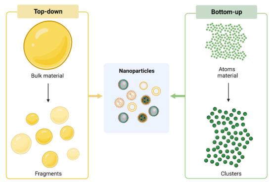
1.1. Synthesis of Nanoparticles

Nanoparticles (NPs) used in cancer therapy can be obtained from organic and inorganic materials or by a combination of them. Besides the “raw” material employed, NPs can differ in size, structure and shape. These features can be tuned and depend on the approach used in the synthesis method employed. NPs can be obtained by using the top-down or the bottom-up approach. The top-down method starts with bulk material to obtain smaller units by disruption or decomposition that are then converted into NPs. Conversely, the bottom-up approach involves atom materials that are progressively “clustered” and then converted into NPs (Figure 1).
Figure 1. NPs synthesis. Current approaches employed for NPs synthesis: top down (on the left) and bottom-up (on the right). Created with BioRender.com.
In both approaches, the synthesis is achieved using physical methods (such as mechanical milling, thermal decomposition, spinning), laser methods (ablation, pyrolysis) or by using chemical techniques [1]. Research is moving towards novel methods and sources of synthesis, such as biosynthesis via bacteria, fungi and plants. The green synthesis is an eco-friendly and low-cost method that reduces the risk and the toxicity connected with NPs synthesis and application [2][3]. The combination of organic and inorganic materials along with post-synthesis functionalization make them suitable as drug delivery systems (DDS). Indeed, chemical properties of NPs ensure the stability of drugs and biomolecules as well as the evasion from uptake, enzymatic and immune clearance. Surface modification of NPs helps to overcome biological barriers and local drug-loading. Functionalization of NPs with antibodies or aptamers specifically addresses drugs to tumor cells avoiding normal bystander cells.

1.2. Mechanisms of Action

A crucial aspect of NPs is their ability to specifically target cancer cells, which improves therapeutic effectiveness while avoiding damage to normal cells. Two are the main mechanisms used by NPs to reach target sites: passive targeting and active targeting.

1.3. Passive Targeting

For the first time, in 1986, Hiroshi Maeda and colleagues described the pathophysiological phenomenon that occurs in solid tumor vasculature known as the “enhanced permeability and retention effect” (EPR effect) [4]. This mechanism describes the intrinsic ability of macromolecules to reach and accumulate in the solid tumors’ interstitium, based on tumor pathophysiological characteristics such as: (i) neovascularization, characterized by deficient basement membranes and fenestrated structures of endothelial tubes, (ii) upregulation of inflammatory factors and (iii) lack of efficient drainage of lymphatic systems, that together sustain the delivery, accumulation and retention of molecules into solid tumor tissues [5][6]. Passive targeting exploits the EPR effect on the delivery and retention of drugs at target site. However, in clinical settings, this strategy has not always worked as well as hoped due to a variety of factors, including the tumor type, location, blood perfusion status, physical-chemical characteristics of delivered agents and difficulty in predicting the distribution of drugs.

1.4. Active Targeting

The concept of active targeting is based on the direct interaction between ligands and receptors. This interaction is exploited by the active targeting of NPs on cancer cells, with high affinity and precision, reducing, on one hand, the cytotoxic effects on non-target cells, and, on the other hand, favoring the endocytosis by tumor cells [7][8]. NPs can be functionalized through the conjugation of ligands on their surfaces. They can be “decorated” with (i) monoclonal antibodies or their fragments against receptors or surface molecules over-expressed in target cells, (ii) proteins or peptide-based molecules, (iii) nucleic acids, and (iv) small molecules. After the ligand-receptor binding, NPs are internalized via receptor-mediated endocytosis and they can successfully release the drugs inside target cells [9]. To overcome the limitations of drug delivery in brain tumors, linked to the presence of brain barrier (BBB), many receptors have been examined for their role in crossing BBB. To improve NPs’ ability to transport molecules across the barrier, NPs have been functionalized against receptors expressed on BBB cells. Here, researcherswe report the best-studied and promising receptors for brain tumor delivery, including the epidermal growth factor receptor (EGFR), transferrin receptor (TfR), insulin receptor (IGFR) and lipoproteins (Figure 2).
Figure 2. NPs target therapy against brain cancer stem-cells. Targeting: NPs platforms can be decorated with receptors that are overexpressed both by BBB and cancer cells (EGFR, TfR, IGFR, lipoproteins). Stem cell surface exposed markers (such as CD133, CD14) can be added to specifically reach brain cancer stem cells (CSCs). Cargoes: besides chemotherapeutics, NPs can deliver nucleic acids such as siRNA and miRNA in order to affect several aspects of cancer biology (such as modulation oncogenes/oncosuppressors). Using these strategies, NPs allow us to simultaneously go beyond BBB and specifically attack brain CSCs. Created with BioRender.com.

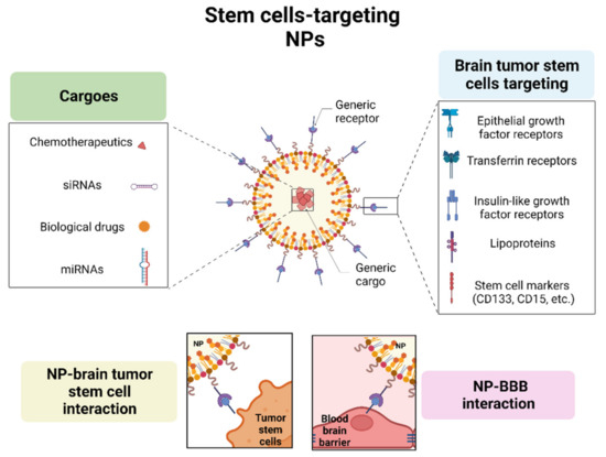
1.4.1. Epidermal Growth Factor Receptor

EGFR is a tyrosine kinase receptor (RTK) that interacts with a variety of EGFR family ligands regulating various aspects of cell growth and development. In the context of human brain tumors, EGFR is overexpressed in a subset of GBM and MB and EGFR targeting is a promising approach for the NPs’ delivery [10][11].
EGFR-decorated NPs (EGFR-NPs) can be used as carriers for chemotherapy agents that are not able to cross the BBB, in physiological conditions (such as temozolomide, TMZ), and that are subsequently internalized by target cells via receptor-mediated endocytosis. Several studies reported examples of EGFR-NPs strategies in glioma tumors. Liu and colleagues developed drug-loaded NPs functionalized with dual-targeting of EGFR (expressed on tumor cells) and of low-density lipoprotein receptor-relative protein-1 (LRP1) (expressed on the BBB endothelial). The dual targeting exhibited enhanced BBB penetrability and tumor targeting effects, in both in vitro and in vivo glioma models [12]. Another study [13] reported the efficiency of EGFR-NPs for the delivery of photosensitizer silicon phthalocyanine (Pc 4) against glioma tumors. Additionally, Whittle et al. described a phase I clinical trial on patients affected by recurrent GBM treated with weekly administration of novel nano cellular compounds loaded with a chemotherapeutic agent and functionalized with anti-EGFR antibodies, demonstrating no dose-limiting toxicity in fourteen patients [14].
Beyond the drug delivery, EGFR is an attractive biomarker also for brain tumor imaging, acting as both a diagnostic and therapeutic agent. This method could be used for the real-time tracking of NPs in crossing the BBB, in the accumulation of NPs within target tissues and in providing shape contour-defining imaging of the tumor. This strategy also aids in the detection of distant metastasis and residual disease following incomplete surgical resection. Additionally, the measurement of the dilution of NPs could be used as a marker of the cell proliferation rate in the different areas of the tumor. Among imaging agents, fluorescent molecules are gaining popularity in clinical diagnostics [15][16]. Hadjipanayis et al. reported a study in which magnetic NPs conjugated to an EGFR deletion mutant (EGFRvIII) antibody are used for GBM detection (via magnetic resonance imaging (MRI)) and targeted therapy [17] (see also Table 1).

1.4.2. Transferrin Receptor

The Tf receptor (TfR) is a transmembrane glycoprotein with two subunits linked by a disulfide bridge, each of which can bind to a molecule of transferrin, and functions to transport iron into cells. TfRs are widely expressed in the body, including red blood cells, hepatocytes, monocytes, erythrocytes, intestinal cells and normal brain cells (endothelial, neurons and glial cells) but also in brain cancers, making TfR an excellent candidate for the design of targeted therapy in brain tumors [28][29]. One of the advantages of using Tf-conjugated nanoparticles (Tf-NPs) is the ability to cross the BBB. However, one of the limitations is that the supplied Tf, carried by Tf-NPs, must compete with the endogenous Tf in plasma, and the BBB TfRs are >99.9% saturated with endogenous Tf [25][30]. Another limitation is that the exogenous Tf could lead to an overdose of the iron transport into the brain, NPs conjugated to TfR-targeted antibodies are preferable to Tf-NPs because they bind a different site on TfR [26]. GBM cells overexpress TfRs to respond to the increased demand of iron to sustain tumor growth, the expression of TfRs in this tumor is indeed almost 100-fold higher compared to healthy normal astrocytes. Many studies reported the use of Tf in drug delivery approaches for GBM therapy [19][20][21][22][23][24]. TfR is also over-expressed on the surfaces of GBM cancer stem cells (GSCs), so it could be considered a common target for both stem and bulk tumor cells. Based on these observations, Sun and colleagues developed a nano-strategy, using TMZ-loaded Tf-NPs, able to effectively penetrate the BBB and target both tumor compartments: glioma stem cells and non-stem cells [31]. Also, Kim et colleagues have developed anti-cancer NPs, using TfR as a common target, in both CSC and non-CSC populations [32] (see also Table 1).

1.4.3. Insulin Receptor

Insulin was the first molecular “Trojan horse” described able to cross BBB and to deliver somatostatin (Pardridge, W.M. Chimeric Peptides for Neuropeptide Delivery through the blood–brain barrier. US Patent 4,801,575, 31 January 1989). Subsequently, Shilo and colleagues developed insulin-targeted NPs and demonstrated, in in vivo models (male BALB/c mice), the ability to cross the BBB five times more than controls [33]. Furthermore, 83-14 monoclonal antibody to the human insulin receptor was used to functionalize NPs which had a greater ability to cross the BBB with respect to the anti-TfR antibody [34]. The anti-insulin receptor antibody 83-14 was also successfully used by Dieu et al., who demonstrated in in vitro experiments the specific endocytosis of these NPs by brain endothelial cells [35]. In another study, insulin or an anti-insulin receptor monoclonal antibody (29B4) linked to NPs were found to be capable of transporting non-penetrating drugs across the BBB [36] (see also Table 1).

1.4.4. Lipoprotein

The low-density lipoprotein (LDL) receptor (LDLR) is over-expressed in tumors (cells and blood vessels) and in the BBB, which allows the use of LDL-NPs for brain-targeted therapy. Various in vivo and in vitro studies reported an increased diffusion of LDLR ligands-functionalized NPs: LDL or Apolipoprotein E and B (ApoE and ApoB) [37][38]. A study led by Grafals-Ruiz et al. highlighted the potential for the use of ApoE-conjugated-NPs in GBM tumors, in in vitro (U87 GBM cells) and in vivo models (GBM syngeneic mice) [27]. Also, synthetic nano-LDL particles were proposed as effective drug delivery vehicles for GBM [39][40]. Additionally, NPs conjugated to Angiopep-2, which is a ligand that binds to the LDL low-density lipoprotein receptor-related protein (LRP), was proposed as an excellent option for drug delivery, as described by Kadari et al. in human (U87MG) and mouse (GL261) glioma cell lines and mouse GBM models [41]. Of note, high-density lipoprotein nanoparticles (HDL NPs) have intrinsic anti-tumoral activity in targeting the cholesterol signaling pathway. HDL-mimetic NPs were successfully used by Bell et al. in Sonic Hedgehog (SHH)-driven MB, demonstrating the high-affinity of HDL NPs in binding the HDL scavenger receptor type B1, SCARB1, depriving cells of natural HDL and cholesterol cargo. This strategy resulted in a promising approach, highly dependent on cellular cholesterol levels, to target stem cell compartments [42] (see also Table 1).

2. Immunotherapy and Nanoparticles in Pediatric Brain Tumors

The complex interplay between cancer and immune cells regulates tumor development: indeed, immune system activity has a key role in controlling disease initiation and progression and at the same time the immune microenvironment is highly influenced by tumor signaling. For these reasons, immunotherapy represents a powerful therapeutic approach for many types of cancer but has shown less benefit against pediatric brain tumors. The presence of the BBB as well as the high tumor heterogeneity and a suppressive immune microenvironment have limited the development of effective immunotherapeutic approaches. To this purpose, combining immunotherapy with nanotechnology could provide novel opportunities to improve pediatric brain cancer therapy. Different types of nano-immunoconjugates have been developed to reduce immunosuppression or improve immune activation in brain tumor sites [43][44]. Checkpoint inhibitors, such as anti-CTLA-4 and anti-PD-1 [45] or anti-PDL-1 [46] have been covalently attached to NPs to activate an immune response in GBM; also cytokines have been loaded into NPs to treat GBM [47]. These approaches, despite being proven to be effective in modulating the immune system to treat brain cancers, can cause an over-activation of immune cells and off-target inflammation, making it necessary to develop alternative ways to affect immune cells. To this purpose, the therapeutic delivery of NPs encapsulating RNA seems to be a promising solution. Different types of RNA have been encapsulated in NPs and tested in several pediatric brain tumors, as follows: (i) mRNA encoding for transcriptions factors that induce inflammatory gene expression in immune cells in GBM [48]; (ii) siRNA to reduce immunosuppression by downregulating anti-inflammatory signals in GBM [49][50] and DIPG [51]; (iii) tumor-derived RNA that quickly activate a tumor-specific immune response in MB [52] and DIPG [53]. This last strategy seems to be the most translatable at a clinical level, as shown by the Food and Drug Administration Investigational New Drug (FDA-IND) approval for the first-in-human trials (IND#BB-19304) in pediatric patients with HGGs using RNA-NPs [PNOC020 study, NCT04573140]. Despite the fact that further investigations are needed to allow the use of nano-immunoconjugates in a clinical setting, this trial has paved the way for the development of NP-based personalized immunotherapy for pediatric brain cancer treatment.

3. Advanced Pre-Clinical Models to Study Nanoparticles in Pediatric Brain Tumors

The BBB represents one of the main challenges in brain cancer therapy due to its low permeability to the majority of drugs. As a result, pre-clinical studies are mainly based on the identification of strategies to enhance BBB-crossing and brain tumor accumulation of drugs. 2D cell cultures have been widely used to study drug responses in vitro, but they are not able to fully recapitulate the tumor features and microenvironment and to properly evaluate the therapeutic potential of anti-cancer compounds. For this reason, recent pre-clinical studies have been focused on the development of 3D culture models to better mimic the tumor’s complexity as well as BBB–brain tumor interactions and to properly predict the in vivo treatment responses. Perini G et al. [54] used a 3D brain cancer model to study the efficacy of liposomal TMZ pre-coated with a protein corona made of human plasma proteins in inhibiting tumor growth. The 3D culture was derived from the U87 human GBM cell line and spheroid size analysis and cell viability assays were performed to evaluate the anticancer activity of these NPs. The results showed that the treatment of 3D GBM culture causes a notable reduction of tumor size, in line with a considerable decrease in cell viability, demonstrating a marked inhibition of tumor growth. To mimic the BBB–GBM interaction in vitro, Straehla JP et al. [55] designed a microfluidic device of vascularized GBM using GBM spheroids, derived from the co-culture of the patient-derived xenograft (PDX) GBM22 cell line with pericytes (PCs), in direct contact with a self-assembled and perfusable vascular network made of induced pluripotent stem cells (IPS), human endothelial cells, astrocytes and pericytes. In this model, GBM spheroids grew rapidly in close contact with their surrounding BBB vasculature, similar to what can be observed in vivo in HGG patients and in animal models of GBM. The authors generated a NP composed of a liposomal core, coated with a first layer of poly-(l-arginine) and a second layer of propargyl-modified poly-(l-aspartic acid) (pPLD), and functionalized with angiopep-2 (AP2) to target the GBM vessels via an interaction with the overexpressed BBB receptor LRP1. After demonstrating that targeted NPs are able to cross the BBB vessels near GBM tumor, the DNA-damaging agent CDDP was encapsulated in NPs and the therapeutical potential of these NPs was tested. The authors showed that NPs were able to accumulate in GBM spheroids and to increase the apoptosis of cancer cells with low damage to the surrounding healthy blood vessels. These results were confirmed in vivo in an ortothoptic, intracranial murine tumor model where treatment with these NPs decreased tumor growth, demonstrating the reliability of this in vitro model as a pre-clinical model. Due to the crucial role of CSC population in the initiation and progression of cancer, Kim JS et al. [56] designed a strategy to improve NPs delivery to specifically target stem cell compartment in SHH MB in vitro, in vivo and ex vivo. After proving the ability of these NPs to cross the BBB and to accumulate in MB cells as well as the therapeutic effect on MB stem cells both in vitro (DAOY and PZp53 cell line) and in vivo (SmoA1-GFP-MB-bearing mouse model and patched (PTC) knockout model), they generated an ex vivo SmoA1 organotypic slice culture to better mimic the BBB–MB interactions and to evaluate the anti-cancer effect of these NPs in the tumor microenvironment. In slice cultures treated with eHNP-A1-CD15, the observed co-localization in the perivascular space of CD15 and NPs demonstrated that this treatment was able to specifically target the stem cell population. Furthermore, the staining for the cell death marker cleaved-caspase 3 (CC3) showed higher CC3 staining in slice culture treated with LDE225-loaded NPs, demonstrating the therapeutic efficacy of these compounds on MB-SHH.

References

  1. Baig, N.; Kammakakam, I.; Falath, W. Nanomaterials: A Review of Synthesis Methods, Properties, Recent Progress, and Challenges. Mater. Adv. 2021, 2, 1821–1871.
  2. Shafey, A.M.E. Green Synthesis of Metal and Metal Oxide Nanoparticles from Plant Leaf Extracts and Their Applications: A Review. Green Process. Synth. 2020, 9, 304–339.
  3. Bhardwaj, B.; Singh, P.; Kumar, A.; Kumar, S.; Budhwar, V. Eco-Friendly Greener Synthesis of Nanoparticles. Adv. Pharm. Bull. 2020, 10, 566–576.
  4. Matsumura, Y.; Maeda, H. A New Concept for Macromolecular Therapeutics in Cancer Chemotherapy: Mechanism of Tumoritropic Accumulation of Proteins and the Antitumor Agent Smancs. Cancer Res. 1986, 46, 6387–6392.
  5. Benjamin, L.E.; Golijanin, D.; Itin, A.; Pode, D.; Keshet, E. Selective Ablation of Immature Blood Vessels in Established Human Tumors Follows Vascular Endothelial Growth Factor Withdrawal. J. Clin. Investig. 1999, 103, 159–165.
  6. Wu, J. The Enhanced Permeability and Retention (EPR) Effect: The Significance of the Concept and Methods to Enhance Its Application. J. Pers. Med. 2021, 11, 771.
  7. Bazak, R.; Houri, M.; El Achy, S.; Kamel, S.; Refaat, T. Cancer Active Targeting by Nanoparticles: A Comprehensive Review of Literature. J. Cancer Res. Clin. Oncol. 2015, 141, 769–784.
  8. Salahpour Anarjan, F. Active Targeting Drug Delivery Nanocarriers: Ligands. Nano-Struct. Nano Objects 2019, 19, 100370.
  9. Bayda, S.; Adeel, M.; Tuccinardi, T.; Cordani, M.; Rizzolio, F. The History of Nanoscience and Nanotechnology: From Chemical-Physical Applications to Nanomedicine. Molecules 2019, 25, 112.
  10. Yao, Y.; Zhou, Y.; Liu, L.; Xu, Y.; Chen, Q.; Wang, Y.; Wu, S.; Deng, Y.; Zhang, J.; Shao, A. Nanoparticle-Based Drug Delivery in Cancer Therapy and Its Role in Overcoming Drug Resistance. Front. Mol. Biosci. 2020, 7, 193.
  11. Sigismund, S.; Avanzato, D.; Lanzetti, L. Emerging Functions of the EGFR in Cancer. Mol. Oncol. 2018, 12, 3–20.
  12. Liu, C.; Zhao, Z.; Gao, H.; Rostami, I.; You, Q.; Jia, X.; Wang, C.; Zhu, L.; Yang, Y. Enhanced Blood-Brain-Barrier Penetrability and Tumor-Targeting Efficiency by Peptide-Functionalized Poly(Amidoamine) Dendrimer for the Therapy of Gliomas. Nanotheranostics 2019, 3, 311–330.
  13. Cheng, Y.; Meyers, J.D.; Agnes, R.S.; Doane, T.L.; Kenney, M.E.; Broome, A.-M.; Burda, C.; Basilion, J.P. Addressing Brain Tumors with Targeted Gold Nanoparticles: A New Gold Standard for Hydrophobic Drug Delivery? Small Weinh. Bergstr. Ger. 2011, 7, 2301–2306.
  14. Whittle, J.R.; Lickliter, J.D.; Gan, H.K.; Scott, A.M.; Simes, J.; Solomon, B.J.; MacDiarmid, J.A.; Brahmbhatt, H.; Rosenthal, M.A. First in Human Nanotechnology Doxorubicin Delivery System to Target Epidermal Growth Factor Receptors in Recurrent Glioblastoma. J. Clin. Neurosci. Off. J. Neurosurg. Soc. Australas. 2015, 22, 1889–1894.
  15. Schmitt, R.R.; Mahajan, S.D.; Pliss, A.; Prasad, P.N. Small Molecule Based EGFR Targeting of Biodegradable Nanoparticles Containing Temozolomide and Cy5 Dye for Greatly Enhanced Image-Guided Glioblastoma Therapy. Nanomed. Nanotechnol. Biol. Med. 2022, 41, 102513.
  16. Meola, A.; Rao, J.; Chaudhary, N.; Sharma, M.; Chang, S.D. Gold Nanoparticles for Brain Tumor Imaging: A Systematic Review. Front. Neurol. 2018, 9, 328.
  17. Hadjipanayis, C.G.; Machaidze, R.; Kaluzova, M.; Wang, L.; Schuette, A.J.; Chen, H.; Wu, X.; Mao, H. EGFRvIII Antibody-Conjugated Iron Oxide Nanoparticles for Magnetic Resonance Imaging-Guided Convection-Enhanced Delivery and Targeted Therapy of Glioblastoma. Cancer Res. 2010, 70, 6303–6312.
  18. Ertas, Y.N.; Abedi Dorcheh, K.; Akbari, A.; Jabbari, E. Nanoparticles for Targeted Drug Delivery to Cancer Stem Cells: A Review of Recent Advances. Nanomaterials 2021, 11, 1755.
  19. Liu, G.; Mao, J.; Jiang, Z.; Sun, T.; Hu, Y.; Jiang, Z.; Zhang, C.; Dong, J.; Huang, Q.; Lan, Q. Transferrin-Modified Doxorubicin-Loaded Biodegradable Nanoparticles Exhibit Enhanced Efficacy in Treating Brain Glioma-Bearing Rats. Cancer Biother. Radiopharm. 2013, 28, 691–696.
  20. Ren, W.; Chang, J.; Yan, C.; Qian, X.; Long, L.; He, B.; Yuan, X.; Kang, C.; Betbeder, D.; Sheng, J.; et al. Development of Transferrin Functionalized Poly(Ethylene Glycol)/Poly(Lactic Acid) Amphiphilic Block Copolymeric Micelles as a Potential Delivery System Targeting Brain Glioma. J. Mater. Sci. Mater. Med. 2010, 21, 2673–2681.
  21. Xiao, W.; Wang, Y.; Zhang, H.; Liu, Y.; Xie, R.; He, X.; Zhou, Y.; Liang, L.; Gao, H. The Protein Corona Hampers the Transcytosis of Transferrin-Modified Nanoparticles through Blood-Brain Barrier and Attenuates Their Targeting Ability to Brain Tumor. Biomaterials 2021, 274, 120888.
  22. Sheykhzadeh, S.; Luo, M.; Peng, B.; White, J.; Abdalla, Y.; Tang, T.; Mäkilä, E.; Voelcker, N.H.; Tong, W.Y. Transferrin-Targeted Porous Silicon Nanoparticles Reduce Glioblastoma Cell Migration across Tight Extracellular Space. Sci. Rep. 2020, 10, 2320.
  23. Luo, M.; Lewik, G.; Ratcliffe, J.C.; Choi, C.H.J.; Mäkilä, E.; Tong, W.Y.; Voelcker, N.H. Systematic Evaluation of Transferrin-Modified Porous Silicon Nanoparticles for Targeted Delivery of Doxorubicin to Glioblastoma. ACS Appl. Mater. Interfaces 2019, 11, 33637–33649.
  24. Liu, D.-Z.; Cheng, Y.; Cai, R.-Q.; Wang Bd, W.-W.; Cui, H.; Liu, M.; Zhang, B.; Mei, Q.-B.; Zhou, S.-Y. The Enhancement of SiPLK1 Penetration across BBB and Its Anti Glioblastoma Activity in Vivo by Magnet and Transferrin Co-Modified Nanoparticle. Nanomed. Nanotechnol. Biol. Med. 2018, 14, 991–1003.
  25. Pardridge, W.M. A Historical Review of Brain Drug Delivery. Pharmaceutics 2022, 14, 1283.
  26. Sharma, G.; Lakkadwala, S.; Modgil, A.; Singh, J. The Role of Cell-Penetrating Peptide and Transferrin on Enhanced Delivery of Drug to Brain. Int. J. Mol. Sci. 2016, 17, 806.
  27. Grafals-Ruiz, N.; Rios-Vicil, C.I.; Lozada-Delgado, E.L.; Quiñones-Díaz, B.I.; Noriega-Rivera, R.A.; Martínez-Zayas, G.; Santana-Rivera, Y.; Santiago-Sánchez, G.S.; Valiyeva, F.; Vivas-Mejía, P.E. Brain Targeted Gold Liposomes Improve RNAi Delivery for Glioblastoma. Int. J. Nanomed. 2020, 15, 2809–2828.
  28. Shen, Y.; Li, X.; Dong, D.; Zhang, B.; Xue, Y.; Shang, P. Transferrin Receptor 1 in Cancer: A New Sight for Cancer Therapy. Am. J. Cancer Res. 2018, 8, 916–931.
  29. Rosager, A.M.; Sørensen, M.D.; Dahlrot, R.H.; Hansen, S.; Schonberg, D.L.; Rich, J.N.; Lathia, J.D.; Kristensen, B.W. Transferrin Receptor-1 and Ferritin Heavy and Light Chains in Astrocytic Brain Tumors: Expression and Prognostic Value. PLoS ONE 2017, 12, e0182954.
  30. Pardridge, W.M.; Chou, T. Mathematical Models of Blood-Brain Barrier Transport of Monoclonal Antibodies Targeting the Transferrin Receptor and the Insulin Receptor. Pharmaceuticals 2021, 14, 535.
  31. Sun, T.; Wu, H.; Li, Y.; Huang, Y.; Yao, L.; Chen, X.; Han, X.; Zhou, Y.; Du, Z. Targeting Transferrin Receptor Delivery of Temozolomide for a Potential Glioma Stem Cell-Mediated Therapy. Oncotarget 2017, 8, 74451–74465.
  32. Kim, S.-S.; Rait, A.; Rubab, F.; Rao, A.K.; Kiritsy, M.C.; Pirollo, K.F.; Wang, S.; Weiner, L.M.; Chang, E.H. The Clinical Potential of Targeted Nanomedicine: Delivering to Cancer Stem-like Cells. Mol. Ther. J. Am. Soc. Gene Ther. 2014, 22, 278–291.
  33. Shilo, M.; Motiei, M.; Hana, P.; Popovtzer, R. Transport of Nanoparticles through the Blood-Brain Barrier for Imaging and Therapeutic Applications. Nanoscale 2014, 6, 2146–2152.
  34. Wu, D.; Yang, J.; Pardridge, W.M. Drug Targeting of a Peptide Radiopharmaceutical through the Primate Blood-Brain Barrier in Vivo with a Monoclonal Antibody to the Human Insulin Receptor. J. Clin. Investig. 1997, 100, 1804–1812.
  35. Dieu, L.-H.; Wu, D.; Palivan, C.G.; Balasubramanian, V.; Huwyler, J. Polymersomes Conjugated to 83-14 Monoclonal Antibodies: In Vitro Targeting of Brain Capillary Endothelial Cells. Eur. J. Pharm. Biopharm. Off. J. Arb. Pharm. Verfahr. EV 2014, 88, 316–324.
  36. Ulbrich, K.; Knobloch, T.; Kreuter, J. Targeting the Insulin Receptor: Nanoparticles for Drug Delivery across the Blood-Brain Barrier (BBB). J. Drug Target. 2011, 19, 125–132.
  37. Pawar, S.; Koneru, T.; McCord, E.; Tatiparti, K.; Sau, S.; Iyer, A.K. LDL Receptors and Their Role in Targeted Therapy for Glioma: A Review. Drug Discov. Today 2021, 26, 1212–1225.
  38. Hayavi, S.; Halbert, G.W. Synthetic Low-Density Lipoprotein, a Novel Biomimetic Lipid Supplement for Serum-Free Tissue Culture. Biotechnol. Prog. 2005, 21, 1262–1268.
  39. Nikanjam, M.; Blakely, E.A.; Bjornstad, K.A.; Shu, X.; Budinger, T.F.; Forte, T.M. Synthetic Nano-Low Density Lipoprotein as Targeted Drug Delivery Vehicle for Glioblastoma Multiforme. Int. J. Pharm. 2007, 328, 86–94.
  40. Nikanjam, M.; Gibbs, A.R.; Hunt, C.A.; Budinger, T.F.; Forte, T.M. Synthetic Nano-LDL with Paclitaxel Oleate as a Targeted Drug Delivery Vehicle for Glioblastoma Multiforme. J. Control. Release Off. J. Control. Release Soc. 2007, 124, 163–171.
  41. Kadari, A.; Pooja, D.; Gora, R.H.; Gudem, S.; Kolapalli, V.R.M.; Kulhari, H.; Sistla, R. Design of Multifunctional Peptide Collaborated and Docetaxel Loaded Lipid Nanoparticles for Antiglioma Therapy. Eur. J. Pharm. Biopharm. Off. J. Arb. Pharm. Verfahr. EV 2018, 132, 168–179.
  42. Bell, J.B.; Rink, J.S.; Eckerdt, F.; Clymer, J.; Goldman, S.; Thaxton, C.S.; Platanias, L.C. HDL Nanoparticles Targeting Sonic Hedgehog Subtype Medulloblastoma. Sci. Rep. 2018, 8, 1211.
  43. Poot, E.; Maguregui, A.; Brunton, V.G.; Sieger, D.; Hulme, A.N. Targeting Glioblastoma through Nano- and Micro-Particle-Mediated Immune Modulation. Bioorg. Med. Chem. 2022, 72, 116913.
  44. Guido, C.; Baldari, C.; Maiorano, G.; Mastronuzzi, A.; Carai, A.; Quintarelli, C.; De Angelis, B.; Cortese, B.; Gigli, G.; Palamà, I.E. Nanoparticles for Diagnosis and Target Therapy in Pediatric Brain Cancers. Diagnostics 2022, 12, 173.
  45. Galstyan, A.; Markman, J.L.; Shatalova, E.S.; Chiechi, A.; Korman, A.J.; Patil, R.; Klymyshyn, D.; Tourtellotte, W.G.; Israel, L.L.; Braubach, O.; et al. Blood-Brain Barrier Permeable Nano Immunoconjugates Induce Local Immune Responses for Glioma Therapy. Nat. Commun. 2019, 10, 3850.
  46. Meng, L.; Wang, C.; Lu, Y.; Sheng, G.; Yang, L.; Wu, Z.; Xu, H.; Han, C.; Lu, Y.; Han, F. Targeted Regulation of Blood-Brain Barrier for Enhanced Therapeutic Efficiency of Hypoxia-Modifier Nanoparticles and Immune Checkpoint Blockade Antibodies for Glioblastoma. ACS Appl. Mater. Interfaces 2021, 13, 11657–11671.
  47. Voth, B.L.; Pelargos, P.E.; Barnette, N.E.; Bhatt, N.S.; Chen, C.H.J.; Lagman, C.; Chung, L.K.; Nguyen, T.; Sheppard, J.P.; Romiyo, P.; et al. Intratumor Injection of CCL21-Coupled Vault Nanoparticles Is Associated with Reduction in Tumor Volume in an in Vivo Model of Glioma. J. Neurooncol. 2020, 147, 599–605.
  48. Zhang, F.; Parayath, N.N.; Ene, C.I.; Stephan, S.B.; Koehne, A.L.; Coon, M.E.; Holland, E.C.; Stephan, M.T. Genetic Programming of Macrophages to Perform Anti-Tumor Functions Using Targeted MRNA Nanocarriers. Nat. Commun. 2019, 10, 3974.
  49. Azambuja, J.H.; Schuh, R.S.; Michels, L.R.; Gelsleichter, N.E.; Beckenkamp, L.R.; Iser, I.C.; Lenz, G.S.; de Oliveira, F.H.; Venturin, G.; Greggio, S.; et al. Nasal Administration of Cationic Nanoemulsions as CD73-SiRNA Delivery System for Glioblastoma Treatment: A New Therapeutical Approach. Mol. Neurobiol. 2020, 57, 635–649.
  50. Kadiyala, P.; Gregory, J.V.; Lowenstein, P.R.; Lahann, J.; Castro, M.G. Targeting Gliomas with STAT3-Silencing Nanoparticles. Mol. Cell. Oncol. 2021, 8, 1870647.
  51. Lenzen, A.; Cole, L.; Lauing, K.L.; Zhai, L.; Ladomersky, E.; Lulla, R.R.; Hashizume, R.; Stegh, A.; Wainwright, D.A. Immu-24. Immunotherapeutic Nanotechnology Targeting Ido1 for Pediatric Diffuse Intrinsic Pontine Glioma. Neuro Oncol. 2018, 20, i103.
  52. Sayour, E.; Mendez-Gomez, H.; Grippin, A.; De Leon, G.; Stover, B.; Flores, C.; Pham, C.; Mitchell, D. Mbrs-02. Personalized Immunotherapy with Translatable Rna Nanoparticles Targeting Medulloblastoma. Neuro Oncol. 2018, 20, i128–i129.
  53. Mendez-Gomez, H.; McGuiness, J.; Grippin, A.; Weidert, F.; Carrera-Justiz, S.; Mitchell, D.; Sayour, E. Immu-13. Customizable Multi-Lamellar Rna-Nanoparticles for Pediatric Glioma. Neuro Oncol. 2021, 23, i29–i30.
  54. Perini, G.; Giulimondi, F.; Palmieri, V.; Augello, A.; Digiacomo, L.; Quagliarini, E.; Pozzi, D.; Papi, M.; Caracciolo, G. Inhibiting the Growth of 3D Brain Cancer Models with Bio-Coronated Liposomal Temozolomide. Pharmaceutics 2021, 13, 378.
  55. Straehla, J.P.; Hajal, C.; Safford, H.C.; Offeddu, G.S.; Boehnke, N.; Dacoba, T.G.; Wyckoff, J.; Kamm, R.D.; Hammond, P.T. A Predictive Microfluidic Model of Human Glioblastoma to Assess Trafficking of Blood-Brain Barrier-Penetrant Nanoparticles. Proc. Natl. Acad. Sci. USA 2022, 119, e2118697119.
  56. Kim, J.S.; Shin, D.H.; Kim, J.-S. Dual-Targeting Immunoliposomes Using Angiopep-2 and CD133 Antibody for Glioblastoma Stem Cells. J. Control. Release Off. J. Control. Release Soc. 2018, 269, 245–257.
More
Video Production Service