Bioapplications of Amphiphilic Janus Dendrimers: Comparison
Please note this is a comparison between Version 1 by Ioana Maria Carmen Ienascu and Version 3 by Jessie Wu.

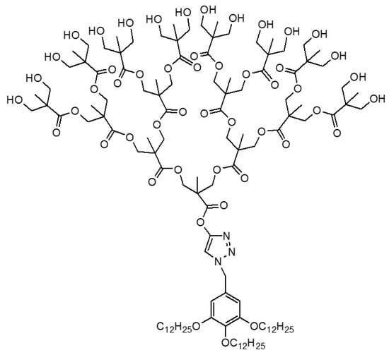
Amphiphilic Janus dendrimers (JDs) can be defined as dendritic macromolecules made of two dendrons with opposite polarities, which either differ from each other by their terminal groups, or their structures differ entirely. Amphiphilic JDs are named following a more or less general scheme that encompass in this order, the lipophilic dendrons, the core if exists, and the hydrophilic dendrons. The unique architectures of amphiphilic Janus dendrimers, with multifunctional terminal groups, different structures of branches in a single molecule (beside the capacity of self-assembly in aqueous media forming dendrimersomes, which in turn benefit from properties like predictable size and thickness, stability and biocompatibility), constitute premises for a wide range of biomedical applications where conventional dendrimers have failed.

  • Janus dendrimers
  • dendrimersomes
  • glycodendrimersomes
  • stabilizing agents
  • nanocarriers
  • gene delivery

1. Janus Dendrimers as Stabilizing Agents

1.1. Drug Suspensions

The formulation of pharmaceutical nanosuspensions requires the presence of stabilizers, which act via electrostatic or steric interactions and enhance the dissolution of poorly water-soluble drug molecules. Amphiphilic Janus dendrimers can be adsorbed onto drug particles and provide steric stabilization against aggregation and recrystallization, offering a versatile platform for general use as stabilizing agents for drug suspensions [1].
Thus, four new amphiphilic Janus dendrimers with hydroxyl-terminated bis-MPA dendrons and dodecyloxy chains were obtained and tested for their ability to stabilize colloidal drug suspensions through steric stabilization. Janus dendrimers (JDs) adsorb onto indomethacine surfaces, forming hydrophobic interactions, and the adsorption kinetics are strongly related to the number of hydrophobic alkyl chains. These experiments proved the potential of such G4 JDs as stabilizers in nanomilling (Figure 1) and other steps of dry-state processing of drugs [1].
Figure 1.
G4 JDs as stabilizers in media milling and dry-state processing of pharmaceuticals
[1]
.

1.2. Au and Ag Nanoparticles

Silver and gold NPs are very important for nanoscience and nanotechnology due to their numerous applications in catalysis, materials science, optical biosensors, and nanomedicine [2][3][4].
Janus metallodendrimers incorporate metal-containing moieties into the structure of JDs and benefit from the exceptional combination of organic and inorganic components, with outstanding catalytic, electronic, magnetic and radioactive properties [2].
Thus, ferrocene (Fc)-containing JDs can be used as neutral single-electron transfer agents for the reduction of Au(III) and Ag(I) in the synthesis of AuNPs [5][6][7][8][9][10][11][12][13] and AgNPs [11][12][13][14][15][16].
Few experiments regarding the preparation of silver and gold NPs using water-soluble Fc-containing dendrimers as both, stabilizers and reductants, in water systems involving no external reductant are available [2][17]. The use of external reducing agents, such as NaBH4, along with Fc-containing dendrimers for obtaining AgNPs and AuNPs, are highlighted in some works [2][17][18].
Hence, two novel amphiphilic triazolylferrocenyl with TEG termini JD were obtained by the click reaction between azidomethylferrocene and TEG-terminated acetylenes, and tested as neutral ligands for the AgNPs-1 and AuNPs-1 stabilization in water in the presence of NaBH4 as a reducing agent for Ag(I) and Au(III). In the reaction between JDs and Ag(I) or Au(III), the significant impact of the electronic properties of the ferrocenyl-core linker on the viability of the redox reaction, and the ability of this kind of JDs to manage such redox processes was proven. The authors also highlighted the potential of ferrocenyl-containing JDs with varied types of redox active units for extending the spectrum of properties in this field [17].
Redox-responsive Fc-containing polymers could also be used as drug carriers, ensuring manageable processes of delivery and release of the drugs due to the reversible conversion between hydrophobic Fc and hydrophilic ferrocenium groups via chemical and electrochemical reduction, offering them the capacity to form reversible self-assembly systems [2][19][20].

2. Janus Dendrimers as Biological Membrane Mimics

The understanding of various cellular processes needs some artifacts like biological reconstitution of the cell membranes, thus a significant interest in designing synthetic analogs and models of the membrane components is granted.
Two important components of cell membranes are phospholipids and glycolipids. Phospholipids can be mimicked by amphiphilic JDs which have the ability to self-assemble in different liquid media into stable dendrimersomes (DSs) [21][22][23] with membrane thickness similar to phospholipids [21]. Janus glycodendrimers (JGDs) obtained by conjugation of sugars with JDs, were able to self-assemble in buffer solutions [21][24][25][26] into glycodendrimersomes (GDSs) which mimic glycolipids.
Therefore, dendrimersomes and glycodendrimersomes could successfully substitute liposomes if rwesearchers consider their special features like tunable size [27], structural organization [24][28], enhanced stability [23][28], biocompatibility, similar membrane to living cells [21] and functional surfaces [24].
Based on the studies on the coassembly into hybrid vesicles of block copolymers, natural phospholipids and membrane proteins [29][30][31][32][33], DSs and GDSs have also been joined with phospholipids, membrane proteins, bacterial cell membranes, etc., forming hybrid architectures with unique morphologies that lend themselves to interesting biological applications.

2.1. Amphiphilic Janus Dendrimers (JDs)

In order to stabilize the cell membranes and vesicles that have originated from human cells, Yadavalli and coworkers (2019) used DSs obtained from appropriate JDs containing hydrophobic 3,5-di-dodecyloxybenzoic ester dendrons and hydrophilic 3,4,5-tris-triethylene glycol benzoic ester dendrons. They also proved the value of such DSs as potential cell-targeting agents by combining them with bacterial membrane vesicles holding a bacterial adhesin protein (YadA), generating a cell-like hybrid vesicles recognized by the living human cells. Using DSs, the development of synthetic and hybrid protocell capsules to perform cell-like functions such as recognition, signaling, and delivery, seems tangible [23].
Natural polyphenols and phenolic acids possessing dodecyloxy chains were employed as building blocks for designing new amphiphilic JDs as cell membrane mimics, with bilayer thickness comparable to the natural phospholipids having 16 or 18 carbons in the alkyl groups. Unilamellar DSs were obtained in higher yields and shorter reaction times via self-assembly of such programmable JDs, and have the potential to act as significant tools for synthetic cell biology, encapsulation, and delivery [34].
A new type of zwitterionic JD, consisting of a zwitterionic phosphocholine hydrophilic headgroup and a 3,5-substituted dihydroxybenzoate-based hydrophobic dendron, was introduced by Joseph et al. [35]. This type of JDs self-assembles in water into zwitterionic dendrimersomes (z-DSs) that closely mimic the thickness, lateral mobility, and flexibility of natural cell membranes.
The combination of the two key features of cell-like DSs assembled from JDs, i.e., high flexibility and stability, confers them the ability to engulf bacteria. Kostina et al. (2019) synthesized new JDs which self-assemble into unilamellar vesicles with a biomimetic thickness, high flexibility and stability conferred by dodecane hydrophobic chains, and ultra-low adhesion to bacteria owed to the tri(ethylene oxide) hydrophilicity. These cell-like DSs proved superior to natural liposomes and synthetic polymersomes in terms of living bacteria endocytosis, emphasizing the opportunities of using the dendrimersomes in biomedical field [36].
Kostina et al. (2021) studied the influence of variation of amphiphile topology in the bilayer of some photoactive/stable JDs on shape changes of such cell membrane models, and demonstrated the utility of DSs to simulate and explain some cellular processes occurring in living cells [37].
Torre et al., 2019 considered the rebuilding of synthetic cells starting from compartmentation, encapsulation, and surface ornamentation of unilamellar and onion-like DSs self-assembled from amphiphilic JDs. They propose an approach for the modularly binding of proteins, nucleic acids, and hydrophobic target compounds to the edge of the DSs, demonstrating their value as adaptable synthetic biological membranes [22].

2.2. Janus glycodendrimers (JGDs)

Janus glycodendrimers (JGDs), synthetic macromolecules mimicking glycolipids, were proved to self-assemble into nanovesicles presenting carbohydrates on their external surface, like the glycocalyx layer of eukaryotic cells, bacteria, and viruses.
Glycodendrimersomes (GDSs) are feasible mimics of biological membranes, i.e., glycans, that can serve to explain the structure and function of glycans. GDSs are self-assembled nanovesicles with cell surface-like programmable structures comprising carbohydrates moieties, which can be combined with lectins to discover the complexity of glycan-lectin specificity. Clinically, GDs could be used to target the nanoparticles to lectins in vivo and to specifically scavenge a lectin at sites of damaging action [24]. Xiao and coworkers (2018) synthesized Janus glycodendrimers with various sugar headgroups and coupled them with homodimeric, heterodimeric, and chimera galectins, to disclose the degree of cross-linking of the biomimetic nanoscale vesicles having both charged and uncharged ligands, hence contributing to elucidate the nature of these vital interactions [24].
Other exceptional Janus amphiphilic glycopeptide dendrimers for the biomimicry of glycans exhibited strong and specific recognitions with C-type mannose-specific lectin [38].
Rodriguez-Emmenegger (2019) obtained GDs acting as cell-membrane mimics, with hierarchical morphologies resembling bicomponent rafts starting from JGDs containing Lac and triethylene glycol. Their structures encode biological recognition and permit to reduce sugar–sugar interactions, allowing stronger binding to proteins, which recommend them for applications in cellular biology and nanomedicine [25].
Some JGDs were obtained by an isothiocyanate–amine coupling reaction among isothiocyanate-containing sequence-defined JDs, and linear or branched carbohydrates containing oligosaccharide and hydrophobic amino-pentyl units. These structures containing thiourea groups, which enhances the hydrophilic character of the N-pentyl linker, self-assemble into nanovesicles with lamellar surface, that mimic the recognition assemblies of the cell-surface of glycans and viral glycoproteins and displayed the potential of elucidating the ability of viruses to camouflage in order to avoid recognition [39].
Similar results, regarding the important influence of the thiourea interconnecting group placed amid the hydrophobic and hydrophilic dendrons of JDSs to their self-assembly, were obtained by Murphy et al. (2021). They also proved the stabilization of glycolipid-rich rafts and association of sulfatide-rich regions with specific glycoproteins [26].
Various pathogenic bacterial strains act by infecting the host cells through multivalent host-guest connections [40]. In order to find optimized carbohydrates-containing dendrimers nanostructures known for their ability to capture bacterial strains [28], Krishnan et al. (2020) designed carbohydrate-based Janus dendrimers that spontaneously self-assemble into high aspect ratio 2D sheets. The conjugation between the hydrophilic dendron containing galactose and hydrophobic tetraphenylethylene (TPE) dendron formed two JDs that proved to be multivalent ligands for bacteria able to capture and agglutinate them and to inhibit the bacterial proliferation [41].
Kostina et al. (2021) utilized glycodendrimersomes synthetic vesicles formed by the self-assembly of mannose-JDs, whose membrane simulate the surface of a cell, to examine the carbohydrate biological activity [42].

3. Janus Dendrimers as Nanocarriers

The limitations of free drugs, for example poor water-solubility, toxicity related to a non-selective biodistribution and multi drug resistance, could be prevented using vesicular assemblies as drug delivery systems. As against micelles, vesicles benefit from the ability to encapsulate both hydrophobic and hydrophilic cargos [43].
The suitable self-assembly comportment of amphiphilic JDs has turned into a powerful tactic to obtain nanocarriers [17][34], with strong advantages for drug-delivery applications [43][44][45][46][47].
Both dyes and drugs have been encapsulated in dendrimersomes. Hydrophobic molecules were added to an organic solution of the JD and used for thin film hydration or oil-in-water approaches, resulting in the inclusion of the molecule in the hydrophobic bilayer of DSs. On the other hand, hydrophilic molecules were encapsulated in the hydrophilic cavity of DSs by addition to the water or buffer solution, used forward for hydration in the thin film approach or to the organic phase in the oil-in-water approach. The excess amount was removed via dialysis or size-exclusion chromatography [21].
Thus, the encapsulation of dyes, such as calcein in DSs self-assembled JDs containing bis-MPA-based polyester hydrophilic dendrons G1-G3 and (3,4,5)-trisubstituted Percec-type hydrophobic dendrons [48], carboxyfluorescein in DSs coassembled from JD comprising bis-MPA-based polyester hydrophilic dendrons G2 and (3,5)-disubstituted Percec-type hydrophobic dendrons [49], fluorescein and Nile red in DSs self-assembled from JD bearing hydrophobic photodegradable polyester dendron G3 with o-nitrobenzyl units and hydrophilic (3,4,5) Percec-type dendron G1 with tri(ethylene glycol) monomethyl ether chains [50], were earlier reported. The dye-loaded DSs proved impermeability and stability in time [48], degradation and triggered release upon irradiation with UV light [50], or fluorescence color changes as a function of pH [51].
Until 2018, the most researches regarding the use of JDs as drug nanocarriers refer to the encapsulation of anticancer drugs. Therefore, the inclusion of doxorubicin in DSs self-assembled from either JDs with bis-MPA-based polyester hydrophilic G2 dendrons and (3,5)-disubstituted Percec-type hydrophobic dendrons [48], or JD bearing a hydrophilic poly(amidoamine) dendron and two hydrophobic C18 alkyl chains [52] and Plitidepsin in DSs co-assembled from JD comprising two bis-MPA dendrimers G1-3 of different polarity, the hydrophilic one with free terminal OH groups, and the hydrophobic one functionalized with aliphatic chains derived from stearic acid [53] were reported. The drug-DSs assemblies showed good stability and impermeability [48][53], pH-dependent drug release [48][51], prevention of systemic toxicity [52], or encapsulating ability dependent on the hydrophobic/hydrophilic ratio of dendrons [53].
More recently, DSs were used as extraordinary carriers for various small molecules and macromolecules, being a concrete promise as therapeutic entities for different diseases treatment.
Unique amphiphilic Janus glycopeptide dendrimers, with precise and varied molecular structures containing β-cyclodextrin as core grafted with hydrophilic saccharides as dendrons and hydrophobic peptides as arms, were designed and synthesized by Bi and colleagues (2022). They were self-assembled into different controllable nanostructures such as glycospheres, worm-like micelles, and fibers conditioned by the repeating unit fraction of saccharides and phenylalanine. These nanomaterials were successfully encapsulated with Nile red dye and fluorofenidone, an anti-inflammatory agent [38].
Redox-sensitive DSs, containing disulfide-linked cholesterol-bearing PEGylated dendrimers, were also able to entrap both the lipophilic Nile red and the hydrophilic dye rhodamine. The redox-triggered release of the encapsulated lipophilic dye from the cholesterol-based DSs was assessed using glutathione as a reducing agent [54]. Rhodamine B was also encapsulated in a triazolylferrocenyl JD micelles and released based on the redox response of the Fc moiety [2]. On the other hand, hydrophobic fluorescent probe molecules, N-phenyl-1-naphthylamine and coumarin-153, were entrapped in DSs obtained by spontaneous self-assembly of a disulfide-linked octadecyl chain modified PEGylated dendrimer, which showed a redox-responsive disassembly at a glutathione concentration similar to the one of the intracellular media [55].
Because the release of loaded molecules from cell-like nanocarriers can be accomplished by optical modulation of membrane properties, and based on the chemical flexibility of JDs, Li et al. (2020) designed an ideal self-assembling light-responsive dendrimersome vesicle platform, starting from JDs containing ortho-nitrobenzyl photolabile core between the lipophilic and hydrophilic dendrons (Figure 2). Supramolecular structures such as unilamellar, multilamellar, and onion-like vesicles, after milliseconds to seconds of illumination, photocleave, disassemble, and reassemble. If loaded with small molecules (Bodipy, a dye, and doxorubicin, an anticancer drug) or macromolecules (His-tagged red fluorescent protein and E. coli dihydrofolate reductase with an N-terminal fusion to glutathione-S-transferase and green fluorescence protein), they photocleave and release up to 90% of their cargo, proving the feasibility of such JDs in an optically controlled recruitment and release of encapsulated components [56].
Figure 2. Light-responsive dendrimersome vesicle platform. (a) Example of photocleavable Janus dendrimer; (b) Representation of light-induced release of attached protein cargo of the outer JD vesicle layer. Redrawn from [56].
Najafi et al. (2020) designed new JDs containing poly(propylene imine) G1-5 as hydrophobic dendron and PAMAM with NH2 end groups as hydrophilic dendron used for increasing drug solubility in water of tetracycline, an antibiotic, and dexamethasone, and an anti-inflammatory agent. The role of hydrophobic dendron was to encapsulate the hydrophobic drug, while the solubility in water was achieved by the hydrophilic one. The improvement of the solubility was dependent on concentration and generation of the used dendrimer [46].
Special attention has been paid in recent years to the use of JDs as carriers for antivirals. Though the existing antiviral agents are efficacious against some viral infection, side effects and acquired resistance can limit their use.
New, broad-spectrum antimicrobial agents are immediately required to efficiently treat some infection diseases [57]. There are some anti-HCV medicines, i.e., camptothecin [47], iopanoic acid, tiratricol [58], acting as bioactive inhibitors of viral NS3 protease, unstable at physiological pH [47] or with no precise activity in inhibition of viral replication in cell-based tests due to cell internalization problems [58]. These issues can be resolved using JDs nanocarriers owing exceptional properties, positive input on drug pharmacokinetics favoring drug bioavailability, drug targeting and cellular internalization [47][58][59][60].
Amphiphilic JDs obtained via combination of 2,2′-bis(hydroxymethyl)propionic acid (bis-MPA) and 2,2′-bis(glyciloxy)propionic acid (bis-GMPA) functionalized with hydrophilic ammonium or stearic acid lipophilic groups were used to encapsulate iopanoic acid and tiratricol antiviral drugs. The antiviral combinations proved the cytotoxicity absence in normal cells and inhibitory effects on HCV replication [58]. Another anti-HCV drug, camptothecin (CPT), was earlier encapsulated either in JDs based on different generations bis-MPA dendrons terminated in ammonium and stearic acid groups [47], or in bis-GMPA dendrimers having inner amide groups [61]. CPT-nanocarriers proved to be less toxic and the solubility of CPT was increased [47][61].
The importance of JDs in the antiviral fight has become evident in recent years. JDs can act not only as guests for inclusion of some antiviral agents, but also as anchor for antiviral peptide sequences. The last approach can initiate the development of a novel strategy for obtaining therapeutically relevant drugs to fight against many global threatening viral infections. Some examples in this regard can be found in Section 4.
Besides, JDs can be used as prodrugs for anti-inflammatory agents. Very recently, a (bis-MPA) Janus-type dendrimer, having multiple naproxen ends in one dendron and multiple ibuprofen ends in the other dendron, was designed and tested for its antiproliferative activity against leukemia and colorectal cancer cells. The antitumoral activity of this ibuprofen-naproxen-Janus conjugate, superior to the one of free drugs and similar to the one of acknowledged anticancer cisplatin, and the lack of toxicity observed for normal cells recommend this kind of conjugates for biomedical application as drug delivery vehicles [62].
The properties of JDs, including unique and specific structures, bioactivity, targeting, and controllable cargo release, could provide a new pattern for the development of smart and structure–controllable nanomaterials with great potential for biological applications, such as target delivery and release of therapeutic and bioactive drugs.

4. Janus Dendrimers as Protein Recruitment Enhancers

Synthetic polymers [63][64][65][66] and nanoscale supramolecular assemblies like dendrimers [67][68][69] were studied as delivery systems for therapeutic peptides that target intracellular proteins. Despite much research in this area, there are still many challenges, like the difficulty of formulation which influences the physicochemical properties and bioactivity of peptides and the lack of robustness shown by some of these carriers in the delivery of peptides with different size, hydrophobicity, and charge. Hence, the development of a facile and robust approach for delivering peptides that can overcome many extracellular and intracellular impediments remains a challenging task [70].
Decoration of the dendrimersomes surface with proteins in order to enhance the interactions with living cells is a trend, especially since the stability of DSs better tolerates the introduction of functionality compared to liposomes [21].
Choi and co-workers (2019) developed some Janus peptide dendrimers (JPDs) self-assembled into 3D structures analogous to those of globular proteins, where only the dendrimer generation dictates the morphology. The co-assembly of two JPD building blocks gives rare and fascinating architectures, proving the relevance of JPD systems as materials with high avidity for the desired cell types and possibly any target receptors [71].
Remarkable results were obtained by Falanga et al. (2021) who designed a polyamide-based Newkome-type JD scaffold for postsynthesis bioconjugation with peptides derived from the envelope fusion-glycoproteins (gH, gB) of Herpes simplex virus type 1 (HSV-1). The JD structure mimics proteins and ensures biocompatibility and biodegradability of the novel engineered antiviral nanotherapeutics. It is capable of improving the viral inhibition mechanism by multivalent binding, which is concretized in the appearance of irreversible local distortion and loss of infectivity at a much lower peptide concentration. Such JD can be coupled, as needed, with the required peptides for the prevention and treatment of multiple viral infections [72].
Wang et al. (2019) designed and obtained new zwitterionic Janus dendrimers with specific functionalities for effective binding/repelling of targeted proteins. Besides the capability to form protein loaded assemblies via interaction and encapsulation of proteins through hybrid electrostatic and hydrophobic forces, these JD-nanocarriers showed the ability to resist the nonspecific serum protein binding in a biological medium due to the zwitterionic dendrons. JDs containing glycerylphosphorylcholine (GPC) were superior in enhancing the intracellular uptake of protein cancer therapeutics against zwitterionic carboxybetain (CB)-JD and PEGylated JDs. Benefits like biocompatibility, efficient lysosomal escape, controlled in vivo release of insulin and the improved blood sugar control in mice, recommend this JD class for application in therapeutic protein delivery [73].
Without endangering the high lateral mobility for advanced functions and the stability of DSs, the periphery of the DSs can be functionalized with a nitrilotriacetic acid-conjugated Janus dendrimer (NTA-JD), resulting in supramolecular structures with great capacity of binding His-tagged protein cargo [22][74] and a DNA aptamer [22].
Besides NTA-JD, TrisNTA-JD can also be used to co-assemble phospholipids and cholesterol with JDs and block copolymers to give liposomes, dendrimersomes, and polymersomes, respectively. The great protein-binding activity of such hybrid liposomes recommends them as novel resourceful tools for biological reconstitution, synthetic cell biology and nanomedicine [74].

5. Vectors for Gene Delivery

Gene therapy, a promising therapy for various diseases such as genetic disorders, viral infection and cancers, depends on the adaptable targeting gene delivery systems. Non-viral vectors of gene delivery, composed of biocompatible materials, are less likely to induce an immune response compared to viral vectors, and have to protect the foreign genetic molecule to remain stable within the host cells [75]. Besides non-viral vectors such as liposomes and polymers, which facilitate gene delivery by the formation of lipoplexes and polyplexes, respectively [75], dendrimers that form dendriplexes with nucleic acids stood out due to their characteristic multivalency, precise structure, and significant binding capability together with high efficiency for the internalization into target cells [76].

5.1. DNA

One of the most frequently used gene therapy techniques consists of recombinant DNA technology, in which the gene of interest, or a healthy gene is inserted into a vector able to carry therapeutic DNA and to selectively deliver the extrachromosomal material to target cells, without side effects to healthy tissues. The main focus of this technique is the optimization of delivery vectors [77].
In this regard, disulfide-linked cholesterol-bearing PEGylated G3 diaminobutyric-polypropylenimine dendrimers formed DSs which proved the ability to condense DNA instantly, being stable for at least 24 h. These vesicles resulted in an enhanced cellular uptake of DNA, and increased the gene transfection on the PC-3 prostate cancer cells [54]. Further study reports for the first time the prodrug camptothecin-bearing PEGylated polypropylenimine dendrimer, which creates spontaneous, redox-sensitive, 7 days stable cationic DSs, with a supplementary ability to bind via electrostatic interactions to negatively charged phosphodiester groups of DNA, leading to improved gene expression in the prostate cancer cell line. Together, camptothecin and DNA were co-localized in the target cells nuclei [78]. Another example of dendriplexes is the one obtained from self-assembled disulfide-linked octadecyl chain modified PEGylated dendrimer, which has the ability to condense DNA, forming positively charged vesicular-shaped dendriplexes with enhanced cellular uptake of DNA on PC-3 and DU145 prostate cancer cell lines [55].
Based on these facts, it can be stated that the DSs are auspicious gene delivery systems for potential applications in cancer therapies, that can combine DNA and chemotherapeutics for optimized efficiency.

5.2. Messenger RNA

Messenger RNA (mRNA) is an emerging class of therapeutic agents used for prophylaxis and treatment of various diseases. Due to the recent triumph of the two highly effective mRNA COVID-19 vaccines, the enormous power of mRNA technology to reform life science and medical research is evident [79]. Issues referring to stability and immunogenicity of mRNA, in vivo distribution and the ability to pass various biological membranes and target selective organs, were widely debated [79][80]. The selective organ targeting strategy was employed for lipid nanoparticles used in mRNA delivery and might cover further nucleic acid therapeutics, providing the approach for a target organ delivery [80]. This key obstacle for clinical applications of nucleic acid therapeutics was addressed recently in the work of Zhang et al. (2021) [81][82]. They designed a synthetic delivery system for mRNA based on one-component multifunctional sequence-defined ionizable amphiphilic Janus dendrimer. Thus, 54 well-defined ionizable amphiphilic JDs were synthesized via an accelerated modular-orthogonal approach. The hydrophilic dendron hold sequence-defined configurations of selected ionizable amines, dimethylamino-acetate/propanoate/butanoate and piperidine/methylpiperazine-butanoate, while the hydrophobic dendron hold linear and branched alkyl radicals of different length. These JDs were used to obtain dendrimersome containing mRNA nanoparticles (mRNA-DNPs). Via a simple injection in acetate buffer, 98% of mRNA was encapsulated in the inner of the DNPs. Several in vitro active mRNA-DNPs, stable at 5 °C for more than 135 days even in serum, exhibited higher transfection efficiency at lower ionizable amine concentration and organ specificity, with the highest luminescence intensity observed in the lung [81]. After some changes in the structure of hydrophilic dendron, like removal of its almost entire hydrophilic part but keeping ionizable amine, and replacement of the interconnecting moiety at the hydrophobic dendron from amide to ester, new supramolecular architectures were obtained. The protonated ionizable amines also acted as binding ligands for mRNA, and modification of the mRNA delivery from lung to spleen and/or liver was achieved. Thus, such simple one-component ionizable amphiphilic Janus dendrimer formulation, easy to transform when needed, and with appropriate size and delivery efficacy, represent promising vectors for mRNA delivery for future biomedical applications [80].

5.3. Small Interference RNAs

Small interference RNAs (siRNAs) constitute one of the most important progresses able to regulate gene expression, showing promising therapeutic results for a good number of diseases, particularly in cancer. The advance of effective siRNA delivery systems in nanomedicine is based on the successful siRNA delivery in vivo, so the development of novel systems with exceptional stability, high reproducibility and lower toxicity is mandatory, due to the critical role of delivery systems in the clinical application [83].
siRNA lipid-based nano-formulations are already scaled up [84], but their instability and toxicity limit their clinical value [85]. Besides, cationic polymers, which are stable and biocompatible agents, can only be synthesized with low reproducibility [86]. Thus, using JDs as siRNA delivery systems seems to offer the opportunity of combining conveniently synthesis and high reproducibility with negligible cytotoxicity, excellent biocompatibility and rapid intracellular siRNA release from nanoparticles improving gene silencing efficacy and safety of such gene delivery vectors [87].
On the other hand, JDs have arisen as promising siRNA carriers due to their well-defined structure, appropriate multivalency, and great ability to carry a large cargo loaded within a nanosized volume [21].
Cationic Janus dendrimers merge the multivalency of dendrimer vectors with the self-assembly of lipid vectors, providing robust and versatile nanoassemblies for siRNA delivery [87][88][89][90][91][92].
Du and colleagues developed an original amphiphilic Janus dendrimer comprising a cationic hydrophilic dendrimer with three amino groups, and a hydrophobic dendrimer with two fatty chains bearing disulfide bonds. They self-assembled into a redox-sensitive dendrimersome able to efficiently bind the siRNA by electrostatic interactions, carry the siRNA into cancer cells and release of siRNA by disulfide bond cleavage in the redox environment of tumor cells [87].
Very promising results were obtained by Dong and coworkers, who report the first example of a targeting strategy employed for self-assembling dendrimer-mediated siRNA delivery. Small amphiphilic Janus dendrimer-based delivery system was equipped with the dual targeting warhead RGDK peptide, which successfully directed the specific delivery to PC-3 prostate cancer cells within the tumor, enhanced gene silencing and spared the other cells from toxicity [89].

References

  1. Selin, M.; Nummelin, S.; Deleu, J.; Ropponen, J.; Viitala, T.; Lahtinen, M.; Koivisto, J.; Hirvonen, J.; Peltonen, L.; Kostiainen, M.A.; et al. High-Generation Amphiphilic Janus-Dendrimers as Stabilizing Agents for Drug Suspensions. Biomacromolecules 2018, 19, 3983–3993.
  2. Zhao, L.; Ling, Q.; Liu, X.; Hang, C.; Zhao, Q.; Liu, F.; Gu, H. Multifunctional triazolylferrocenyl Janus dendron: Nanoparticle stabilizer, smart drug carrier and supramolecular nanoreactor. Appl. Organometal. Chem. 2018, 32, e4000.
  3. Zhao, P.; Li, N.; Astruc, D. State of the art in gold nanoparticle synthesis. Coord. Chem. Rev. 2013, 257, 638–665.
  4. Chernousova, S.; Epple, M. Silver as Antibacterial Agent: Ion, Nanoparticle, and Metal. Angew. Chem. Int. Ed. 2013, 52, 1636–1653.
  5. Ciganda, R.; Gu, H.; Hernandez, R.; Escobar, A.; Martínez, A.; Yates, L.; Moya, S.; Ruiz, J.; Astruc, D. Electrostatic Assembly of Functional and Macromolecular Ferricinium Chloride-Stabilized Gold Nanoparticles. Inorg. Chem. 2017, 56, 2784–2791.
  6. Rapakousiou, A.; Deraedt, C.; Gu, H.; Salmon, L.; Belin, C.; Ruiz, J.; Astruc, D. Mixed-Valent Click Intertwined Polymer Units Containing Biferrocenium Chloride Side Chains Form Nanosnakes that Encapsulate Gold Nanoparticles. J. Am. Chem. Soc. 2014, 136, 13995–13998.
  7. Deraedt, C.; Rapakousiou, A.; Wang, Y.; Salmon, L.; Bousquet, M.; Astruc, D. Multifunctional Redox Polymers: Electrochrome, Polyelectrolyte, Sensor, Electrode Modifier, Nanoparticle Stabilizer, and Catalyst Template. Angew. Chem. Int. Ed. 2014, 53, 8445–8449.
  8. Li, N.; Zhao, P.; Igartua, M.E.; Rapakousiou, A.; Salmon, L.; Moya, S.; Ruiz, J.; Astruc, D. Stabilization of AuNPs by monofunctional triazole linked to ferrocene, ferricenium, or coumarin and applications to synthesis, sensing, and catalysis. Inorg. Chem. 2014, 53, 11802–11808.
  9. Wang, Y.; Salmon, L.; Ruiz, J.; Astruc, D. Metallodendrimers in three oxidation states with electronically interacting metals and stabilization of size-selected gold nanoparticles. Nat. Commun. 2014, 5, 3489.
  10. Ciganda, R.; Irigoyen, J.; Gregurec, D.; Hernández, R.; Moya, S.; Wang, C.; Ruiz, J.; Astruc, D. Liquid–Liquid Interfacial Electron Transfer from Ferrocene to Gold(III): An Ultrasimple and Ultrafast Gold Nanoparticle Synthesis in Water under Ambient Conditions. Inorg. Chem. 2016, 55, 6361–6363.
  11. Gu, H.; Ciganda, R.; Castel, P.; Vax, A.; Gregurec, D.; Irigoyen, J.; Moya, S.; Salmon, L.; Zhao, P.; Ruiz, J.; et al. Redox-Robust Pentamethylferrocene Polymers and Supramolecular Polymers, and Controlled Self-Assembly of Pentamethylferricenium Polymer-Embedded Ag, AgI, and Au Nanoparticles. Chem. Eur. J. 2015, 21, 18177–18186.
  12. Rapakousiou, A.; Deraedt, C.; Irigoyen, J.; Wang, Y.; Pinaud, N.; Salmon, L.; Ruiz, J.; Moya, S.; Astruc, D. Synthesis and Redox Activity of “Clicked” Triazolylbiferrocenyl Polymers, Network Encapsulation of Gold and Silver Nanoparticles and Anion Sensing. Inorg. Chem. 2015, 54, 2284–2299.
  13. Wang, H.; Wang, X.; Winnik, M.A.; Manners, I.J. Redox-Mediated Synthesis and Encapsulation of Inorganic Nanoparticles in Shell-Cross-Linked Cylindrical Polyferrocenylsilane Block Copolymer Micelles. J. Am. Chem. Soc. 2008, 130, 12921–12930.
  14. Jia, L.; Tong, L.; Liang, Y.; Petretic, A.; Guerin, G.; Manners, I.; Winnik, M.A. Templated Fabrication of Fiber-Basket Polymersomes via Crystallization-Driven Block Copolymer Self-Assembly. J. Am. Chem. Soc. 2014, 136, 16676–16682.
  15. Sui, X.; Feng, X.; Di Luca, A.; van Blitterswijk, C.A.; Moroni, L.; Hempenius, M.A.; Vancso, G.J. Poly(N-isopropylacrylamide)–poly(ferrocenylsilane) dual-responsive hydrogels: Synthesis, characterization and antimicrobial applications. Polym. Chem. 2013, 4, 337–342.
  16. Sui, X.; Shui, L.; Cui, J.; Xie, Y.; Song, J.; van den Berg, A.; Hempenius, M.A.; Vancso, G.J. Redox-responsive organometallic microgel particles prepared from poly(ferrocenylsilane)s generated using microfluidics. Chem. Commun. 2014, 50, 3058–3060.
  17. Liu, Y.; Mu, S.; Liu, X.; Ling, Q.; Hang, C.; Ruiz, J.; Astruc, D.; Gu, H. Ferrocenyl Janus mixed-dendron stars and their stabilization of Au and Ag nanoparticles. Tetrahedron 2018, 74, 4777–4789.
  18. Enciso, A.E.; Doni, G.; Nifosì, R.; Palazzesi, F.; Gonzalez, R.; Ellsworth, A.A.; Coffer, J.L.; Walker, A.V.; Pavan, G.M.; Mohamed, A.A.; et al. Facile synthesis of stable, water soluble, dendron-coated gold nanoparticles. Nanoscale 2017, 9, 3128.
  19. Shi, P.; Qu, Y.; Liu, C.; Khan, H.; Sun, P.; Zhang, W. Redox-Responsive Multicompartment Vesicles of Ferrocene-Containing Triblock Terpolymer Exhibiting On–Off Switchable Pores. ACS Macro Lett. 2016, 5, 88–93.
  20. Morsbach, J.; Elbert, J.; Rüttiger, C.; Winzen, S.; Frey, H.; Gallei, M. Polyvinylferrocene-Based Amphiphilic Block Copolymers Featuring Functional Junction Points for Cross-Linked Micelles. Macromolecules 2016, 49, 3406–3414.
  21. Sherman, S.E.; Xiao, Q.; Percec, V. Mimicking complex biological membranes and their programmable glycan ligands with dendrimersomes and glycodendrimersomes. Chem. Rev. 2017, 117, 6538–6631.
  22. Torre, P.; Xiao, Q.; Buzzacchera, I.; Sherman, S.E.; Rahimi, K.; Kostina, N.Y.; Rodriguez-Emmenegger, C.; Möller, M.; Wilson, C.J.; Klein, M.L.; et al. Encapsulation of hydrophobic components in dendrimersomes and decoration of their surface with proteins and nucleic acids. Proc. Natl. Acad. Sci. USA 2019, 116, 15378–15385.
  23. Yadavalli, S.S.; Xiao, Q.; Sherman, S.E.; Hasley, W.D.; Klein, M.L.; Goulian, M.; Percec, V. Bioactive cell-like hybrids from dendrimersomes with a human cell membrane and its components. Proc. Natl. Acad. Sci. USA 2019, 116, 744–752.
  24. Xiao, Q.; Ludwig, A.-K.; Romanò, C.; Buzzacchera, I.; Sherman, S.E.; Vetro, M.; Vértesy, S.; Kaltner, H.; Reed, E.H.; Möller, M.; et al. Exploring functional pairing between surface glycoconjugates and human galectins using programmable glycodendrimersomes. Proc. Natl. Acad. Sci. USA 2018, 115, E2509–E2518.
  25. Rodriguez-Emmenegger, C.; Xiao, Q.; Kostina, N.Y.; Sherman, S.E.; Rahimi, K.; Partridge, B.E.; Li, S.; Sahoo, D.; Reveron Perez, A.M.; Buzzacchera, I.; et al. Encoding biological recognition in a bicomponent cell-membrane mimic. Proc. Natl. Acad. Sci. USA 2019, 116, 5376–5382.
  26. Murphy, P.V.; Romero, A.; Xiao, Q.; Ludwig, A.-K.; Jogula, S.; Shilova, N.V.; Singh, T.; Gabba, A.; Javed, B.; Zhang, D.; et al. Probing sulfatide-tissue lectin recognition with functionalized glycodendrimersomes. Iscience 2021, 24, 101919.
  27. Peterca, M.; Percec, V.; Leowanawat, P.; Bertin, A. Predicting the Size and Properties of Dendrimersomes from the Lamellar Structure of Their Amphiphilic Janus Dendrimers. J. Am. Chem. Soc. 2011, 133, 20507–20520.
  28. Zhang, S.; Moussodia, R.O.; Sun, H.J.; Leowanawat, P.; Muncan, A.; Nusbaum, C.D.; Chelling, K.M.; Heiney, P.A.; Klein, M.L.; André, S.; et al. Mimicking biological membranes with programmable glycan ligands self-assembled from amphiphilic Janus glycodendrimers. Angew. Chem. Int. Ed. 2014, 53, 10899–10903.
  29. Mukherjee, A.; Waters, A.K.; Kalyan, P.; Achrol, A.S.; Kesari, S.; Yenugonda, V.M. Lipid-polymer hybrid nanoparticles as a next-generation drug delivery platform: State of the art, emerging technologies, and perspectives. Int. J. Nanomed. 2019, 14, 1937–1952.
  30. Sivadasan, D.; Sultan, M.H.; Madkhali, O.; Almoshari, Y.; Thangavel, N. Polymeric lipid hybrid nanoparticles (plns) as emerging drug delivery platform—A comprehensive review of their properties, preparation methods, and therapeutic applications. Pharmaceutics 2021, 13, 1291.
  31. Shah, S.; Famta, P.; Raghuvanshi, R.S.; Singh, S.B.; Srivastava, S. Lipid polymer hybrid nanocarriers: Insights into synthesis aspects, characterization, release mechanisms, surface functionalization and potential implications. Colloids Interface Sci. Commun. 2022, 46, 100570.
  32. Thoma, J.; Belegrinou, S.; Rossbach, P.; Grzelakowski, M.; Kita-Tokarczyk, K.; Meier, W. Membrane protein distribution in composite polymer-lipid thin films. Chem. Commun. 2012, 48, 8811–8813.
  33. Xia, T.; Hao, W.; Shang, Y.; Xu, S.; Liu, H. Incorporation of Amphipathic Diblock Copolymer in Lipid Bilayer for Improving pH Responsiveness. Int. J. Polym. Sci. 2016, 2016, 5879428.
  34. Buzzacchera, I.; Xiao, Q.; Han, H.; Rahimi, K.; Li, S.; Kostina, N.Y.; Toebes, B.J.; Wilner, S.E.; Möller, M.; Rodriguez-Emmenegger, C.; et al. Screening libraries of amphiphilic Janus dendrimers based on natural phenolic acids to discover monodisperse Unilamellar Dendrimersomes. Biomacromolecules 2019, 20, 712–727.
  35. Joseph, A.; Wagner, A.M.; Garay-Sarmiento, M.; Aleksanyan, M.; Haraszti, T.; Söder, D.; Georgiev, V.N.; Dimova, R.; Percec, V.; Rodriguez-Emmenegge, C. Zwitterionic Dendrimersomes: A Closer Xenobiotic Mimic of Cell Membranes. Adv. Mater. 2022, 34, 2206288.
  36. Kostina, N.Y.; Rahimi, K.; Xiao, Q.; Haraszti, T.; Dedisch, S.; Spatz, J.P.; Schwaneberg, U.; Klein, M.L.; Percec, V.; Möller, M.; et al. Membrane-mimetic Dendrimersomes engulf living bacteria via endocytosis. Nano Lett. 2019, 19, 5732–5738.
  37. Kostina, N.Y.; Wagner, A.M.; Haraszti, T.; Rahimi, K.; Xiao, Q.; Klein, M.L.; Percec, V.; Rodriguez-Emmenegger, C. Unraveling topology-induced shape transformations in dendrimersomes. Soft Matter 2021, 17, 254–267.
  38. Bi, F.; Zhang, J.; Wei, Z.; Yu, D.; Zheng, S.; Wang, J.; Li, H.; Hua, Z.; Zhang, H.; Yang, G. Dynamic Glycopeptide Dendrimers: Synthesis and Their Controllable Self-Assembly into Varied Glyco-Nanostructures for the Biomimicry of Glycans. Biomacromolecules 2022, 23, 128–139.
  39. Xiao, Q.; Delbianco, M.; Sherman, S.E.; Perez, A.M.R.; Bharate, P.; Pardo-Vargas, A.; Rodriguez-Emmenegger, C.; Kostina, N.Y.; Rahimi, K.; Söder, D.; et al. Nanovesicles displaying functional linear and branched oligomannose self-assembled from sequence-defined Janus glycodendrimers. Proc. Natl. Acad. Sci. USA 2020, 117, 11931–11939.
  40. Gupta, A.; Mumtaz, S.; Li, C.-H.; Hussain, I.; Rotello, V.M. Combatting antibiotic-resistant bacteria using nanomaterials. Chem. Soc. Rev. 2019, 48, 415–427.
  41. Krishnan, N.; Perumal, D.; Atchimnaidu, S.; Harikrishnan, K.S.; Golla, M.; Kumar, N.M.; Kalathil, J.; Krishna, J.; Vijayan, D.K.; Varghese, R. Galactose-Grafted 2D Nanosheets from the Self-Assembly of Amphiphilic Janus Dendrimers for the Capture and Agglutination of Escherichia coli. Chem. Eur. J. 2020, 26, 1037–1041.
  42. Kostina, N.Y.; Söder, D.; Haraszti, T.; Xiao, Q.; Rahimi, K.; Partridge, B.E.; Klein, M.L.; Percec, V.; Rodriguez-Emmenegger, C. Enhanced concanavalin A binding to preorganized mannose nanoarrays in glycodendrimersomes revealed multivalent interactions. Angew. Chem. Int. Ed. 2021, 60, 8352–8360.
  43. Taabache, S.; Bertin, A. Vesicles from Amphiphilic Dumbbells and Janus Dendrimers: Bioinspired Self-Assembled Structures for Biomedical Applications. Polymers 2017, 9, 280.
  44. Caminade, A.-M.; Laurent, R.; Delavaux-Nicot, B.; Majoral, J.-P. “Janus” dendrimers: Syntheses and properties. New J. Chem. 2012, 36, 217–226.
  45. Sikwal, D.R.; Kalhapure, R.S.; Govender, T. An emerging class of amphiphilic dendrimers for pharmaceutical and biomedical applications: Janus amphiphilic dendrimers. Eur. J. Pharm. Sci. 2017, 97, 113–134.
  46. Najafi, F.; Salami-Kalajahi, M.; Roghani-Mamaqani, H. Synthesis of amphiphilic Janus dendrimer and its application in improvement of hydrophobic drugs solubility in aqueous media. Eur. Polym. J. 2020, 134, 109804.
  47. Lancelot, A.; Clavería-Gimeno, R.; Velázquez-Campoy, A.; Abian, O.; Serrano, J.L.; Sierra, T. Nanostructures based on ammonium-terminated amphiphilic Janus dendrimers as camptothecin carriers with antiviral activity. Eur. Polym. J. 2017, 90, 136–149.
  48. Percec, V.; Wilson, D.A.; Leowanawat, P.; Wilson, C.J.; Hughes, A.D.; Kaucher, M.S.; Hammer, D.A.; Levine, D.H.; Kim, A.J.; Bates, F.S.; et al. Self-assembly of Janus dendrimers into uniform dendrimersomes and other complex architectures. Science 2010, 328, 1009–1014.
  49. Filippi, M.; Martinelli, J.; Mulas, G.; Ferraretto, M.; Teirlinck, E.; Botta, M.; Tei, L.; Terreno, E. Dendrimersomes: A new vesicular nano-platform for MR-molecular imaging applications. Chem. Commun. 2014, 50, 3453–3456.
  50. Nazemi, A.; Gillies, E.R. Dendrimersomes with photodegradable membranes for triggered release of hydrophilic and hydrophobic cargo. Chem. Commun. 2014, 50, 11122–11125.
  51. Zhang, X.; Rehm, S.; Safont-Sempere, M.M.; Wurthner, F. Vesicular Perylene Dye Nanocapsules as Supramolecular Fluorescent pH Sensor Systems. Nat. Chem. 2009, 1, 623–629.
  52. Wei, T.; Chen, C.; Liu, J.; Liu, C.; Posocco, P.; Liu, X.; Cheng, Q.; Huo, S.; Liang, Z.; Fermeglia, M.; et al. Anticancer drug nanomicelles formed by self-assembling amphiphilic dendrimer to combat cancer drug resistance. Proc. Natl. Acad. Sci. USA 2015, 112, 2978–2983.
  53. Fedeli, E.; Lancelot, A.; Serrano, J.L.; Calvo, P.; Sierra, T. Self-Assembling Amphiphilic Janus Dendrimers: Mesomorphic Properties and Aggregation in Water. New J. Chem. 2015, 39, 1960–1967.
  54. Laskar, P.; Somani, S.; Altwaijry, N.; Mullin, M.; Bowering, D.; Warzecha, M.; Keating, P.; Tate, R.J.; Leung, H.Y.; Dufès, C. Redox-sensitive, cholesterol-bearing PEGylated poly(propylene imine)-based dendrimersomes for drug and gene delivery to cancer cells. Nanoscale 2018, 10, 22830–22847.
  55. Laskar, P.; Somani, S.; Mullin, M.; Tate, R.J.; Warzecha, M.; Bowering, D.; Keating, P.; Irving, C.; Leung, H.Y.; Dufès, C. Octadecyl chain-bearing PEGylated poly(propyleneimine)-based dendrimersomes: Physicochemical studies, redox-responsiveness, DNA condensation, cytotoxicity and gene delivery to cancer cells. Biomater. Sci. 2021, 9, 1431–1448.
  56. Li, S.; Xia, B.; Javed, B.; Hasley, W.D.; Melendez-Davila, A.; Liu, M.; Kerzner, M.; Agarwal, S.; Xiao, Q.; Torre, P.; et al. Direct visualization of vesicle disassembly and reassembly using photocleavable dendrimers elucidates cargo release mechanisms. ACS Nano 2020, 14, 7398–7411.
  57. García-Serradilla, M.; Risco, C.; Pacheco, B. Drug repurposing for new, efficient, broad spectrum antivirals. Virus Res. 2020, 264, 22–31.
  58. San Anselmo, M.; Lancelot, A.; Egido, J.E.; Clavería-Gimeno, R.; Casanova, Á.; Serrano, J.L.; Hernández-Ainsa, S.; Abian, O.; Sierra, T. Janus Dendrimers to Assess the Anti-HCV Activity of Molecules in Cell-Assays. Pharmaceutics 2020, 12, 1062.
  59. Patil, A.; Mishra, V.; Thakur, S.; Riyaz, B.; Kaur, A.; Khursheed, R.; Patil, K.; Sathe, B. Nanotechnology Derived Nanotools in Biomedical Perspectives: An Update. Curr. Nanosci. 2019, 15, 137–146.
  60. Su, C.; Liu, Y.; Li, R.; Wu, W.; Fawcett, J.P.; Gu, J. Absorption, distribution, metabolism and excretion of the biomaterials used in Nanocarrier drug delivery systems. Adv. Drug Deliv. Rev. 2019, 143, 97–114.
  61. Lancelot, A.; González-Pastor, R.; Clavería-Gimeno, R.; Romero, P.; Abian, O.; Martín-Duque, P.; Serrano, J.L.; Sierra, T. Cationic poly(ester amide) dendrimers: Alluring materials for biomedical applications. J. Mater. Chem. B 2018, 6, 3956–3968.
  62. Castillo-Rodríguez, I.O.; Hernández-Alducin, P.A.; Pedro-Hernández, L.D.; Barajas-Mendoza, I.; Ramírez-Ápan, T.; Martínez-García, M. Antileukemia and Anticolorectal Cancer Activity of Janus Dendrimer Conjugates with Naproxen and Ibuprofen. ChemistrySelect 2023, 8, e20220422.
  63. Lv, J.; Fan, Q.; Wang, H.; Cheng, Y. Polymers for Cytosolic Protein Delivery. Biomaterials 2019, 218, 119358.
  64. Lv, S.; Sylvestre, M.; Prossnitz, A.N.; Yang, L.F.; Pun, S.H. Design of Polymeric Carriers for Intracellular Peptide Delivery in Oncology Applications. Chem. Rev. 2021, 121, 11653–11698.
  65. Cheng, Y. Design of Polymers for intracellular protein and peptide delivery. Chin. J. Chem. 2021, 39, 1443–1449.
  66. Zhang, Z.; Gao, X.; Li, Y.; Lv, J.; Cheng, Y. Catechol-based polymers with high efficacy in cytosolic protein delivery. CCS Chem. 2022, 1–11.
  67. Ren, L.; Lv, J.; Wang, H.; Cheng, Y. A Coordinative Dendrimer Achieves Excellent Efficiency in Cytosolic Protein and Peptide Delivery. Angew. Chem. Int. Ed. 2020, 59, 4711–4719.
  68. Lv, J.; Liu, C.; Lv, K.; Wang, H.; Cheng, Y. Boronic acid-rich dendrimer for efficient intracellular peptide delivery. Sci. China Mater. 2020, 63, 620–628.
  69. Ren, L.; Gao, Y.; Cheng, Y. A manganese (II)-based coordinative dendrimer with robust efficiency in intracellular peptide delivery. Bioact. Mater. 2022, 9, 44–53.
  70. Rong, G.; Wang, C.; Hu, J.; Li, Y.; Cheng, Y. Benzaldehyde-tethered fluorous tags for cytosolic delivery of bioactive peptides. J. Control. Release 2022, 351, 703–712.
  71. Choi, S.-J.; Kwon, S.H.; Lim, Y.-B. 3D2 Self-Assembling Janus Peptide Dendrimers with Tailorable Supermultivalency. Adv. Funct. Mater. 2019, 29, 1808020.
  72. Falanga, A.; Del Genio, V.; Kaufman, E.A.; Zannella, C.; Franci, G.; Weck, M.; Galdiero, S. Engineering of Janus-Like Dendrimers with Peptides Derived from Glycoproteins of Herpes Simplex Virus Type 1: Toward a Versatile and Novel Antiviral Platform. Int. J. Mol. Sci. 2021, 22, 6488.
  73. Wang, L.; Shi, C.; Wang, X.; Guo, D.; Duncan, T.M.; Luo, J. Zwitterionic Janus Dendrimer with distinct functional disparity for enhanced protein delivery. Biomaterials 2019, 215, 119233.
  74. Xiao, Q.; Rivera-Martinez, N.; Raab, C.J.; Bermudez, J.G.; Good, M.C.; Klein, M.L.; Percec, V. Co-assembly of liposomes, Dendrimersomes, and Polymersomes with amphiphilic Janus dendrimers conjugated to Mono- and Tris-Nitrilotriacetic Acid (NTA, TrisNTA) enhances protein recruitment. Giant 2022, 9, 100089.
  75. Sung, Y.K.; Kim, S.W. Recent advances in the development of gene delivery systems. Biomater. Res. 2019, 23, 8.
  76. Apartsin, E.; Venyaminova, A.; Majoral, J.-P.; Caminade, A.-M. Dendriplex-Impregnated Hydrogels with Programmed Release Rate. Front. Chem. 2022, 9, 780608.
  77. Gonçalves, G.A.R.; Paiva, R.M.A. Gene therapy: Advances, challenges and perspectives. Einstein 2017, 15, 369–375.
  78. Laskar, P.; Somani, S.; Campbell, S.J.; Mullin, M.; Keating, P.; Tate, R.J.; Irving, C.; Leung, H.Y.; Dufès, C. Camptothecin-based dendrimersomes for gene delivery and redoxresponsive drug delivery to cancer cells. Nanoscale 2019, 11, 20058–20071.
  79. Huang, X.; Kong, N.; Zhang, X.; Cao, Y.; Langer, R.; Tao, W. The landscape of mRNA nanomedicine. Nat. Med. 2022, 28, 2273–2287.
  80. Chen, J.; Zhu, D.; Liu, X.; Peng, L. Amphiphilic Dendrimer Vectors for RNA Delivery: State-of-the-Art and Future Perspective. Acc. Mater. Res. 2022, 3, 484.
  81. Zhang, D.; Atochina-Vasserman, E.N.; Maurya, D.S.; Huang, N.; Xiao, Q.; Ona, N.; Liu, M.; Shahnawaz, H.; Ni, H.; Kim, K.; et al. One-Component Multifunctional Sequence-Defined Ionizable Amphiphilic Janus Dendrimer Delivery Systems for mRNA. J. Am. Chem. Soc. 2021, 143, 12315–12327.
  82. Zhang, D.; Atochina-Vasserman, E.N.; Maurya, D.S.; Liu, M.; Xiao, Q.; Lu, J.; Lauri, G.; Ona, N.; Reagan, E.K.; Ni, H.; et al. Targeted Delivery of mRNA with One-Component Ionizable Amphiphilic Janus Dendrimers. J. Am. Chem. Soc. 2021, 143, 17975–17982.
  83. Mahmoodi Chalbatani, G.; Dana, H.; Gharagouzloo, E.; Grijalvo, S.; Eritja, R.; Logsdon, C.D.; Memari, F.; Miri, S.R.; Rad, M.R.; Marmari, V. Small interfering RNAs (siRNAs) in cancer therapy: A nano-based approach. Int. J. Nanomed. 2019, 2019, 3111–3128.
  84. Yonezawa, S.; Koide, H.; Asai, T. Recent advances in siRNA delivery mediated by lipid-based nanoparticles Adv. Drug Deliv. Rev. 2020, 154–155, 64–78.
  85. Krishnamurthy, S.; Vaiyapuri, R.; Zhang, L.; Chan, J.M. Lipid-coated polymeric nanoparticles for cancer drug delivery. Biomater. Sci. 2015, 3, 923–936.
  86. Yang, X.-Z.; Dou, S.; Wang, Y.-C.; Long, H.-Y.; Xiong, M.-H.; Mao, C.-Q.; Yao, Y.-D.; Wang, J. Single-step assembly of cationic lipid-polymer hybrid nanoparticles for systemic delivery of siRNA. ACS Nano 2012, 6, 4955–4965.
  87. Du, X.-J.; Wang, Z.-Y.; Wang, Y.-C. Redox-sensitive dendrimersomes assembled from amphiphilic Janus dendrimers for siRNA delivery. Biomater. Sci. 2018, 6, 2122–2129.
  88. Chen, C.; Posocco, P.; Liu, X.; Cheng, Q.; Laurini, E.; Zhou, J.; Liu, C.; Wang, Y.; Tang, J.; Col, V.; et al. Mastering dendrimer self-assembly for efficient siRNA delivery: From conceptual design to in vivo efficient gene silencing. Small 2016, 12, 3667–3676.
  89. Dong, Y.; Yu, T.; Ding, L.; Laurini, E.; Huang, Y.; Zhang, M.; Weng, Y.; Lin, S.; Chen, P.; Marson, D.; et al. A Dual Targeting Dendrimer-Mediated siRNA Delivery System for Effective Gene Silencing in Cancer Therapy. J. Am. Chem. Soc. 2018, 140, 16264–16274.
  90. Yu, T.; Liu, X.; Bolcato-Bellemin, A.; Wang, Y.; Liu, C.; Erbacher, P.; Qu, F.; Rocchi, P.; Behr, J.; Peng, L. An amphiphilic dendrimer for effective delivery of small interfering RNA and gene silencing in vitro and in vivo. Angew. Chem. Int. Ed. 2012, 51, 8478–8484.
  91. Liu, X.; Zhou, J.; Yu, T.; Chen, C.; Cheng, Q.; Sengupta, K.; Huang, Y.; Li, H.; Liu, C.; Wang, Y.; et al. Adaptive amphiphilic dendrimer-based nanoassemblies as robust and versatile siRNA delivery systems. Angew. Chem. Int. Ed. 2014, 53, 11822–11827.
  92. Liu, X.; Wang, Y.; Chen, C.; Tintaru, A.; Cao, Y.; Liu, J.; Ziarelli, F.; Tang, J.; Guo, H.; Rosas, R.; et al. A fluorinated bola-amphiphilic dendrimer for on-demand delivery of siRNA, via specific response to reactive oxygen species. Adv. Funct. Mater. 2016, 26, 8594–8603.
More