Protein Glutathionylation and Glutaredoxin in Neurodegenerative Diseases: Comparison
Please note this is a comparison between Version 2 by Amina Yu and Version 1 by Latha Diwakar.

The brain is highly sensitive to oxidative stress due to its high oxygen consumption, abundance of unsaturated fatty acids which are prone to oxidation, and low antioxidant levels. It is a metabolically active and a high energy demanding organ that relies heavily on mitochondria for its energy needs. Majority of oxygen consumed by mitochondria during oxidative phosphorylation is coupled to ATP synthesis while ~4% contributes to the generation of superoxides which are further metabolized to reactive oxygen species (ROS). ROS modify proteins causing functional and structural damage to biomolecules. Prolonged exposure to reactive oxygen species (ROS) also damages DNA, mitochondrial membranes, and lipids, impairing its metabolic functions including synthesis of ATP, fatty acid oxidation and metabolism of essential biomolecules.

  • oxidative stress
  • glutathione
  • glutaredoxin
  • protein thiol
  • ischemia

1. Brain Glutathione

Glutathione (GSH) has critical roles in maintaining the brain redox status and protecting brain cells from oxidative damage [29][1]. It is the most abundant non-protein thiol that can directly scavenge free radicals to detoxify oxidants. GSH exists in two forms in the cell, reduced GSH which undergoes oxidation during ROS scavenging and serves as a substrate for thiol oxidoreductase enzymes, and the oxidized form, GSSG. GSH synthesis is catalyzed by a two-step enzymatic reaction: γ-glutamylcysteine synthetase (γ-GCL/GCS) combines glutamate and cysteine to generate γGlu-Cys which is merged with glycine by GSH synthetase (GS) to form tripeptide, GSH. The rate of GSH synthesis depends on the activity of the rate limiting enzyme γ-GCL and the availability of amino acid cysteine [30][2]. The cytosolic and mitochondrial pool of GSH is present in cells. Mitochondrial GSH concentration is 1–2% of the cytosolic GSH and is maintained by the uptake of cytosolic GSH. GSH synthesis is an energy-driven process and any mitochondrial dysfunction that reduces ATP synthesis affects the total GSH pool [31][3]. Moreover, GSH turnover in the brain is slower, ~72 h compared with the liver which has a turnover time of 4 h.
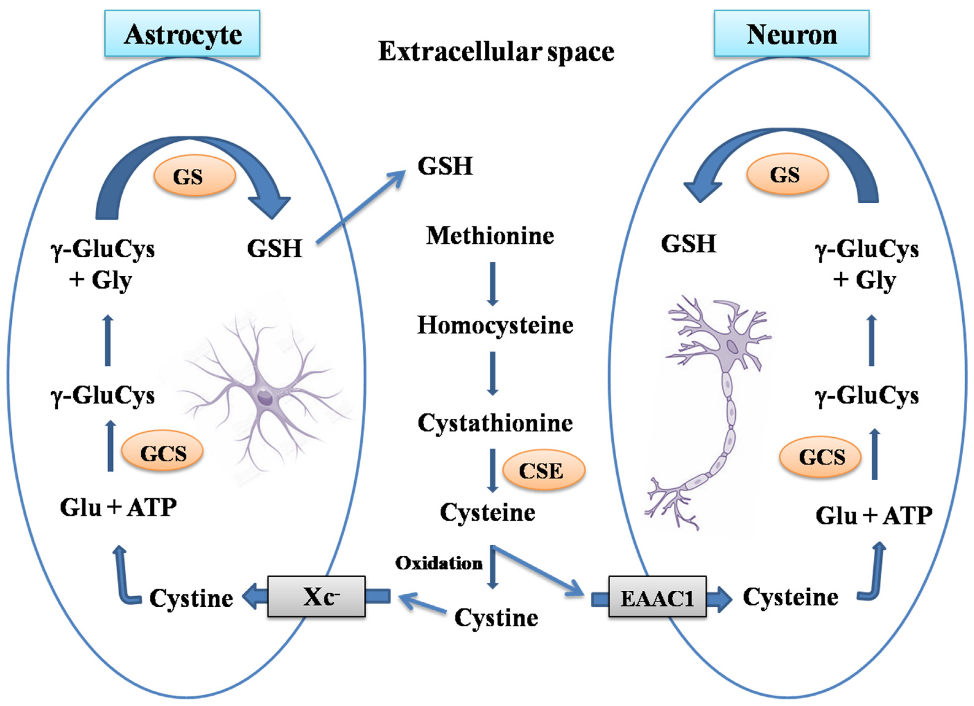
Astrocytes have higher GSH concentration compared to neurons, and both have a differential preference for the precursors of GSH synthesis. Secretion of GSH into the extracellular space by astrocytes provides GSH precursors to neurons [32] (Figure 1)[4]. In addition, cysteine transport to the cell also occurs differently in neurons and astrocytes. Neurons use excitatory aminoacid transporter (EAATs) to take up cysteine from extracellular space, while cystine/glutamate transporter (Xc-) mediates transport in astrocytes (Figure 21). Of the five EAATs known to exist, excitatory amino acid carrier 1 (EAAC1) is involved in the neuronal uptake of cysteine [33][5]. Neurons use extracellular cysteine for GSH synthesis and cannot use oxidized form cystine, unlike astrocytes. Transsulfuration pathways provide intracellular cysteine for GSH synthesis. The majority of GSH synthesized in the liver utilizes the cysteine formed from transsulfuration. The activity of cystathionine-γ-lyase (the rate-limiting enzyme in transsulfuration pathway) in the brain is approximately 1% of the corresponding liver activity [34][6]. However, astrocytes also depend on transsulfuration pathways for intracellular cysteine availability, while it is nearly absent in neurons. Thus, the availability of cysteine is very critical to neurons that exclusively depend on the extracellular cysteine for GSH synthesis [35][7].
Figure 21. GSH synthesis in astrocytes and neurons. The Transsulfuration pathway synthesizes homocysteine from the dietary amino acid methionine to produce cystathionine. Further, cystathionine-γ-lyase (CSE) converts cystathionine into cysteine in extracellular to neurons and astrocytes. Neurons utilize extracellular cysteine for GSH synthesis, unlike astrocytes which uses the oxidized form cystine. Neurons uses excitatory amino acid transporter (EAATs) to take up cysteine, while a cystine/glutamate transporter (Xc-) mediates the transport of cystine in astrocytes. γ-Glutamylcysteine synthetase (GCS), rate limiting enzyme in GSH synthesis catalyzes the formation of γ-glutamylcysteine dipeptide utilizing ATP. γ-GluCys is then combined with glycine to form GSH by glutathione synthetase (GS) in both neurons and astrocytes. GSH formed in astrocytes is released into extracellular space where it is cleaved into individual aminoacids by a set of enzymes.
Replenishing GSH in the brain could be a promising treatment strategy for neurodegenerative disorders. GSH does not cross blood-brain-barrier and its systemic administration is not effective to increase the concentration in the brain as most of the GSH are metabolized in blood. Several studies have investigated the effect of GSH precursors in a mouse model in restoring GSH. Treatment with GSH precursors has been shown to protect from Aβ induced neurotoxicity [36][8] and cognitive impairment in AD mouse models. N-acetyl-cysteine, a membrane-permeable cysteine precursor with a potent antioxidant activity helps to reduce oxidative stress and cognitive decline promoting neuronal survival [37][9]. NAC also promotes the reduction of GSSG to GSH. Oral administration of γ-glutamylcysteine (γ-GC) to APP/PS1 mice increased the GSH content and ratio of GSH/GSSG restoring spatial memory deficit. In vitro treatment of γ-GC attenuate the BSO induced GSH reduction and protects from oligomeric Aβ [38][10]. Recently, a drug delivery system using ultrasound combined with microbubbles containing anti-miR- 96-5p successfully increased the EAAC1 and GSH levels in mice hippocampus. MicroRNA miR-96-5p increases the levels of GTRAP3-18, an inhibitor of cysteine transporter EAAC1 [39][11]. Although several studies reported beneficial effects of restoring GSH in a mouse model, they have shown minimal benefits to patients. Further studies need to be performed to effectively augment the redox system to combat oxidative stress-mediated dysfunctions in the brain.

2. Glutaredoxin

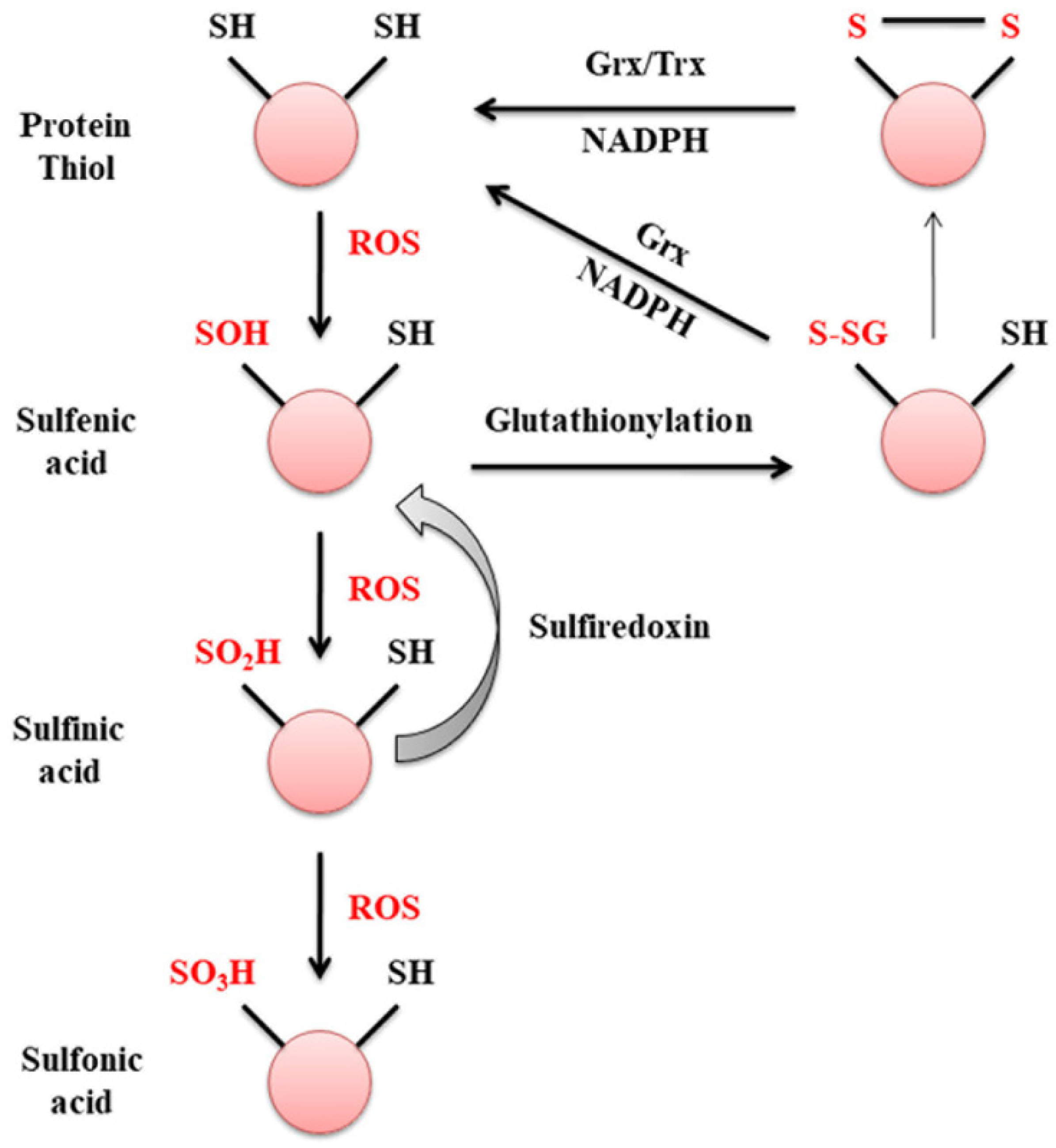
Glutaredoxins are ubiquitously expressed thiol oxidoreductases that regulate protein thiol homeostasis by reversing protein S-glutathionylation using GSH as a substrate. Grx1 regulates proteins involved in cell survival and death including Akt, cjun, ion transporters, NFκB, intracellular signaling molecule Ras and transcription factor nuclear factor-1 (NF1) [22][12]. A major function of Grx1 in the cell is to maintain protein thiol status (Pr-SH) or sulfhydryl homeostasis during oxidative damage [40][13]. Protein thiols undergo oxidation to sulfenic acid, an intermediate in the oxidation pathway that readily reacts to form intramolecular disulfide bonds. Sulfenylated cysteines can be glutathionylated by glutaredoxins and reduced back to active protein thiols. Further oxidation of sulfenic acid generates sulfinic acid which can be reduced by sulfiredoxin (Figure 32). The formation of sulfonic acid is irreversible, where it inhibits protein functions and targets them for degradation. There are no known enzymes present to catalyze the reduction of sulfonic acids due to the low pH needed for its reduction [18][14]. Thus, Grx1 induces the formation of reversible protein S-glutathionylation to protect proteins from irreversible modifications by ROS.
Figure 32. Oxidation of protein thiols to sulfonic acids. During oxidative stress, ROS such as hydrogen peroxide can catalyze the two-electron oxidation of sulfur present in amino acid cysteine. Protein thiols are oxidized to sulfenic acid, which can react with GSH to form protein disulfide preventing further oxidation. Pr-SSG formed is reduced back to active thiols by glutaredoxin or thioredoxin using reducing equivalent NADPH. Sulfinic acid formed by subsequent oxidation of sulfenic acid is still reversible by the reductase enzyme, sulferedoxin. Further oxidation of sulfinic acid results in an irreversible formof sulfonic acid, which targets the protein for degradation.
The glutaredoxin-mediated reduction of protein mixed disulfides to protein thiols is critical in the brain where glutathionylation of protein is more prominent. In the brain, under conditions of oxidative stress such as that seen during reperfusion following ischemia or in animal models of MPTP, there is a predominant formation of Pr-SS-Pr. During recovery following these insults, Pr-SSG is reduced back to Pr-SH by Grx1, thus restoring protein function.
Glutaredoxin was first purified by Mieyal et al. from human erythrocytes as thioltransferase specific for GSH containing mixed disulfides [41][15] and cloned in Escherichia coli [42][16]. In rat brains, a constitutive expression of glutaredoxin exhibits regional and cellular variability in its localization. Glutaredoxin activity is detected higher in the hippocampus where they could recover more rapidly from oxidative damage compared to other regions with less activity in the cerebellum and striatum [43][17]. A similar study carried out using human autopsy brain samples detected higher glutaredoxin activity in the hippocampus and cerebellum. Glutaredoxins are localized predominantly in neurons in the cerebral cortex and hippocampus, purkinje and granule cell layers of the cerebellum, and granular cell layers of the dentate gyrus in the human brain [44][18].

3. Glutaredoxin and Mitochondrial Dysfunction
Maintaining protein thiol homeostasis is important for mitochondrial functions. Loss of GSH and formation of Pr-SSG in the brain contributes to various dysfunctions including loss of activity of mitochondrial enzymes, structural damage to mitochondrial membranes and deregulation of cell survival pathways. Mitochondria are prone to oxidative damage as ROS generated during oxidative phosphorylation can in turn releases more reactive oxygen species that can damage the macromolecules essential for its normal function. The percentage of total cell GSH present in mitochondria is 10–15% [45][19].
The response of brain mitochondria to oxidative stress is very different from the liver mitochondria. In the brain, the GSH lost during oxidative stress is recovered as Pr-SSG and only less than 5% of GSH is recovered as GSSG [46][20]. However, in the liver, most of the GSH lost is recovered as GSSG, and a reduced state of protein thiols are maintained. The excess GSSG formed is effluxed out of the cell to prevent oxidative modification of proteins [47][21].
Mitochondrial enzymes are particularly vulnerable to oxidative damage, complex Ⅰ being the most affected as it increases the oxidation of the GSH pool. Protein thiol groups on 75 and 51 KDa subunits of complex Ⅰ undergo glutathionylation and forms mixed disulfide with GSH [48][22] (Figure 2). Thiol modifications on complex Ⅰ inhibit its activity and increase ROS production that can be reversed by either Grx1 or the disulfide reducing agent dithiothreitol [49][23]. Downregulation of Grx1 in the brain regions of Swiss albino mice results in a significant loss of complex Ⅰ activity signifying the role of glutaredoxins in mitochondrial function [50][24]. ROS-mediated inhibition of activity is specific to complex Ⅰ while other complexes in the electron transport chain remain unaffected.
Glutaredoxin 2 (Grx2) was first identified and cloned by Holmgren et al., which is 34% identical to cytosolic Grx1 [51][25]. It has three isoforms: Grx2a is localized to mitochondria, and Grx2b and Grx2c are both localized to the nucleus and cytosol [52][26]. Grx2a, a mitochondrial isoform of glutaredoxin regulates the glutathionylation of mitochondrial enzymes. Constitutive expression of Grx2 is observed in mouse and human brains and is localized to neurons and glia cells including the neurons of substantia nigra. Grx2 expression is transiently upregulated in MPTP-treated mice and a partial loss in complex Ⅰ activity due to Grx2 downregulation [53][27]. Theoverexpression of Grx2 prevents apoptosis by inhibiting cytochrome c release and caspase activation [54][28] and could ameliorate the toxic effect of MPP+ in mitochondria [53][27].
Further, overexpression of Grx2 attenuatesthe mutant superoxide dismutase (SOD1) mediated degeneration of motor neurons. MutSOD1 accumulates in the mitochondria of motor neurons, and impairs respiratory complexes and ATP production. Grx2 is required for normal mitochondrial function and viability, whereas overexpression of Grx1 is shown to be beneficial in the cytosol and does not preserve mitochondrial dynamics or apoptosis induced by mutSOD1 [55][29].
Increased Pr-SSG formation due to loss of Grx1 activity in the cytosol is known to impair mitochondrial dynamics in the brain. Pr-SSG formed by the oxidation of sulfhydryl groups of cysteine increases the permeability of the inner mitochondrial membrane and opens upa mitochondrial transition pore, resulting in the uncoupling of oxidative phosphorylation and ATP hydrolysis. In addition, the oxidation of voltage-dependent anion channel (VDAC), a mitochondrial outer membrane protein results in the loss of mitochondrial membrane potential (MMP Figure 43). The redox state of vicinal thiol groups in VDAC plays a critical role in the tuning of the voltage sensor of the transition pore and increases its permeability upon oxidation [56][30]. Moreover, VDAC is present in the outer membrane of mitochondria and is exposed to cytosolic oxidative stress as well. shRNA mediated downregulation of Grx1 results in the oxidation of VDAC but not adenosine nucleotide translocase (ANT), an inner mitochondrial membrane protein. Exposure of Neuro2A cells to β-N-oxalyl amino-L-alanine (L-BOAA), an excitatory amino acid implicated in neurolathyrism also leads to MMP loss, which is alleviated by overexpression of Grx1 [57][31]. MMP loss results in cristae unfolding in mitochondria facilitating the release of apoptotic factor cytochrome c leading to cell death [58][32]. Thus, Grx1 functions to maintain mitochondrial integrity during oxidative stress and the downregulation of Grx1 results in mitochondrial dysfunction through oxidative modification of the thiol group present in the outer membrane protein VDAC.
Figure 43. Oxidative stress leads to complex Ⅰ inhibition and mitochondrial membrane potential loss. Protein thiols present in VDAC undergo oxidation leading to increased permeability of transition pore and loss of mitochondrial membrane potential. MMP loss facilitates the release of apoptotic factors such as cytochrome c promoting cell death.
Overall, glutaredoxin acts as a potential neuroprotective mediator to maintain mitochondrial function during oxidative stress, excitatory amino acid toxicity and MPTP mediated neurotoxicity. The role of glutaredoxins will be discussed under each of the disease conditions that follow.

4. Excitotoxicity and Glutaredoxin

Neurolathyrism is a human neurological disorder caused by the ingestion of plant toxin, 3-oxalylamino-L-alanine (L-BOAA) present in Lathyrus sativus. Neurolathyrism affects motor neurons, and anterior horn cells and results in the loss of axons in the pyramidal tract of the lumbar spinal cord in humans. It has been more commonly seen in men, while women are protected from the disease [59][33].
Animal models of L-BOAA toxicity showed GSH depletion and loss of mitochondrial complex Ⅰ activity [60][34] that can be restored by thiol reducing agent, dithiothreitol. GSH depletion has a direct effect on oxidative phosphorylation where it inhibits the complex Ⅰ and reduces ATP production [61][35]. Upregulation of glutaredoxin mRNA and activation of the AP1 transcription factor is observed within a short time after the systemic administration of L-BOAA. The AP1 transcription factor binding site is present upstream of Grx1 gene and mediates the transcription upon insult through the activation of Jun N-terminal kinase (JNK). Grx1 is critical for the recovery of complex Ⅰ in the motor cortex after excitotoxic insult [62][36]. Systemic administration of L-BOAA results in the phosphorylation and translocation of cJun to the nucleus, accompanied by activation of AP1 and subsequent increase in glutaredoxin transcription. Overexpression of glutaredoxin could protect against mitochondrial dysfunction during excitotoxicity in motor neurons [63][37].
A gender specific effect of L-BOAA is seen in human as well as in animal models. L-BOAA mediated GSH depletion and mitochondrial dysregulation are identified only in male [61][35] and ovariectomized female mice. Higher levels of glutaredoxin are seen in the lumbosacral cord, striatum and the mid-brain regions of female mice which reduces upon ovariectomy, indicating estrogen regulation of Grx1 expression. Ovariectomy downregulates the glutaredoxin expression, GSH levels and complex Ⅰ activity, rendering the female mice vulnerable to L-BOAA toxicity. Treatment of SH-SY5Y cells with estrogen upregulated Grx1 levels and protects from L-BOAA-mediated mitochondrial membrane potential loss and cell death. Therefore, estrogen-mediated Grx1 could probably protect brain regions against excitatory aminoacid toxicity exerted by L-BOAA [63][37].

5. Ischemic Reperfusion Injury

GSH homeostasis in the brain during cerebral ischemia has been studied. Reactive oxygen species formed during reperfusion injury following ischemia cause damage to the brain which generally occurs in conditions such as stroke, head trauma and cardiac arrest in humans [64][38]. Reperfusion following ischemia is detrimental, and causes maximum damage to tissues, in part due to oxidative stress. A major consequence of reperfusion damage is the depletion of GSH accompanied by an increase in free radicals and lipid peroxidation, indicative of oxidative stress.
Reperfusion following severe ischemia, induced by bilateral carotid artery occlusion and systemic hypotension results in massive loss of GSH along with an increased mortality rate [65][39]. Partial ischemia induced by bilateral carotid artery occlusion for 30 min reduces blood flow to 50%. Total GSH level significantly reduces during reperfusion for 1 h following partial ischemia. Concurrent with the increase in GSSG, there is an increase in malondialdeheyle, indicative of lipid peroxidation. Even though the brain is vulnerable to oxidative stress, it is capable of recovering from moderate oxidative damage, potentially depending on the extent of damage and may vary from region to region. GSH homeostasis or GSH/GSSG ratios are restored after 24 h of reperfusion following partial ischemia [66][40].
Maintenance of thiol homeostasis is important during reperfusion as it can leads to the oxidation of protein thiols, damage to membrane structures and loss of activity of critical enzymes with active thiol groups such as Na+/K+-ATPase. Thus, the administration of thiol antioxidants such as α-lipoic acid was shown to restore and maintain thiol homeostasis to recover from reperfusion injury. α-lipoic acid shows a remarkable neuroprotective effect during severe ischemic-reperfusion injury with the mortality rate in rats reduced from 78% to 26% accompanied by attenuation of brain GSH loss [65][39].

References

  1. Aoyama, K.; Nakaki, T. Glutathione in Cellular Redox Homeostasis: Association with the Excitatory Amino Acid Carrier 1 (EAAC1). Molecules 2015, 20, 8742–8758.
  2. Lu, S.C. Regulation of Glutathione Synthesis. Mol. Asp. Med. 2009, 30, 42–59.
  3. Vali, S.; Mythri, R.B.; Jagatha, B.; Padiadpu, J.; Ramanujan, K.S.; Andersen, J.K.; Gorin, F.; Bharath, M.M.S. Integrating Glutathione Metabolism and Mitochondrial Dysfunction with Implications for Parkinson’s Disease: A Dynamic Model. Neuroscience 2007, 149, 917–930.
  4. Wang, X.F.; Cynader, M.S. Astrocytes Provide Cysteine to Neurons by Releasing Glutathione. J. Neurochem. 2000, 74, 1434–1442.
  5. Aoyama, K.; Watabe, M.; Nakaki, T. Modulation of Neuronal Glutathione Synthesis by EAAC1 and Its Interacting Protein GTRAP3-18. Amino Acids 2012, 42, 163–169.
  6. Diwakar, L.; Ravindranath, V. Inhibition of Cystathionine-γ-Lyase Leads to Loss of Glutathione and Aggravation of Mitochondrial Dysfunction Mediated by Excitatory Amino Acid in the CNS. Neurochem. Int. 2007, 50, 418–426.
  7. Johnson, W.M.; Wilson-Delfosse, A.L.; Mieyal, J.J. Dysregulation of Glutathione Homeostasis in Neurodegenerative Diseases. Nutrients 2012, 4, 1399–1440.
  8. Braidy, N.; Zarka, M.; Jugder, B.E.; Welch, J.; Jayasena, T.; Chan, D.K.Y.; Sachdev, P.; Bridge, W. The Precursor to Glutathione (GSH), γ-Glutamylcysteine (GGC), Can Ameliorate Oxidative Damage and Neuroinflammation Induced by Aβ40 Oligomers in Human Astrocytes. Front. Aging Neurosci. 2019, 10, 177.
  9. Skvarc, D.R.; Dean, O.M.; Byrne, L.K.; Gray, L.; Lane, S.; Lewis, M.; Fernandes, B.S.; Berk, M.; Marriott, A. The Effect of N-Acetylcysteine (NAC) on Human Cognition—A Systematic Review. Neurosci. Biobehav. Rev. 2017, 78, 44–56.
  10. Liu, Y.; Chen, Z.; Li, B.; Yao, H.; Zarka, M.; Welch, J.; Sachdev, P.; Bridge, W.; Braidy, N. Supplementation with γ-Glutamylcysteine (γ-GC) Lessens Oxidative Stress, Brain Inflammation and Amyloid Pathology and Improves Spatial Memory in a Murine Model of AD. Neurochem. Int. 2021, 144, 104931.
  11. Kinoshita, C.; Kikuchi-Utsumi, K.; Aoyama, K.; Suzuki, R.; Okamoto, Y.; Matsumura, N.; Omata, D.; Maruyama, K.; Nakaki, T. Inhibition of MiR-96-5p in the Mouse Brain Increases Glutathione Levels by Altering NOVA1 Expression. Commun. Biol. 2021, 4, 1–12.
  12. Gorelenkova Miller, O.; Mieyal, J.J. Critical Roles of Glutaredoxin in Brain Cells—Implications for Parkinson’s Disease. Antioxid. Redox Signal. 2019, 30, 1352–1368.
  13. Shelton, D.M.; Chock, P.B.; Mieyal, J.J. S-Glutathionylation As a Mechanism of Redox Signal Transduction and Regulation of Protein Translocation, and the Central Role of Glutaredoxin. Antioxid. Redox Signal. 2005, 7, 348–367.
  14. Van Bergen, L.A.H.; Roos, G.; De Proft, F. From Thiol to Sulfonic Acid: Modeling the Oxidation Pathway of Protein Thiols by Hydrogen Peroxide. J. Phys. Chem. A 2014, 118, 6078–6084.
  15. Mieyal, J.J.; Starke, D.W.; Chung, J.S.; Gravina, S.A.; Dothey, C. Thioltransferase in Human Red Blood Cells: Purification and Properties. Biochemistry 1991, 30, 6088–6097.
  16. Chrestensen, C.A.; Eckman, C.B.; Starke, D.W.; Mieyal, J.J. Cloning, Expression and Characterization of Human Thioltransferase (Glutaredoxin) in E. coli. FEBS Lett. 1995, 374, 25–28.
  17. Balijepalli, S.; Tirumalai, P.S.; Swamy, K.V.; Boyd, M.R.; Mieyal, J.J.; Ravindranath, V. Rat Brain Thioltransferase: Regional Distribution, Immunological Characterization, and Localization by Fluorescent in Situ Hybridization. J. Neurochem. 1999, 72, 1170–1178.
  18. Balijepalli, S.; Boyd, M.R.; Ravindranath, V. Human Brain Thioltransferase: Constitutive Expression and Localization by Fluorescence in Situ Hybridization. Mol. Brain Res. 2000, 85, 123–132.
  19. Griffith, O.W.; Meister, A. Origin and Turnover of Mitochondrial Glutathione. Proc. Natl. Acad. Sci. USA 1985, 82, 4668–4672.
  20. Ravindranath, V.; Reed, D.J. Glutathione Depletion and Formation of Glutathione-Protein Mixed Disulfide Following Exposure of Brain Mitochondria to Oxidative Stress. Biochem. Biophys. Res. Commun. 1990, 169.
  21. Olafsdottir, K.; Reed, D.J. Retention of Oxidized Glutathione by Isolated Rat Liver Mitochondria during Hydroperoxide Treatment. BBA Gen. Subj. 1988, 964, 377–382.
  22. Taylor, E.R.; Hurrell, F.; Shannon, R.J.; Lin, T.K.; Hirst, J.; Murphy, M.P. Reversible Glutathionylation of Complex I Increases Mitochondrial Superoxide Formation. J. Biol. Chem. 2003, 278, 19603–19610.
  23. Beer, S.M.; Taylor, E.R.; Brown, S.E.; Dahm, C.C.; Costa, N.J.; Runswick, M.J.; Murphy, M.P. Glutaredoxin 2 Catalyzes the Reversible Oxidation and Glutathionylation of Mitochondrial Membrane Thiol Proteins: Implications for Mitochondrial Redox Regulation and Antioxidant Defense. J. Biol. Chem. 2004, 279, 47939–47951.
  24. Kenchappa, R.S.; Ravindranath, V. Glutaredoxin Is Essential for Maintenance of Brain Mitochondrial Complex I: Studies with MPTP. FASEB J. 2003, 17, 717–719.
  25. Lundberg, M.; Johansson, C.; Chandra, J.; Enoksson, M.; Jacobsson, G.; Ljung, J.; Johansson, M.; Holmgren, A. Cloning and Expression of a Novel Human Glutaredoxin (Grx2) with Mitochondrial and Nuclear Isoforms. J. Biol. Chem. 2001, 276, 26269–26275.
  26. Lönn, M.E.; Hudemann, C.; Berndt, C.; Cherkasov, V.; Capani, F.; Holmgren, A.; Lillig, C.H. Expression Pattern of Human Glutaredoxin 2 Isoforms: Identification and Characterization of Two Testis/Cancer Cell-Specific Isoforms. Antioxid. Redox Signal 2008, 10, 547–557.
  27. Karunakaran, S.; Saeed, U.; Ramakrishnan, S.; Koumar, R.C.; Ravindranath, V. Constitutive Expression and Functional Characterization of Mitochondrial Glutaredoxin (Grx2) in Mouse and Human Brain. Brain Res. 2007, 1185.
  28. Enoksson, M.; Fernandes, A.P.; Prast, S.; Lillig, C.H.; Holmgren, A.; Orrenius, S. Overexpression of Glutaredoxin 2 Attenuates Apoptosis by Preventing Cytochrome c Release. Biochem. Biophys. Res. Commun. 2005, 327, 774–779.
  29. Ferri, A.; Fiorenzo, P.; Nencini, M.; Cozzolino, M.; Pesaresi, M.G.; Valle, C.; Sepe, S.; Moreno, S.; Carrì, M.T. Glutaredoxin 2 Prevents Aggregation of Mutant SOD1 in Mitochondria and Abolishes Its Toxicity. Hum. Mol. Genet. 2010, 19, 4529–4542.
  30. Petronilli, V.; Costantini, P.; Scorrano, L.; Colonna, R.; Passamonti, S.; Bernardi, P. The Voltage Sensor of the Mitochondrial Permeability Transition Pore Is Tuned by the Oxidation-Reduction State of Vicinal Thiols. Increase of the Gating Potential by Oxidants and Its Reversal by Reducing Agents. J. Biol. Chem. 1994, 269, 16638–16642.
  31. Saeed, U.; Durgadoss, L.; Valli, R.K.; Joshi, D.C.; Joshi, P.G.; Ravindranath, V. Knockdown of Cytosolic Glutaredoxin 1 Leads to Loss of Mitochondrial Membrane Potential: Implication in Neurodegenerative Diseases. PLoS ONE 2008, 3, e2459.
  32. Gottlieb, E.; Armour, S.M.; Harris, M.H.; Thompson, C.B. Mitochondrial Membrane Potential Regulates Matrix Configuration and Cytochrome c Release during Apoptosis. Cell Death Differ. 2003, 10, 709–717.
  33. Ravindranath, V. Neurolathyrism: Mitochondrial Dysfunction in Excitotoxicity Mediated by L-β-Oxalyl Aminoalanine. Neurochem. Int. 2002, 40, 505–509.
  34. Sadashiva Pai, K.; Ravindranath, V. L-BOAA Induces Selective Inhibition of Brain Mitochondrial Enzyme, NADH-Dehydrogenase. Brain Res. 1993, 621, 215–221.
  35. Sriram, K.; Shankar, S.K.; Boyd, M.R.; Ravindranath, V. Thiol Oxidation and Loss of Mitochondrial Complex I Precede Excitatory Amino Acid-Mediated Neurodegeneration. J. Neurosci. 1998, 18, 10287–10296.
  36. Kenchappa, R.S.; Diwakar, L.; Boyd, M.R.; Ravindranath, V. Thioltransferase (Glutaredoxin) Mediates Recovery of Motor Neurons from Excitotoxic Mitochondrial Injury. J. Neurosci. 2002, 22, 8402–8410.
  37. Diwakar, L.; Kenchappa, R.S.; Annepu, J.; Ravindranath, V. Downregulation of Glutaredoxin but Not Glutathione Loss Leads to Mitochondrial Dysfunction in Female Mice CNS: Implications in Excitotoxicity. Neurochem. Int. 2007, 51.
  38. Granger, D.N.; Kvietys, P.R. Reperfusion Injury and Reactive Oxygen Species: The Evolution of a Concept. Redox Biol. 2015, 6, 524–551.
  39. Panigrahi, M.; Sadguna, Y.; Shivakumar, B.R.; Kolluri, S.V.R.; Roy, S.; Packer, L.; Ravindranath, V. α-Lipoic Acid Protects against Reperfusion Injury Following Cerebral Ischemia in Rats. Brain Res. 1996, 717.
  40. Shivakumar, B.R.; Kolluri, S.V.R.; Ravindranath, V. Glutathione Homeostasis in Brain during Reperfusion Following Bilateral Carotid Artery Occlusion in the Rat. Mol. Cell. Biochem. 1992, 111, 125–129.
More
Video Production Service