Stabilization Strategies of Pharmaceutical Proteins: Comparison
Please note this is a comparison between Version 4 by Amina Yu and Version 3 by Amina Yu.

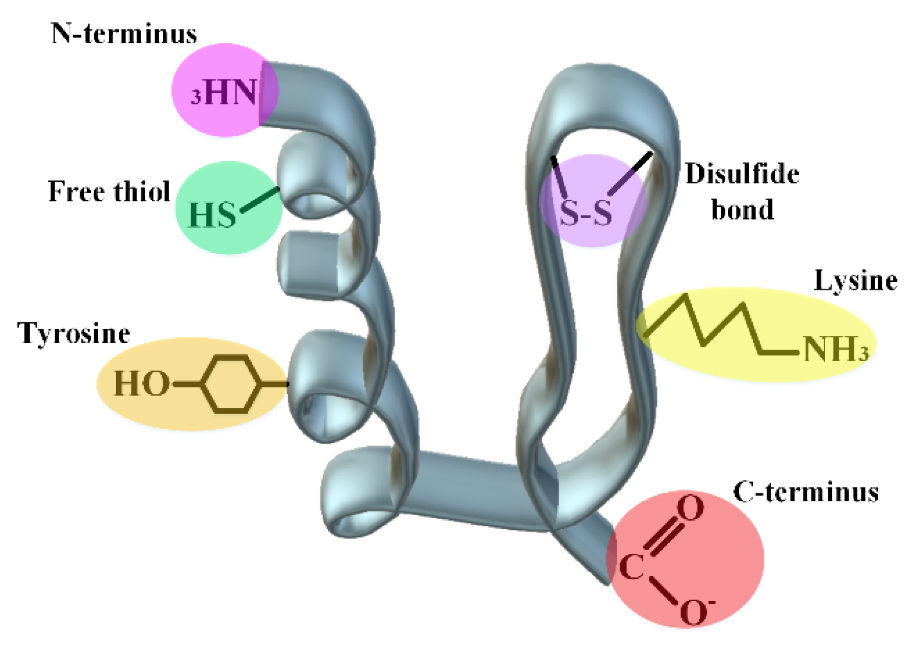
At the beginning of the transcription of a protein, thanks to the intracellular chaperone systems and the biophysical laws governing protein folding, correct folding occurs most of the time. When due to cellular defects and rapid protein expression, protein folding becomes problematic, and several fates may occur for the protein. In cases where misfolding leads to the loss of protein activity (such as enzymes), the corresponding disease will appear directly. As this state continues, the misfolded protein may turn into amorphous clots or aggregates with regular structures, each of which can lead to various neurological diseases or even cancer. In the most optimistic scenario, the misfolded protein enters the proteasome machinery and is initially converted into smaller peptides and finally broken down into building amino acids.

  • protein drugs
  • antibody drugs
  • pharmaceutical proteins and peptides
  • denaturing stresses
  • protein drug characterizations

1. Genetic Engineering: Protein Analogs

With the discovery of genetic engineering and gaining the knowledge that it is possible to change the DNA sequence in our favor, in 1973, the hope was revived to start producing proteins with properties beyond the natural states. From that year onwards, precisely 1982, and with the creation of the first protein drug with a human genetic source (insulin), it was gradually added to the number of recombinant drugs [1]. Today, with all types of analog insulins, all types of stability and speed of activity are accessible to diabetic patients [2]. The genetic manipulation of medicinal proteins and peptides should be aimed at not harming the protein’s activity as much as possible and only stepping in the valley of its stability must be careful. Meanwhile, two general strategies for the production of analog proteins using direct mutation (site-directed mutagenesis) and random mutation of proteins and the production of fusion proteins will be discussed.

1.1. Site-Directed Mutagenesis

There are countless ways to induce a mutation in the genetic structure of the protein to produce a protein analog, which is to create a more resistant or even more active species than the wild form of the protein. For the purposeful creation of protein analog species, one must have sufficient knowledge of the structure of the wild-type protein. Among all changes that lead to the production of analog or mutant proteins, it is worthwhile to refer to substitution, deletion and addition of one or more amino acids in the protein structure. As an example, in author's previous study, two amino acids lysine were added at the end of the B chain of insulin, which led to the aggregation of obtained analogs during subcutaneous injection, which increased the in vivo stability of these insulins [3]. Besides, sometimes, some essential amino acids are also added to native proteins to introduce the glycosylation and acylation sites, for increasing the biological and physical stabilities of the target proteins with the help of sugar and fatty acid molecules.
Studying some successful protein drugs such as interferon (IFN) β1b, human fibroblast growth factor (FGF) and granulocyte colony-stimulating factor (G-CSF), has had the result that the substitution of cysteine residue with serine has led to the production of some analogs with a longer half-life [4][5]. Today, with the help of in silico platforms for estimating and predicting domains sensitive to aggregation (such as hydrophobic domains buried in the structure of proteins), researchers can succeed in mutations that make the resulting proteins more stable against temperature and other physical stresses. Human growth hormone (hGH) and G-CSF can be mentioned among these case studies. In the first case, it was reported that the obtained hGH analogs with 10–14 mutations showed a four- to ten-fold increase in shelf life and an increase in temperature stability. Speaking about G-CSF, it was also reported that in addition to an increase in temperature stability up to 16 °C compared to the wild-type protein, the analog has activity in cell proliferation studies, as well [6][7]. As previously reviewed, the digestion/degradation and cleavage of medicinal proteins in many chemical modifications such as oxidation and deamidation can be the coup de grâce to the proteins. Accordingly, efforts to create mutants resistant to protein digestion can be valuable. For example, it is commonly seen that the site of proteolysis of proteins is evolutionarily institutionalized in the flexible loops. Thus, mutations that increase the flexibility of proteins may cause resistance to proteolytic digestion [7]. As an example, it has been found that the mutation of two arginine amino acids in the flexible region of factor VIIIa protein leads to the resistance of this medicinal protein to the proteases of thrombin, activated protein C, and factor VIIIa [8].

1.2. Fusion Strategies

Usually, the most important purpose of creating a fusion state in medicinal proteins is to increase their half-life in the blood circulation system and significantly improve pharmacokinetic profiles [9]. For this reason, it is often used to increase the size of protein molecules to escape from the renal filtration system. In this achievement, a protein that can circulate in the blood for a long period is usually used (such as albumin or the FC part of antibodies) [10]. As a famous and appreciated example, the etanercept protein that is used medicinally for rheumatoid arthritis disease, today, is obtained from the addition of the extracellular domain of p75 tumor necrosis factor receptor (TNFR) and the Fc part of IgG antibody. The FC part leads to an increase in the life of the protein in the blood circulation by the mechanisms of increasing the size of the fusion protein and possibly by endosomal recycling. On the other hand, because the Fc part is a dimer, the binding rate of this protein to TNF-alpha is between 50 and 1000 times higher than that of monomeric TNF-alpha samples [7]. Regarding albumin, two major mechanisms have been proposed. One is the direct binding of this protein to the medicinal proteins (e.g., different variants of barbourin [11] and hirudin [12] and the other is the binding of peptides that can act as linkers between the medicinal protein and albumin (peptides that have a high binding tendency to albumin); extending an albumin-binding peptide label to the anti-tissue factor D3H44 Fab that increased the half-life nearly 40-fold [13]. In addition to these two natural protein molecules, other molecules have also been used to produce fusion therapeutic proteins. 
Sometimes, the purpose of creating a fusion is to increase the stability of the medicinal protein in bacterial expression systems. Especially for therapeutic peptides, the expression of such small molecules in prokaryotic systems such as E. coli is always accompanied by the challenge of protease digestion in the bacterial cells, which leads to a low yield of the final product. On the other hand, by designing fusion (carrier protein) to the target peptide, post-expression pathways such as purification will take place more easily. As a previous attempt, in the expression system of E. coli, the genes of insulin A and B chains were separately fused with the chaperone protein carrier alpha-B crystallin of the human eye lens, in addition to increasing the efficiency of the expression in the bacterial platform, the purification pathways were also conducted more simply [14]. Recently, a similar study has been carried out to produce proinsulin in the prokaryotic system [15].

2. Covalent Engineering

In this section, the purpose is to explain those changes that are covalently applied to the structure of proteins and peptides to increase their stability. In general, this category of changes includes the connection of synthetic polymer molecules such as polyethylene glycol and some other extracted molecules: sugars and fatty acids.

2.1. Protein–Polymer Conjugates

The challenges to the stability of medicinal proteins and peptides are so deep that they cannot be completely solved by the strategies reviewed so far. In many cases, low-stability proteins need to be conjugated with synthetic polymers to brighten the hope of increasing their stability. One of the most prominent examples of protein-polymer conjugation is the PEGylation of medicinal proteins, which leads to an increase in the life of proteins in the circulatory systems [16]. There are currently more than 40 FDA-approved protein drugs that have been conjugated by PEG [17]. In addition to increasing the life expectancy in plasma, other benefits such as protein stability in the digestive system, control of receptor-ligand binding, and improving the storage stability of medicinal proteins using PEG polymer have been reported. It is necessary to take great care about the selection of the type of protein, the type of polymer and even the conjugation method, to follow the paths that the guest protein does not change from its natural state (fold) to an unnatural form (unfold) [16].
In this section, the types of polymers that have been used for the engineering of medicinal proteins will be discussed. In order to correctly and systematically choose a suitable polymer for conjugation to protein, it is necessary to consider several criteria.
Additionally, in the selection of protein, attention should be paid to the types of functional groups that can react with the selected polymer. These functional groups should not be considered in the sensitive areas or the active site of the protein.  The following figure (Figure 1) shows the types of possible active groups in proteins
Figure 1. Introducing several active groups on the surface and inside a hypothetical protein structure to introduce desired covalent modifications.
In addition to both C- and N-ends of a protein, disulfide bonds, side chains of lysine and tyrosine as well as free thiol can undergo purposely various chemical changes to increase protein stability.
The suitable environment for the reactivity of lysine (pKa~10.5) is more in the neutral to basic ranges, which is not suitable for some proteins that are not stable in this range [18]. Additionally, the amino acid surface accessibility and the charge around it will be effective in its reactivity. In the case of cysteine, because it is less abundant than lysine in the primary structure of proteins, the residue is more popular for targeted chemical modifications. Nevertheless, the changes in this amino acid are a bit riskier and can lead to changes in the protein structure. So, the structural characterization of proteins should be investigated after cysteine modification. In many medicinal proteins and especially antibodies, there are a large number of disulfide bonds, which with their slight reduction, it has been seen that it does not cause severe damage to the tertiary structure of the proteins. The same method is used for the changes of some proteins in the reduced cysteines. For example, in the brentuximab vedotin antibody model, after partial reduction of the disulfide bonds, the obtained structure is then conjugated to the maleimide functionalized anti-cancer drug [18]. This method cannot be useful for small proteins with a small number of disulfide bonds, since the whole structure may collapse by reducing the bonds. For chemical changes in the disulfide bonds of these proteins, replacing natural disulfide bonds with synthetic ones has obtained hopeful results [19]. Additionally, pka for the N-terminus amine of proteins is usually reported 6–8, which is usually used as a mindset for changes in the amine end (pH-dependent reactions). For example, the pegfilgrastim protein drug with the same method has a PEG at the N-terminus amine. Reductive amination in acidic pH usually leads to N-terminus amine PEGylation of proteins with yields above 90% [18][20]. However, it should be kept in mind that connecting the polymer to the N-terminus amine will be advantageous in proteins that are small and have a few lysine residues. Because the amine group located in the side chain of lysine can win by competing with the amine at the end of the protein and reducing the percentage of the final product. Usually, it is difficult to distinguish between the C-terminus of the protein and the carboxyl present in the side chain of amino acids Asp and Glu, which indicates the necessity of using tags at the C-terminus to confirm the reactivity of this end [21][22]. Tyrosine is also a residue that has a low abundance on the surface of proteins and is usually not followed by high popularity in the field of polymer-protein conjugation due to its cross-reactivity with the amino acid histidine in nucleophilic reactions.

2.2. Linker Chemistry

The linker is a very essential part of protein conjugation, especially for ADCs. Linker chemistry can be tuned to impart adequate stability for protein drugs [23]. As mentioned above, PEGylation is an example of using hydrophilic PEG linkers to improve drug efficacy [24]. There are many factors to consider when choosing linkers for ADCs. Nevertheless, one of the most important of them is the stability of the linker throughout its presence in the plasma, while it must break down as soon as it enters the target cell (in the form of a conjugate with ADCs) and leads to the activation of the released anti-cancer drugs [25]. There are cleavable and non-cleavable linkers. The cleavable linkers are cleaved by chemical or biochemical changes in the environment such as acidic changes for hydrozone linkers and glutathione or high pH values for disulfide linkers. Some other cleavable linkers contain enzymatic digestion sites and are sensitive to the enzymes inside the vesicle such as valine-citrulline di-peptides for cathepsin B enzyme. On the other hand, the non-cleavable linkers depend on the complete digestion of the antibody after entering the vesicle [26].
Among the important reasons that led to the removal of Mylotarg from the marketing list in 2010 were items such as the instability of the hydrazine linker and its payload release [27][28]. More recently, Trastuzumab-emtansine (T-DM1) 1 with a non-cleavable linker is the first generation of ADCs approved for the treatment of HER2 (human epidermal growth factor receptor 2)-positive breast cancer. Second-generation ADCs targeting HER2, trastuzumab duocarmazine (SYD985), was characterized to supersede T-DM1 by overcoming all resistance using a cleavable linker and a more potent payload, duocarmycin (DUBA) via different conjugation chemistry. There are, however, no measurable changes observed between the half-life of Adcetris® with a cleavable linker and Kadcyla® with non-cleavable linker at the clinical level [29][30].
The linker property will also determine how the final products act during blood circulation and also touch the sensitivity of the antibody to environmental stresses [31]. Some disulfide-linked ADCs were found to conjugate with blood proteins through cleaved free thiols. Hydrophobic linkers and payloads frequently encourage the aggregation of ADC molecules, e.g., King et al. showed that multiple loading of the BR96 antibody that is simultaneously attached to doxorubicin drug leads to non-covalent dimerization of the antibody, which will eventually lead to hepatotoxicity [32][33]. It has also been seen that the linkers that are more hydrophobic are not able to significantly affect the MDR1+ cells. Instead, linkers with a more polar and amphiphilic nature were able to appear with a greater effect such as mal-PEG4-N-hydroxysuccinimide and N-Hydroxysuccinimidyl-4-(2-pyridyldithio)-2-sulfobutanoate (sulfo-SPDB) [34]. In addition to the MDR1 challenge, it has also been found that reducing the percentage of hydrophobicity by employing hydrophilic linkers containing negatively charged sulfonate groups, polyethylene glycol (PEG) groups, or pyrophosphate diester groups can also help in improving therapeutic index level and pharmacokinetics [35].

2.3. Acylation

This strategy has mostly been used in the case of medicinal peptides; liraglutide and insulin detemir are among the most successful cases of lipidated peptides in the world market. It has been reported that lipidation in the backbone of peptides can give positive characteristics to the obtained product, such as impressive stability against enzymatic digestion, and specificity to the receptor and also to the bioavailability of peptides. In addition to the issues related to the stabilization of medicinal peptides, this process has received much attention in the field of drug delivery of medicinal peptides [36]. This process occurs unsurprisingly in nature. In the living system, various enzymes help a protein or peptide to react with a chain of saturated or unsaturated fatty acids in the form of N- and/or O-links [37]. The binding site of lipids can be the terminal amine and carboxyl, and even in the middle part of the primary protein/peptide structure, which occurs through residues such as serine, threonine, glycine, lysine, and cysteine [38]. It is interesting to note that this addition of lipids can be irreversible (when it is through the connection of the N-terminal of glycine/cysteine) or reversible (when a thioester bond occurs between the fatty acid and thiol group of cysteine) [39][40]. Regarding the medicinal aspects of lipidopeptides, some of these peptides are naturally extracted from microorganism sources such as fungi and bacteria. The use cases of this type of peptides have been proven in anti-cancer, anti-fungal and anti-bacterial fields [41]. However, from another line of view, in silico addition of lipids to peptides is also of special interest [42]. In addition to the fact that the presence of peptides can lead to the appearance of some new features in the peptide, it is reported that the type of spacer between the lipid and the peptide, the type of lipid and its chemical nature (saturated and unsaturated) are also effective in the activity of the lipidopeptide [43][44]

2.4. Cyclization

Cyclization is another solution that has been proposed to increase the stability of medicinal peptides, especially against enzymatic digestions. The idea of this strategy is taken from nature by discovering peptides such as mammalian theta-defensins [45] and plant cyclotides [46]. The best example of an engineered cyclic peptide is a 29 amino acids peptide, kalata-B1, which is resistant to a wide range of environmental stresses such as 8 M urea, acidic environments (0.5 M HCl), 6 M GnHCl, and the presence of various proteases [47]. Taken as a whole, the high stability of these cyclic peptides has caused special attention in recent years to search for faster synthesis methods and the purification of cyclized peptides.
Most cyclic peptides belong to non-synthetic peptides, extracted from microorganisms. This is due to the complex routes of cyclization of peptides and finally their purification to an acceptable level [48].
Based on the nature of the linkage between the two residues in the peptide structure that ultimately ensures the cyclicity, there are two major classes of cyclic peptides: homodetic (involving only a conventional peptide bond) and heterodetic (using a variety of functional groups that required for looping the peptides) [49].

2.5. Nanoparticles; Double-Blade Sword

In most past studies, nanoparticles have been used as a platform for the delivery of protein and peptide drugs. The oral route of using biological agents is the easiest way to use such drugs, but due to the destructive conditions of the digestive system (acidic environment, the presence of many protease enzymes, and a small percentage of surface absorption in the small intestine), this route has always been associated with many challenges. For this reason, the use of nanoparticles has been focused on facilitating the delivery of protein drugs in the alimentary tract. However, in some studies, nanoparticle systems have also been used for the intravenous routes of drugs. In any case, for the use of nanoplatforms for the delivery of protein or peptide drugs, the surface and nanoparticle size are factors that should be taken into consideration [50]. From the surface chemistry point of view, firstly, nanoparticles should ideally have hydrophilic surfaces that can be easily dissolved in living systems and secondly escape from the macrophage system [51].
Given particle size, in living systems of vertebrates, macrophages are responsible for ingesting large foreign particles (size around 0.5 µm). The selected nanoparticles should be small enough to escape from this system on the one hand, and on the other hand, they should be large enough to not quickly enter the outside environment of the vessels through the vascular leakage system. It has been agreed that this size is between 150–200 nanometers in the spleen and liver on the one hand, and on the other hand, the gap junctions between endothelial cells are between 100 and 600 nanometers [52][53]. According to this evidence, choosing the right size is a challenging factor. However, looking at the successful studies in this field, in general, it is understood that the useful nanoparticles that have been used so far have been between 3 and 200 nanometers in size. A wide range of nanoparticles have been used to deliver all kinds of drugs, nevertheless, in the case of protein and peptide drugs, most of the attention has been on polymer nanoparticles and solid lipid nanoparticles. Of these two groups, the latter has also been pursued more seriously due to the controlled release of the cargo and its ability to be engineered for a specific organ [54]

3. Non-Covalent Engineering of Therapeutic Proteins and Peptides; Solvent Engineering Pathways

3.1. Formulation: Solvent Engineering Pathways

Another solution that has been used to preserve the integrity of medicinal proteins and peptides is the use of excipients during storage and even through the use of such drugs. According to the types of these agents, their mechanisms of action are also diverse [55]. Herein, the types of these excipients are presented along with their mechanism to protect proteins. Some of these excipients should be used for a certain amount of window with specific considerations such as salts that can lead to the aggregation of proteins after a critical concentration [56]. Or as another important example, in the category of sugars, usually non-reducing sugars are used because reducing sugars via Millard’s reactions may cause destructive glycosylation of proteins [57].
According to all the information that was reviewed about the stabilization of proteins and peptides, at the end of this section, it is necessary to briefly suggest which types of proteins usually suffer from which stress and what are the proposed solutions to overcome the challenges (Figure 2).
Figure 2. Proposed solutions to the challenges faced by medicinal proteins and peptides.

3.2. Choice of Container

Depending on the type and even the architecture of the protein containers, proteins may react with the containers’ surfaces, be adsorbed without or with a change in the tertiary structure, and lead to protein aggregation that is either reversible or irreversible. For example, it has been detected that rhFVIII proteins undergo tertiary structure changes in contact with hydrophobic silica surfaces, and changing the nature of the surface to a negative charge has led to a slight change in the protein structure [58]. It has also resulted in some cases where no significant structural change occurs in the protein, but a severe aggregation behavior occurs during protein binding to the surfaces. For instance, rhPAF-AH protein does not show a measurable change in contact with the hydrophilic surface of silica, though, at a pH 6.5, it showed a very high aggregation rate [59].
Based on past efforts in this field, scientists have generally agreed that glass containers are not ideal, but suitable, options for transporting and storing these therapeutic macromolecules [60][61]. Some studies have also pointed out that even these glass containers can interact with proteins and cause structural changes in them, which in itself can reduce the stability of the protein and other consequences such as ectopic responses in the patient’s body [61]. It is worth mentioning a few studies on the changes that have been made to the surfaces of glass containers to help stabilize drug proteins. From about 1850, therapeutic glass containers were introduced to world markets. Then, in the 1950s, due to problems such as the reactivity of pharmaceutical proteins and some other chemicals with glassware, efforts began to engineer glassware using beneficial polymers [62]. Although glass vials are mainly used for the storage of medications, in some of them, the presence of glass particles, separated from the bottom of the container, has been reported [63]. Container delamination is an undesirable process in which a thin layer of glass is separated from the main body glass and the separated particles have the potential to destroy the drug [64]. For such cases, the US Pharmaceutical Association (USP) has guided assessment glass delamination called USP <1660> [62]. USP <1660> lists most of the possible factors: the composition of the glass, the conditions under which the container is made, the subsequent displacement of the container and the medicinal product in the container. Thus, assessing the inner surface of glass vials, including examining the surface of the glass using an electron microscope (to analyze the degree of the surface cavity or depth), measuring the total composition that may have been separated from the glass using inductively coupled plasma-mass spectrometry, or examination of visible particles using dynamic light diffraction or scanning electron microscopy-energy scattering (SEM-EDX) is essential [62]. Combining all mentioned studies, for industrial technology, glass vials are preferred over plastic vials due to the standard depyrogenation, operations of washing and possibly terminal autoclaving and in another view, glass containers have less immunogenicity in comparison with plastic counterparts [65]. Although in recent years, different particles with an outer layer of silica have also been invented, which can be an alternative option for current protein drugs containers. However, it is still necessary to go through several confirmation stages to industrialize these candidates, [66][67].

References

  1. Akbarian, M. Insulin therapy: A valuable legacy and its future perspective. Int. J. Biol. Macromol. 2021, 181, 1224–1230.
  2. Akbarian, M.; Ghasemi, Y.; Uversky, V.N.; Yousefi, R. Chemical modifications of insulin: Finding a compromise between stability and pharmaceutical performance. Int. J. Pharm. 2018, 547, 450–468.
  3. Akbarian, M.; Yousefi, R.; Moosavi-Movahedi, A.A.; Ahmad, A.; Uversky, V.N. Modulating insulin fibrillation using engineered B-chains with mutated C-termini. Biophys. J. 2019, 117, 1626–1641.
  4. Arakawa, T.; Prestrelski, S.J.; Narhi, L.O.; Boone, T.C.; Kenney, W.C. Cysteine 17 of recombinant human granulocyte-colony stimulating factor is partially solvent-exposed. J. Protein Chem. 1993, 12, 525–531.
  5. Culajay, J.F.; Blaber, S.I.; Khurana, A.; Blaber, M. Thermodynamic characterization of mutants of human fibroblast growth factor 1 with an increased physiological half-life. Biochemistry 2000, 39, 7153–7158.
  6. Luo, P.; Hayes, R.J.; Chan, C.; Stark, D.M.; Hwang, M.Y.; Jacinto, J.M.; Juvvadi, P.; Chung, H.S.; Kundu, A.; Ary, M.L.; et al. Development of a cytokine analog with enhanced stability using computational ultrahigh throughput screening. Protein Sci. 2002, 11, 1218–1226.
  7. Marshall, S.A.; Lazar, G.A.; Chirino, A.J.; Desjarlais, J.R. Rational design and engineering of therapeutic proteins. Drug Discov. Today 2003, 8, 212–221.
  8. Pipe, S.W.; Kaufman, R.J. Characterization of a genetically engineered inactivation-resistant coagulation factor VIIIa. Proc. Natl. Acad. Sci. USA 1997, 94, 11851–11856.
  9. Sytkowski, A.J.; Lunn, E.D.; Risinger, M.A.; Davis, K.L. An erythropoietin fusion protein comprised of identical repeating domains exhibits enhanced biological properties. J. Biol. Chem. 1999, 274, 24773–24778.
  10. Strohl, W.R. Fusion proteins for half-life extension of biologics as a strategy to make biobetters. Biodrugs 2015, 29, 215–239.
  11. Marques, J.A.; George, J.K.; Smith, I.J.; Bhakta, V.; Sheffield, W.P. A barbourin-albumin fusion protein that is slowly cleared in vivo retains the ability to inhibit platelet aggregation in vitro. Thromb. Haemost. 2001, 86, 902–908.
  12. Syed, S.; Schuyler, P.D.; Kulczycky, M.; Sheffield, W.P. Potent antithrombin activity and delayed clearance from the circulation characterize recombinant hirudin genetically fused to albumin. Blood J. Am. Soc. Hematol. 1997, 89, 3243–3252.
  13. Dennis, M.S.; Zhang, M.; Meng, Y.G.; Kadkhodayan, M.; Kirchhofer, D.; Combs, D.; Damico, L.A. Albumin binding as a general strategy for improving the pharmacokinetics of proteins. J. Biol. Chem. 2002, 277, 35035–35043.
  14. Akbarian, M.; Yousefi, R. Human αB-crystallin as fusion protein and molecular chaperone increases the expression and folding efficiency of recombinant insulin. PLoS ONE 2018, 13, e0206169.
  15. Khosravi, F.; Upadhyay, M.; Kumar, A.; Shahsavani, M.B.; Akbarian, M.; Najafi, H.; Tamaddon, A.M.; Yousefi, R. A novel method for the chaperone aided and efficient production of human proinsulin in the prokaryotic system. J. Biotechnol. 2022, 346, 35–46.
  16. Ko, J.H.; Maynard, H.D. A guide to maximizing the therapeutic potential of protein–polymer conjugates by rational design. Chem. Soc. Rev. 2018, 47, 8998–9014.
  17. Pelegri-O’Day, E.M.; Lin, E.-W.; Maynard, H.D. Therapeutic protein–polymer conjugates: Advancing beyond PEGylation. J. Am. Chem. Soc. 2014, 136, 14323–14332.
  18. Rosen, C.B.; Francis, M.B. Targeting the N terminus for site-selective protein modification. Nat. Chem. Biol. 2017, 13, 697–705.
  19. Kuan, S.L.; Wang, T.; Weil, T. Site-Selective Disulfide Modification of Proteins: Expanding Diversity beyond the Proteome. Chem.–A Eur. J. 2016, 22, 17112–17129.
  20. Kinstler, O.; Molineux, G.; Treuheit, M.; Ladd, D.; Gegg, C. Mono-N-terminal poly (ethylene glycol)–protein conjugates. Adv. Drug Deliv. Rev. 2002, 54, 477–485.
  21. Qi, Y.; Amiram, M.; Gao, W.; McCafferty, D.G.; Chilkoti, A. Sortase-catalyzed initiator attachment enables high yield growth of a stealth polymer from the C terminus of a protein. Macromol. Rapid Commun. 2013, 34, 1256–1260.
  22. Stephanopoulos, N.; Francis, M.B. Choosing an effective protein bioconjugation strategy. Nat. Chem. Biol. 2011, 7, 876–884.
  23. Su, Z.; Xiao, D.; Xie, F.; Liu, L.; Wang, Y.; Fan, S.; Zhou, X.; Li, S. Antibody–drug conjugates: Recent advances in linker chemistry. Acta Pharm. Sin. B 2021, 11, 3889–3907.
  24. Sano, K.; Nakajima, T.; Miyazaki, K.; Ohuchi, Y.; Ikegami, T.; Choyke, P.L.; Kobayashi, H. Short PEG-linkers improve the performance of targeted, activatable monoclonal antibody-indocyanine green optical imaging probes. Bioconjugate Chem. 2013, 24, 811–816.
  25. Kostova, V.; Désos, P.; Starck, J.-B.; Kotschy, A. The chemistry behind ADCs. Pharmaceuticals 2021, 14, 442.
  26. Perez, H.L.; Cardarelli, P.M.; Deshpande, S.; Gangwar, S.; Schroeder, G.M.; Vite, G.D.; Borzilleri, R.M. Antibody–drug conjugates: Current status and future directions. Drug Discov. Today 2014, 19, 869–881.
  27. Petersdorf, S.H.; Kopecky, K.J.; Slovak, M.; Willman, C.; Nevill, T.; Brandwein, J.; Larson, R.A.; Erba, H.P.; Stiff, P.J.; Stuart, R.K.; et al. A phase 3 study of gemtuzumab ozogamicin during induction and postconsolidation therapy in younger patients with acute myeloid leukemia. Blood 2013, 121, 4854–4860.
  28. Abaza, Y.; Fathi, A. Monoclonal Antibodies in Acute Myeloid Leukemia—Are We There Yet? Cancer J. 2022, 28, 37–42.
  29. Younes, A.; Bartlett, N.L.; Leonard, J.P.; Kennedy, D.A.; Lynch, C.M.; Sievers, E.L.; Forero-Torres, A. Brentuximab vedotin (SGN-35) for relapsed CD30-positive lymphomas. N. Engl. J. Med. 2010, 363, 1812–1821.
  30. LoRusso, P.M.; Weiss, D.; Guardino, E.; Girish, S.; Sliwkowski, M.X. Trastuzumab emtansine: A unique antibody-drug conjugate in development for human epidermal growth factor receptor 2–positive cancer. Clin. Cancer Res. 2011, 17, 6437–6447.
  31. Tsuchikama, K.; An, Z. Antibody-drug conjugates: Recent advances in conjugation and linker chemistries. Protein Cell 2018, 9, 33–46.
  32. King, H.D.; Dubowchik, G.M.; Mastalerz, H.; Willner, D.; Hofstead, S.J.; Firestone, R.A.; Lasch, S.J.; Trail, P.A. Monoclonal antibody conjugates of doxorubicin prepared with branched peptide linkers: Inhibition of aggregation by methoxytriethyleneglycol chains. J. Med. Chem. 2002, 45, 4336–4343.
  33. Finbloom, D.S.; Abeles, D.; Rifai, A.; Plotz, P.H. The specificity of uptake of model immune complexes and other protein aggregates by the murine reticuloendothelial system. J. Immunol. 1980, 125, 1060–1065.
  34. Chari, R.V.; Miller, M.L.; Widdison, W.C. Antibody–drug conjugates: An emerging concept in cancer therapy. Angew. Chem. Int. Ed. 2014, 53, 3796–3827.
  35. Lyon, R.P.; Bovee, T.D.; Doronina, S.O.; Burke, P.J.; Hunter, J.H.; Neff-LaFord, H.D.; Jonas, M.; Anderson, M.E.; Setter, J.R.; Senter, P.D. Reducing hydrophobicity of homogeneous antibody-drug conjugates improves pharmacokinetics and therapeutic index. Nat. Biotechnol. 2015, 33, 733–735.
  36. Sunna, A.; Care, A.; Bergquist, P.L. Peptides and Peptide-Based Biomaterials and Their Biomedical Applications; Springer: Berlin/Heidelberg, Germany, 2017.
  37. Resh, M. Fatty acylation of proteins: The long and the short of it. Prog. Lipid Res. 2016, 63, 120–131.
  38. Diallo, I.; Seve, M.; Cunin, V.; Minassian, F.; Poisson, J.-F.; Michelland, S.; Bourgoin-Voillard, S. Current trends in protein acetylation analysis. Expert Rev. Proteomic 2019, 16, 139–159.
  39. Driessen, H.; De Jong, W.; Tesser, G.; Bloemendal, H. The mechanism of N-terminal acetylation of protein. Crit. Rev. Biochem. 1985, 18, 281–325.
  40. Lee, K.K.; Workman, J. Histone acetyltransferase complexes: One size doesn’t fit all. Nat. Rev. Mol. Cell Biol. 2007, 8, 284–295.
  41. Akbarian, M.; Khani, A.; Eghbalpour, S.; Uversky, V.N. Bioactive Peptides: Synthesis, Sources, Applications, and Proposed Mechanisms of Action. Int. J. Mol. Sci. 2022, 23, 1445.
  42. Silva, O.; Alves, E.S.F.; De La Fuente-Núñez, C.; Ribeiro, S.; Mandal, S.M.; Gaspar, D.; Veiga, A.S.; Castanho, M.; de Andrade, C.A.S.; Nascimento, J.M.; et al. Structural studies of a lipid-binding peptide from tunicate hemocytes with anti-biofilm activity. Sci. Rep. 2016, 6, 27128.
  43. Zhang, L.; Bulaj, G. Converting peptides into drug leads by lipidation. Curr. Med. Chem. 2012, 19, 1602–1618.
  44. Grimsey, E.; Collis, D.W.; Mikut, R.; Hilpert, K. The effect of lipidation and glycosylation on short cationic antimicrobial peptides. Biochim. Biophys. Acta-Biomembr. 2020, 1862, 183195.
  45. Tang, Y.-Q.; Yuan, J.; Osapay, G.; Osapay, K.; Tran, D.; Miller, C.J.; Ouellette, A.J.; Selsted, M.E. A cyclic antimicrobial peptide produced in primate leukocytes by the ligation of two truncated α-defensins. Science 1999, 286, 498–502.
  46. Craik, D.J.; Daly, N.L.; Bond, T.; Waine, C. Plant cyclotides: A unique family of cyclic and knotted proteins that defines the cyclic cystine knot structural motif. J. Mol. Biol. 1999, 294, 1327–1336.
  47. Colgrave, M.L.; Craik, D.J. Thermal, chemical, and enzymatic stability of the cyclotide kalata B1: The importance of the cyclic cystine knot. Biochemistry 2004, 43, 5965–5975.
  48. Abdalla, M.A.; McGaw, L.J. Natural cyclic peptides as an attractive modality for therapeutics: A mini review. Molecules 2018, 23, 2080.
  49. Claro, B.; Bastos, M.; Garcia-Fandino, R. Design and applications of cyclic peptides. In Peptide Applications in Biomedicine, Biotechnology and Bioengineering; Elsevier: Amsterdam, The Netherlands, 2018; pp. 87–129.
  50. Kianpour, M.; Akbarian, M.; Uversky, V.N. Nanoparticles for Coronavirus Control. Nanomaterials 2022, 12, 1602.
  51. Albanese, A.; Tang, P.S.; Chan, W.C. The effect of nanoparticle size, shape, and surface chemistry on biological systems. Annu. Rev. Biomed. Eng. 2012, 14, 1–16.
  52. Wisse, E.; Braet, F.; Luo, D.; De Zanger, R.; Jans, D.; Crabbe, E.; Vermoesen, A. Structure and function of sinusoidal lining cells in the liver. Toxicol. Pathol. 1996, 24, 100–111.
  53. Yuan, F.; Dellian, M.; Fukumura, D.; Leunig, M.; Berk, D.A.; Torchilin, V.P.; Jain, R.K. Vascular permeability in a human tumor xenograft: Molecular size dependence and cutoff size. Cancer Res. 1995, 55, 3752–3756.
  54. Batista, P.; Castro, P.M.; Madureira, A.R.; Sarmento, B.; Pintado, M. Recent insights in the use of nanocarriers for the oral delivery of bioactive proteins and peptides. Peptides 2018, 101, 112–123.
  55. Rayaprolu, B.M.; Strawser, J.J.; Anyarambhatla, G. Excipients in parenteral formulations: Selection considerations and effective utilization with small molecules and biologics. Drug Dev. Ind. Pharm. 2018, 44, 1565–1571.
  56. Rubin, J.; Linden, L.; Coco, W.M.; Bommarius, A.S.; Behrens, S. Salt-induced aggregation of a monoclonal human immunoglobulin G1. J. Pharm. Sci. 2013, 102, 377–386.
  57. Zhou, P.; Guo, M.; Liu, D.; Liu, X.; Labuza, T. Maillard-reaction-induced modification and aggregation of proteins and hardening of texture in protein bar model systems. J. Food Sci. 2013, 78, C437–C444.
  58. Joshi, O.; McGuire, J.; Wang, D. Adsorption and function of recombinant factor VIII at solid–water interfaces in the presence of Tween-80. J. Pharm. Sci. 2008, 97, 4741–4755.
  59. Chi, E.Y.; Weickmann, J.; Carpenter, J.F.; Manning, M.C.; Randolph, T.W. Heterogeneous nucleation-controlled particulate formation of recombinant human platelet-activating factor acetylhydrolase in pharmaceutical formulation. J. Pharm. Sci. 2005, 94, 256–274.
  60. Allmendinger, A.; Lebouc, V.; Bonati, L.; Woehr, A.; Kishore, R.S.; Abstiens, K. Glass leachables as a nucleation factor for free fatty acid particle formation in biopharmaceutical formulations. J. Pharm. Sci. 2021, 110, 785–795.
  61. Sharma, B. Immunogenicity of therapeutic proteins. Part 2: Impact of container closures. Biotechnol. Adv. 2007, 25, 318–324.
  62. Yoneda, S.; Torisu, T.; Uchiyama, S. Development of syringes and vials for delivery of biologics: Current challenges and innovative solutions. Expert Opin. Drug Deliv. 2021, 18, 459–470.
  63. Srinivasan, C.; Ma, Y.; Liu, Y.; Wang, Y.; Hengst, L.; Liu, X.; Toth, R.; Rodriguez, J.; Mohammad, A.; Bandaranayake, B.M.; et al. Quality attributes and evaluation of pharmaceutical glass containers for parenterals. Int. J. Pharm. 2019, 568, 118510.
  64. Ditter, D.; Nieto, A.; Mahler, H.-C.; Roehl, H.; Wahl, M.; Huwyler, J.; Allmendinger, A. Evaluation of glass delamination risk in pharmaceutical 10 mL/10R vials. J. Pharm. Sci. 2018, 107, 624–637.
  65. Jenke, D.R.; Jene, J.M.; Poss, M.; Story, J.; Tsilipetros, T.; Odufu, A.; Terbush, W. Accumulation of extractables in buffer solutions from a polyolefin plastic container. Int. J. Pharm. 2005, 297, 120–133.
  66. Stan, M.S.; Cinteză, L.O.; Petrescu, L.; Mernea, M.A.; Călborean, O.; Mihailescu, D.F.; Sima, C.; Dinischiotu, A. Dynamic analysis of the interactions between Si/SiO2 quantum dots and biomolecules for improving applications based on nano-bio interfaces. Sci. Rep. 2018, 8, 5289.
  67. Buchman, J.T.; Elmer, W.H.; Ma, C.; Landy, K.M.; White, J.C.; Haynes, C.L. Chitosan-Coated mesoporous silica nanoparticle treatment of Citrullus lanatus (watermelon): Enhanced fungal disease suppression and modulated expression of stress-related genes. ACS Sustain. Chem. Eng. 2019, 7, 19649–19659.
More
ScholarVision Creations